Functionalization of Micro-And Nano Particles for Selective Attachment to Calcium Biomineral Surfaces

a biomineral surface and micro-and-nano particle technology, applied in the field of biomaterials, can solve the problems of increasing morbidity and cost, reducing the use of biocide, so as to achieve the effect of stone free state, enhanced yellow fluorescent protein, and enhanced cyan fluorescent protein

Inactive Publication Date: 2009-05-28
BOARD OF RGT THE UNIV OF TEXAS SYST
View PDF17 Cites 81 Cited by
  • Summary
  • Abstract
  • Description
  • Claims
  • Application Information

AI Technical Summary

Benefits of technology

[0013]In one aspect, the deployed state is further defined as expansible, wherein the expansible state that has a proximal end and a distal end, and markers are positioned proximate at least one of the distal and proximal ends of the expansible member. In another aspect, the expansible member has a tapered proximal end to facilitate releasable engagement with a distal end of the instrument. In yet another aspect, the expansible member includes a protrusion at the proximal end to facilitate engagement between the instrument and the expansible member. The expansible member may include a material that exhibits an expansion / compression size ratio of approximately 10:1. Examples of expansible member may be made from a biocompatible polymer, plastic, nylon, polyester, or metal. In one example, the expansible member comprises a cavity. The expansible member may also define one or more holes formed therein for passing irrigation therethrough in the deployed state. In one aspect, the device further includes grasping forceps, a collapsible basket, a hook, a net, a lasso, or a sponge. In yet another embodiment, the expansible member expands to fill a cross-sectional area of an anatomical lumen in the expanded, deployed state.
[0019]Another embodiment of the present invention includes a method of identifying a biological particle by contacting a biological particle with a ferrous particle that is able to specifically bind the biological particle and a dye; reacting, adsorbing, or adhering the biological particle with the ferrous particle under physiological conditions, wherein the ferrous or magnetic particle causes the biological particle to become attractable magnetically; directing a magnet to the location of the ferrous particle to move the particle using the magnetic field generated by the magnet; and pointing a light that excites the dye to provide visualization of the particles. In one aspect, the dye comprises a fluorescent dye and / or a visible dye. In another aspect, the dye comprises a fluorescent dye selected from the group of Acridine homodimer and derivatives thereof, Acridine Orange and derivatives thereof, 7-aminoactinomycin D and derivatives thereof, Actinomycin D and derivatives thereof, 9-amino-6-chloro-2-methoxyacridine (ACMA) and derivatives thereof, DAPI and derivatives thereof, Dihydroethidium and derivatives thereof, Ethidium bromide and derivatives thereof, EthD-1 and derivatives thereof, EthD-2 and derivatives thereof, Ethidium monoazide and derivatives thereof, Hexidium iodide and derivatives thereof, bisbenzimide (Hoechst 33258) and derivatives thereof, Hoechst 33342 and derivatives thereof, Hoechst 34580 and derivatives thereof, hydroxystilbamidine and derivatives thereof, LDS 751 and derivatives thereof, Propidium Iodide (PI) and derivatives thereof and Cy-dyes derivatives. Yet another aspect includes a dye selected from a group consisting of blue fluorescent protein (BFP), green fluorescent protein (GFP), photo activatable-GFP(PA-GFP), yellow shifted green fluorescent protein (Yellow GFP), yellow fluorescent protein (YFP), enhanced yellow fluorescent protein (EYFP), cyan fluorescent protein (CFP), enhanced cyan fluorescent protein (ECFP), monomeric red fluorescent protein (mRFP1), kindling fluorescent protein (KFP1), aequorin, autofluorescent proteins (AFPs), JRed, TurboGFP, PhiYFP and PhiYFP-m, tHc-Red (HcRed-Tandem), PS-CFP2 and KFP-Red. The method may also include the step of vacuuming dyed kidney stone dust and / or illuminating the surgical field with polarized light to maximize the visualization of dyed kidney stone dust.

Problems solved by technology

Kidney stone disease is a major health problem affecting 5-10% of the U.S. population and accounts for over $2 billion in annual expenses.
Until the mid 1980s, patients requiring treatment of urinary stones underwent invasive surgical procedures associated with significant surgical morbidity and time lost from work.
However, in up to 30% of cases, residual stone fragments (RSF) remain in the kidney, necessitating secondary surgical procedures and thus increasing morbidity and costs.
Therefore, achieving a stone free state remains a surgical and technological challenge.
Upon fragmentation, innumerable small stone pieces, most less than 2 mm, are generated, making complete clearance of all stone fragments difficult.
Clearance is further hindered by scattering of the tiny fragments throughout the collecting system due to fluid irrigation that is used for visualization, by limited ability to access some calyces due to anatomical or technical limitations of the endoscope, and by suboptimal working conditions such as low visibility due to bleeding.
Furthermore, retrieving the entire burden of stone fragments is time-consuming, and thus increases operative time and morbidity.
However, the interaction at the cellular level between cell and extracellular matrix, as well as encapsulation within tissue has made this approach to capturing cells challenging to implement.

Method used


Image

  • Functionalization of Micro-And Nano Particles for Selective Attachment to Calcium Biomineral Surfaces
  • Functionalization of Micro-And Nano Particles for Selective Attachment to Calcium Biomineral Surfaces
  • Functionalization of Micro-And Nano Particles for Selective Attachment to Calcium Biomineral Surfaces

Examples


example 1

Development of Magnetic Particles

[0051]Magnetic nanoparticles were developed that bond preferentially to the calcium oxalate crystalline structure of most kidney stones. The use of magnetic nanoparticles in medicine was recently reviewed. Typical applications include imaging and tumor treatment. In our case, once magnetic particles are attached, the stone fragment can be attracted to a magnetic tool (wire or stone basket) and moved as needed. Particles of Fe2O3 or Fe3O4 can be used with no known toxic effects on human tissues. Nevertheless, because of some uncertainty of the toxic effects of these particles, an attempt to prevent passage into general circulation is advisable. Therefore, the nanoparticles will be designed to exceed the diameter of capillary vessels (7 to 8 μm, which is the diameter of a red blood cell) and possibly collecting ducts (40 to 200 μm). There are several processes that have been developed to produce magnetic nanoparticles, but in general these particles ar...

example 2

Rendering Biomaterials Magnetic

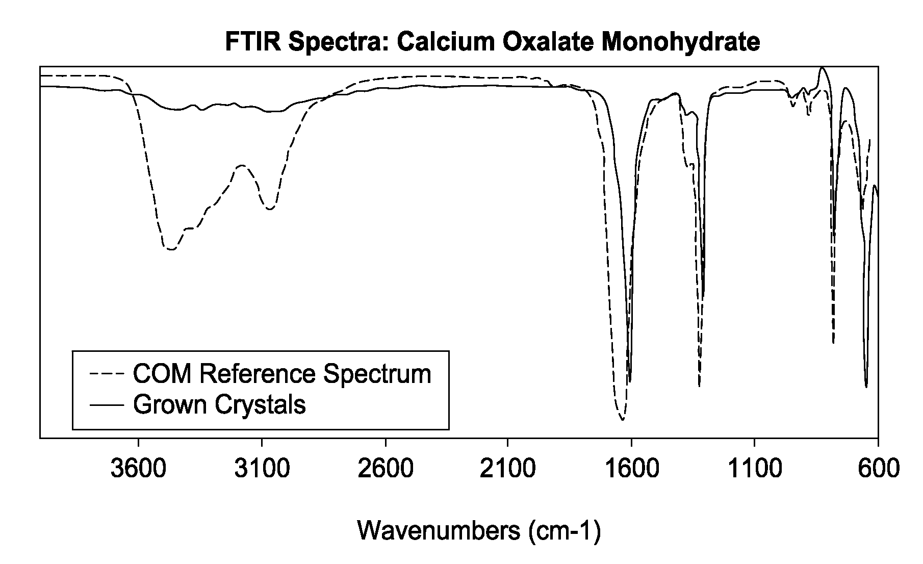
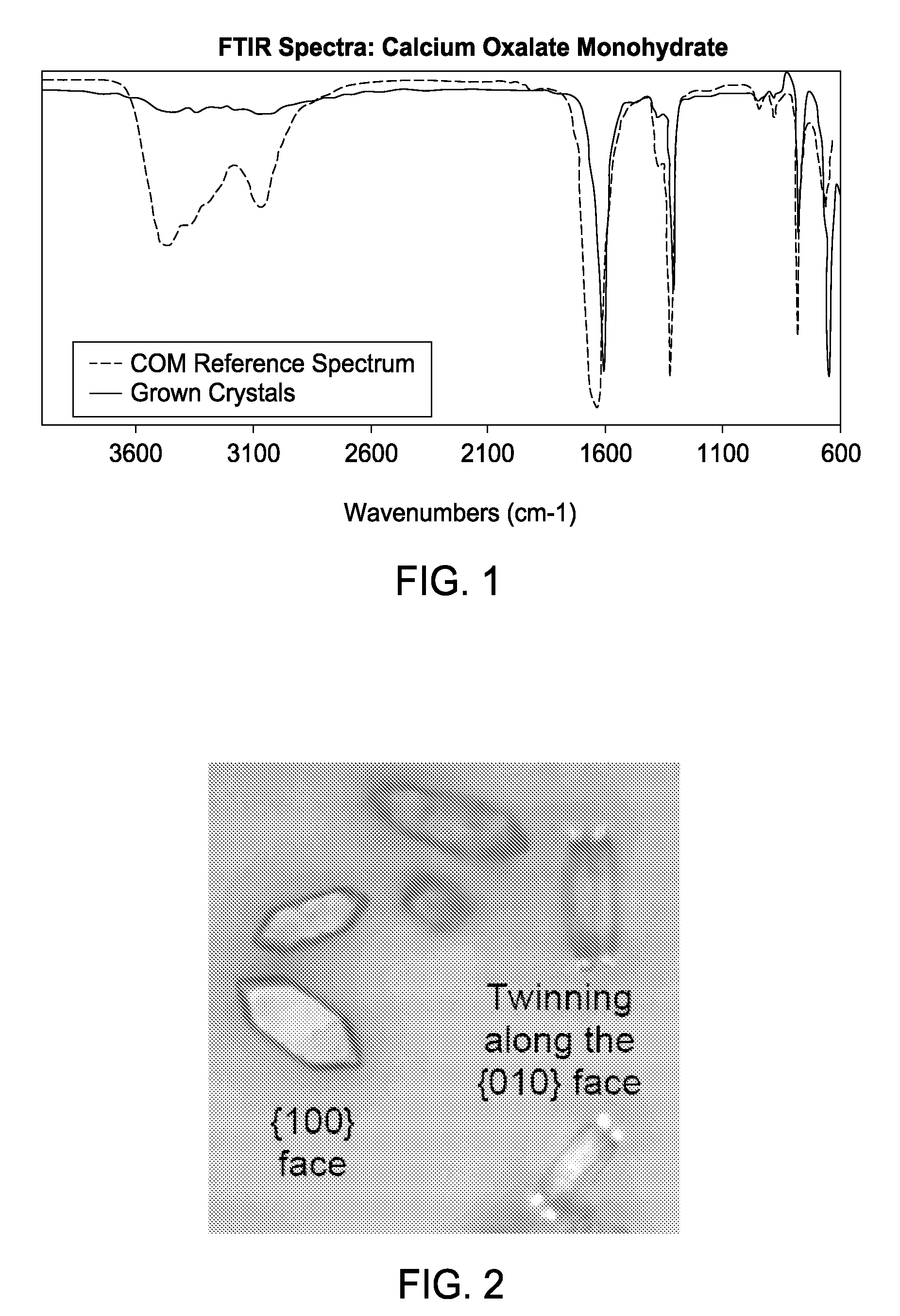
[0057]The results from rendering biomaterials magnetic are shown in FIGS. 1-17. Calcium-based biominerals are the primary component of most pathological biomineralization in humans, including atherosclerosis and kidney stones. The ability to deliver targeted therapeutic agents to a biomineralized surface opens a wide variety of treatment, therapy, and imaging options for doctors and surgeons. The functionality of calcium binding proteins was used to develop the compositions and methods of the present invention to selectively attaching micro- and nano-particles to calcium-rich surfaces such as calcium oxalate. The results show selective attachment of iron oxide micro- and nano-particles to calcium-containing biomineral surfaces, although it is anticipated that a wide variety of particles or therapeutic agents could be functionalized using this technique. The resulting functionalized materials are characterized using Scanning and Transmission Electron Mi...

example 3

Movement of Kidney Stone Fragments by Attachment of Nanocomposite Superparamagnetic Particles

[0068]Large renal or ureteral calculi that are unable to pass spontaneously require endoscopic fragmentation or shock wave lithotripsy (SWL). While endoscopy has the advantage of manual fragment retrieval to improve stone free rates, access to these fragments may be hindered by collecting system anatomy, technical limitations of the endoscope, or poor visibility. Furthermore, retrieving the entire burden of stone fragments, while desirable, is time-consuming. The ability to attract and move these fragments en masse into an easily accessible area of the collecting system could facilitate endoscopic stone surgery as well as diminish costs by reducing time and obviating the need for secondary procedures. Novel nanocomposite paramagnetic particles were developed that selectively adhere to the calcium oxalate crystalline structure of a stone such that introduction of a magnetized instrument and p...


PUM


Abstract

The present invention includes compositions, methods, devices and kits for magnetizing a biological particle by contacting a biological particle with a ferrous or magnetic particle that is able to specifically bind the biological particle and reacting the biological particle with the ferrous or magnetic particle under physiological conditions, wherein the ferrous or magnetic particle causes the biological particle to become attractable magnetically.

Description

CROSS-REFERENCE TO RELATED APPLICATIONS[0001]This application claims priority to U.S. Provisional Application Ser. No. 60 / 990,602, filed Nov. 27, 2007, to U.S. Provisional Application Ser. No. 61 / 052,452, filed May 12, 2008, and to U.S. Provisional Application Ser. No. 61 / 102,154, filed Oct. 2, 2008, the contents of each of which is incorporated by reference herein in its entirety.TECHNICAL FIELD OF THE INVENTION[0002]The present invention relates in general to the field of biomaterials, and more particularly, to compositions, methods, tools and kits for the functionalization of biomaterials and their selective capture under physiologic conditions.STATEMENT OF FEDERALLY FUNDED RESEARCH[0003]None.INCORPORATION-BY-REFERENCE OF MATERIALS FILED ON COMPACT DISC[0004]None.BACKGROUND OF THE INVENTION[0005]Without limiting the scope of the invention, its background is described in connection with the functionalization of biomaterials.[0006]Kidney stone disease is a major health problem affe...

Claims


Application Information

Patent Timeline
Patent Type & Authority Applications(United States)
IPC IPC(8): A61K33/26A61M29/00A61B17/28A61B19/00A61B6/00
CPCA61B17/221A61B2017/00876A61B2019/2253A61K33/26A61K45/06A61K2300/00A61B34/73
Inventor MCLEROY, STACEY L.GNADE, BRUCE E.CADEDDU, JEFFREY A.PEARLE, MARGARET
Owner BOARD OF RGT THE UNIV OF TEXAS SYST
Why Eureka
Try Eureka