
Method and agent for in-situ stabilization of vascular tissue

a technology of vascular tissue and in-situ stabilization, which is applied in the directions of biocide, plant/algae/fungi/lichens, prosthesis, etc., can solve the problems of insufficient oxygen for myocardial muscle cells, inability to withstand high hemodynamic load, and inability to provide adequate oxygen to patients, etc., to achieve normal endothelium function, promote healing, and reduce thrombosis

Inactive Publication Date: 2009-06-18
ENDOLOGIX LLC
View PDF53 Cites 19 Cited by
  • Summary
  • Abstract
  • Description
  • Claims
  • Application Information

AI Technical Summary

Benefits of technology

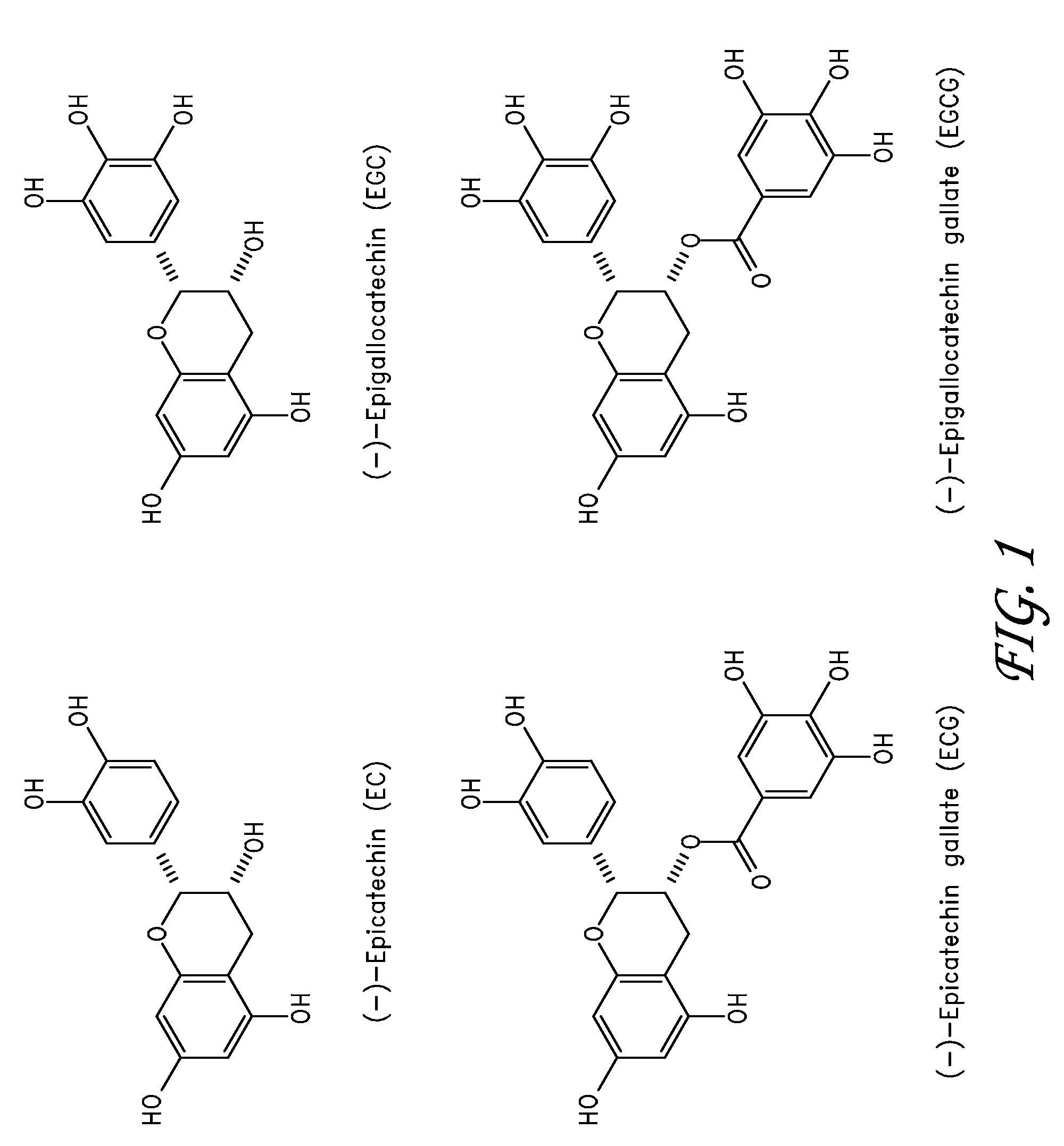
[0015]Catechins can be applied to local vessel injuries to promote healing, restore normal function of the endothelium, reduce thrombosis, stabilize the extra-cellular matrix via cross-linking and inhibition of enzymatic degradation, reduce inflammation, and inhibit smooth muscle cell proliferation. Injuries to the vessel wall can be caused by atherosclerosis, vulnerable plaque, angioplasty, stent placement, atherectomy, surgical anestomosis, and endovascular devices. It will be obvious to one of ordinary skill in the art that catechin can be used for treating a wide range of local vessel injuries or diseases.
[0026]Other embodiments comprise improving the biocompatibility of a medical device, specifically ePTFE vascular graft, by loading the graft material with an anti-thrombogenic and / or anti-hyperplastic agent. ePTFE grafts consist of a matrix of PTFE fibers and nodes. The density of the material and degree of porosity can be controlled during the manufacturing process. In some embodiments, the open space in the ePTFE structure can be used to store a therapeutic agent that modifies the surface kinetics of the ePTFE to prevent or reduce thrombosis. In another embodiment, the open space in the ePTFE structure can be used to store a therapeutic agent that can be released into the adjacent tissue after implantation to reduce smooth muscle cell proliferation and hyperplasia of the blood vessel, or aneurysm growth.
[0027]Additionally, in some embodiments, the agent can also be applied in conjunction with traditional endovascular stent grafts in the treatment of aortic aneurysms. For example, the distal and proximal seal area of the stent graft can be stabilized by applying the agent to the adjacent tissue to prevent dilation of the aorta in the seal zone.

Problems solved by technology

In cases of severe occlusion and high cardiac workload, myocardial muscle cells do not receive sufficient oxygen and die.
Despite the effectiveness of these procedures in treating stenotic lesions, patients still suffer from sudden cardiac events even in the absence of stenotic lesions.
The cap can become too weak to withstand high hemodynamic loads and can ultimately rupture, exposing the highly thrombogenic content of the plaque to the bloodstream.
Thrombi can form rapidly and can cause partial or complete occlusion of the blood vessel.
Vulnerable plaque is difficult to research because current imaging systems are not capable of detecting the plaque in the vessel wall.
Therefore, the investigation into vulnerable plaque has been limited to the biophysical and biochemical analysis of cadavers and retrospective studies of patients who have suffered a sudden cardiac event.
One major shortcoming of stenting is the need for several stents in cases of multiple or diffuse lesions and the high cost of drug-eluding stents.
Degradation of the collagen structure can ultimately lead to aortic rupture and potential patient death.
Additionally, one of the primary failure modes of small-diameter surgical grafts are thrombosis of the lumen and stenosis due to hyperplasia at the anastomosis site.
It is believed that thrombosis is due to the limited hemocompatibility of the graft material in high-shear flows.
The design of the catheter is well suited for the continuous infusion of a therapeutic agent, but lacks control of delivery of a single dose of a therapeutic agent into the vessel wall.

Method used


Image

  • Method and agent for in-situ stabilization of vascular tissue
  • Method and agent for in-situ stabilization of vascular tissue
  • Method and agent for in-situ stabilization of vascular tissue

Examples


experiment 1

[0070]

[0071]A 7 cm×7 cm piece of pericardial tissue was fixed in 40 ml of 0.5% EGCG / phosphate buffered saline (PBS), pH 7.42 in a 100 mm petri dish at 37° C., 82 rpm shaking speed, for 15 min. After 15 minutes, the tissue was quickly rinsed with PBS. Three samples were cut out for immediate shrinkage temperature testing (Group A). Shrinkage temperature can be an indication of the stability of the tissue. Increased shrinkage temperature is associated with an increased resistance to degradation. An additional 7 cm×7 cm piece of pericardial tissue was fixed in 40 ml of 0.5% EGCG / PBS, pH 7.42 in a 100 mm petri dish at 37° C. for 48 hours and quickly rinsed in PBS for 15 minutes. Six samples were cut out for shrinkage temperature testing (Group B). Six samples of fresh pericardial tissue were also cut for shrinkage temperature testing (Group C—Control Group).

[0072]Observation:

[0073]1. Group A samples looked light pink (from opaque white) and more rigid than fresh tissue but still soft. T...

embodiment 74

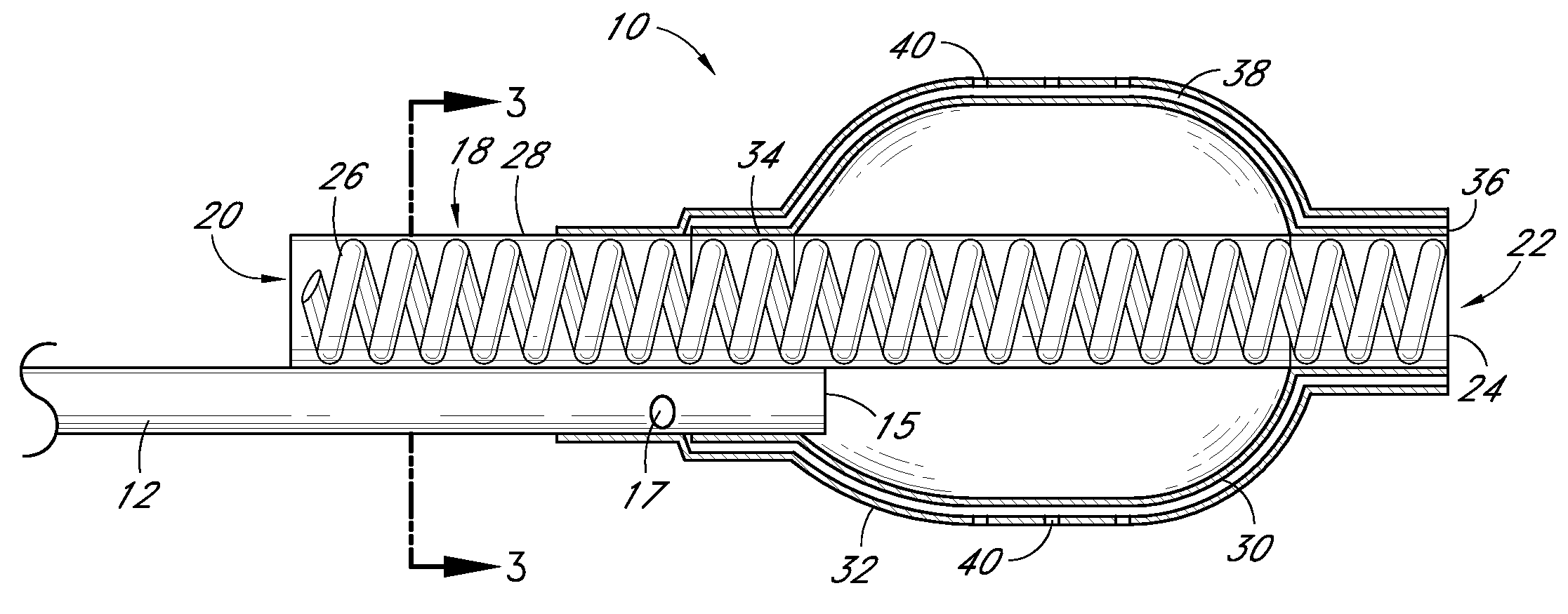
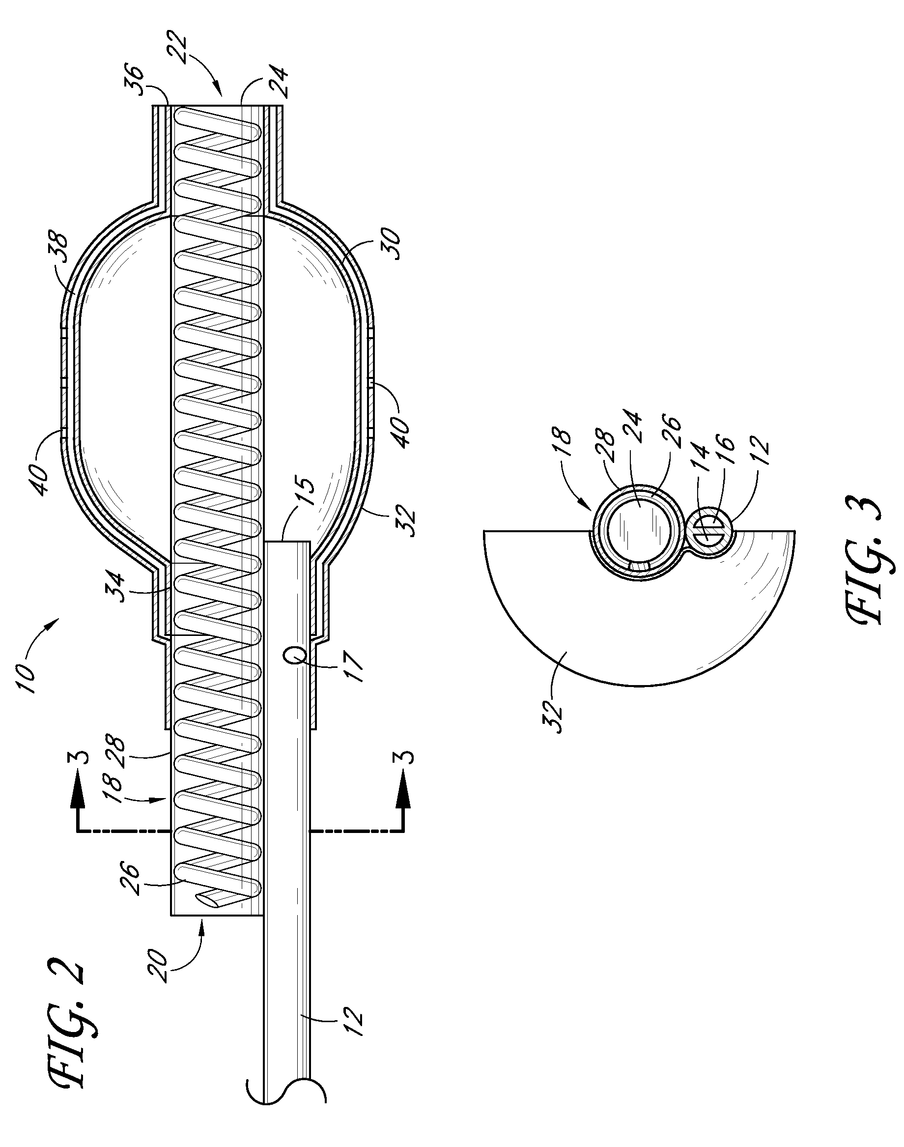
[0113]FIG. 7 is a partial sectional side view of a non-stent arrangement of a catheter. FIG. 8 is a cross-sectional view taken along the lines 8-8 in FIG. 7. FIG. 9 is a cross-sectional view taken along the lines 9-9 in FIG. 7. FIG. 10 is a cross-sectional view taken along the lines 10-10 in FIG. 7. FIG. 11 is a side view of a non-stent arrangement in communication with a fluid delivery and guide-wire entry apparatus. Referring to FIGS. 7-11, there is disclosed a nonperfusion catheter embodiment 74 which, in some embodiments, also does not include a temporary stent. The non-perfusion embodiment 74 can be designed for use in percutaneous coronary transluminal angioplasty and adjunctive site specific intraluminal infusion of pharmacological agents.

[0114]The non-perfusion embodiment 74 can comprise a tubular body 12 which includes an inflation lumen 14, a drug delivery lumen 16, and a guidewire lumen 52. Two concentric balloons, an inner inflation balloon 30, and an outer delivery ball...


PUM

PropertyMeasurementUnit
Fractionaaaaaaaaaa
Fractionaaaaaaaaaa
Fractionaaaaaaaaaa

Abstract

A method for stabilizing an extra cellular matrix layer in the vascular system of the body is disclosed herein. The method can comprise placing a vascular catheter adjacent to the extra cellular matrix layer, delivering a solution containing a bioflavonoid to the extra cellular matrix layer with the vascular catheter, and cross-linking protein in the extra cellular matrix layer. The bioflavonoid can be a catechin, particularly epigallocatechin gallate (EGCG).

Description

PRIORITY INFORMATION[0001]This application also claims priority benefit under 35 U.S.C. § 119(e) of Provisional Application 60 / 987,268 filed Nov. 12, 2007, Provisional Application 60 / 987,261 filed Nov. 12, 2007, Provisional Application 61 / 012,356 filed Dec. 7, 2007, Provisional Application 61 / 127,654 filed May 14, 2008, and Provisional Application 61 / 012,579 filed Dec. 10, 2007, which applications are hereby incorporated by reference as if fully set forth herein.BACKGROUND OF THE INVENTION[0002]1. Field of the Disclosure[0003]The present disclosure relates to therapeutic agents and delivery methods for in-situ stabilization of vascular tissue[0004]2. Background of the Disclosure[0005]Cardiovascular disease is one of the leading causes of death in the developed countries. It is estimated that more than one million people in the United States suffer from a sudden cardiac event each year. For a long time, coronary artery occlusions have been believed to be the main cause of sudden card...

Claims


Application Information

Patent Timeline
IPC IPC(8): A61F2/00A61K31/352A61K33/06
CPCA61K36/00
Inventor SCHRECK, STEFAN G.SCHANKERELI, KEMAL
Owner ENDOLOGIX LLC
Learn More