Nanostructures, methods of synthesizing thereof, and methods of use thereof

a nanostructure and nanoparticle technology, applied in the field of nanostructures, can solve the problems of insufficient concentration of non-tumor targeted nanoparticles at tumor sites, limiting clinical application, and mortality in breast cancer patients

Active Publication Date: 2009-08-06
EMORY UNIVERSITY
View PDF5 Cites 34 Cited by
  • Summary
  • Abstract
  • Description
  • Claims
  • Application Information

AI Technical Summary

Problems solved by technology

Although the feasibility of using nanoparticles for cancer detection and drug delivery has been demonstrated in several laboratories [1-3], a major obstacle limiting its clinical application is that non-tumor targeted nanoparticles are unable to reach sufficient concentration in the tumor site to either produce a strong signal for tumor imaging or to carry optimal amounts of therapeutic agents into tumor cells.
Furthermore, since cancer metastasis is the main cause for the mortality in breast cancer patients.
Although various imaging technologies and modalities have been widely used for management of cancer including diagnosis and treatment monitoring, conventional tumor imaging methods such as magnetic resonance imaging (MRI), X-ray computerized tomography (CT) or even positron emission tomography (PET) have their limitations in both specificity and sensitivity of cancer detection [4-6].
Although different modalities vary in imaging sensitivity and resolution, the technical challenge in improving target specificity and sensitivity is common.
However, both have significant limitations in sensitivity and specificity in delineating tumor and detecting cancer cells in the early stage of development of tumor [5, 44, 45].
However, to develop a promising tumor imaging approach to clinical applications, several important issues have to be addressed in the research laboratory.
However, several obstacles remain to be overcome.
The major challenge is to develop a surface coating material that not only can stabilize the nanoparticles but also to provide active functional groups available for controllable bioconjugation of “probe” ligands.
Since further derivatization is needed, such a weak interaction between ligand and particle may not withstand the required reaction conditions.
Therefore, a heretofore-unaddressed need exists in the art to address the aforementioned deficiencies and inadequacies.

Method used


Image

  • Nanostructures, methods of synthesizing thereof, and methods of use thereof
  • Nanostructures, methods of synthesizing thereof, and methods of use thereof
  • Nanostructures, methods of synthesizing thereof, and methods of use thereof

Examples


example 1

Receptor-Targeted Nanoparticle for In Vivo Targeting and Non Invasive Imaging of Cancer

Experimental Methods

[0090]Breast Cancer Cell Lines: Mouse mammary carcinoma cell line 4T1B was provided by the Barbara Ann Karmanos Cancer Institute, Detroit, Mich. 4T1 cell line stable expressing a firefly luciferase gene was obtained by Duke University, Durham, N.C. Human breast cancer cell line T47D was purchased from American Type Culture Collection (ATCC, Rockville, Md.). 4T1 cells were grown in Dulbecco's modified Eagle's medium containing 10% fetal bovine serum (FBS). T47D cells were grown in RPMI 1640 containing 10% FBS, 100 ug / ml gentamicin, and 0.2 IU insulin / ml.

[0091]Engineering A TF-IO Nanoparticles: A cDNA fragment encoding amino acids 1-135 of mouse uPA, isolated by PCR amplification using a PCR primer pair containing forward (5′-CACCATGGGCAGTGTACTTGGAGCTCC-3′) and reverse (5′-GCTAAGAGAGCAGTCA-3′) primers, was cloned into pETIO 1 / D-TOPO expression vector (Invitrogen, Carlsbad, Calif....

example 2

Peptide Conjugated IO Nanoparticles for Targeted MR Imaging and Therapy of Pancreatic Cancer

Experimental Methods

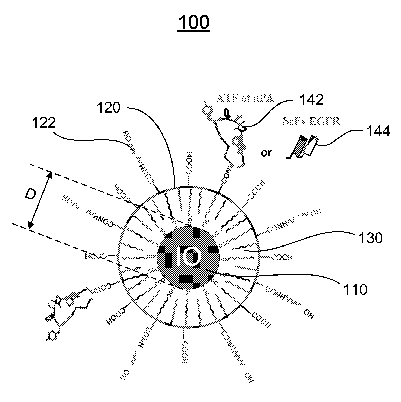
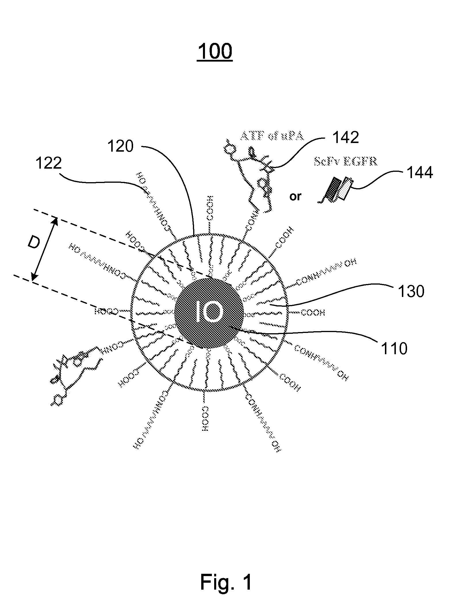
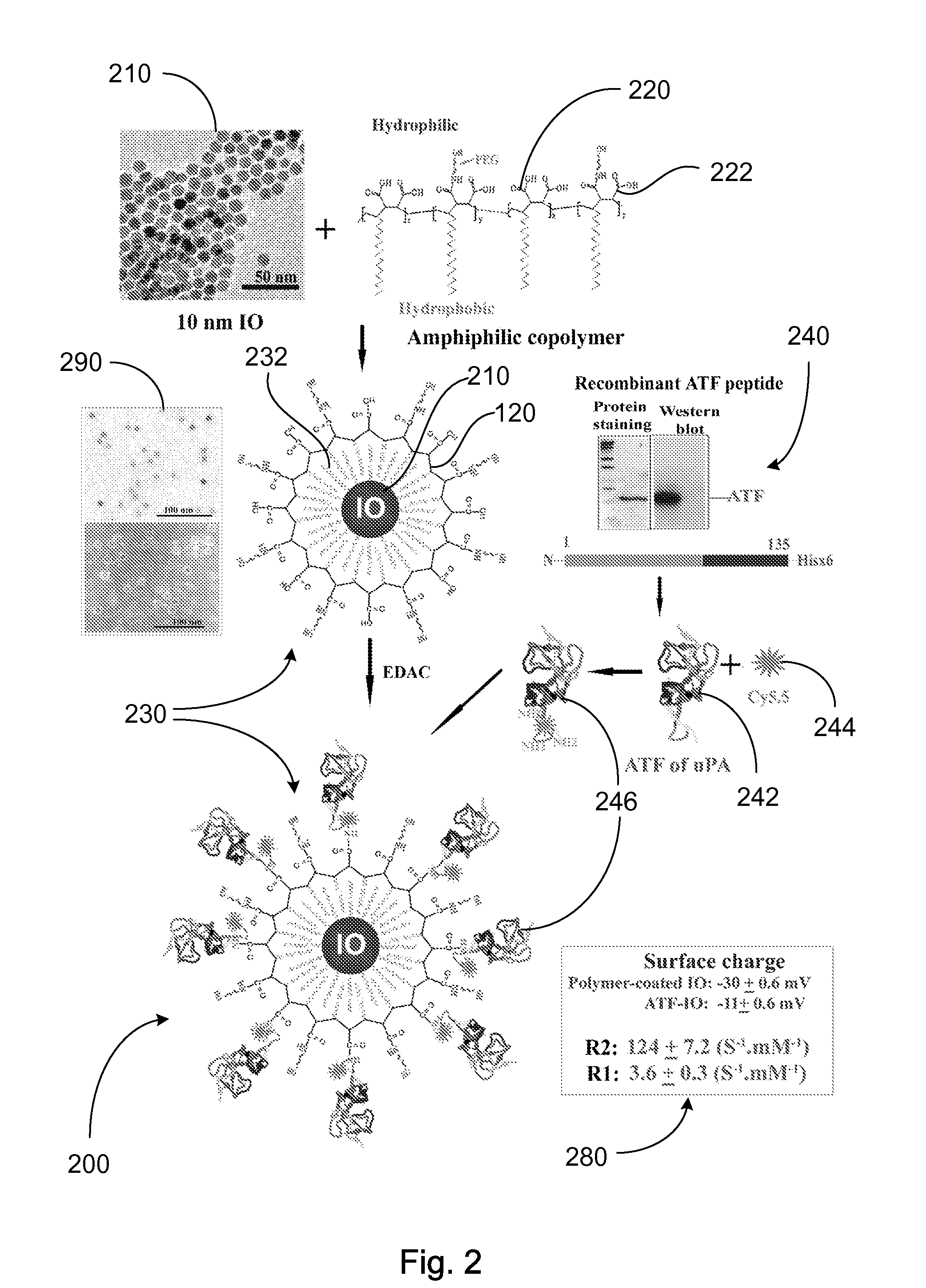
[0120]Preparation of IO Nanoparticles with Functionalized Surface: IO was prepared by heating iron oxide powder and oleic acid in octadecene over 315° C. The size of IO was tuned through changes such as heating time, temperature, and concentration of the iron oxide and oleic acid [14]. IO nanoparticles with highly uniformed core sizes of 9 or 10 nm were used for this example. Amphiphilic polymers were coated to the surface to convert hydrophobic IO nanoparticles to stable, water soluble and biocompatible nanoparticles. The hydrocarbon chains of the polymer intercalate into the inner hydrophobic layer that stabilize IO nanoparticle surface while carboxylic acid groups in the out-layer make the IO nanoparticles hydrophilic and reactive for conjugating proteins, peptides or small molecules.

[0121]Engineering Tumor Targeted-IO Nanoparticles: A 135-amino acids of the ATF of mous...


PUM

PropertyMeasurementUnit
core sizeaaaaaaaaaa
temperatureaaaaaaaaaa
sizeaaaaaaaaaa

Abstract

A nanostructure and methods of synthesizing same. In one embodiment, the nanostructure includes a magnetic iron oxide nanoparticle, a hydrophobic protection structure including at least an amphiphilic copolymer, wherein the hydrophobic protection structure encapsulates the magnetic iron oxide nanoparticle, and at least one amino-terminal fragment (ATF) peptide or epidermal growth factor receptor (EGFR) antibody conjugated to the amphiphilic copolymer.

Description

CROSS-REFERENCE TO RELATED PATENT APPLICATION[0001]This application claims the benefit, pursuant to 35 U.S.C. §119(e), of U.S. provisional patent application Ser. No. 60 / 797,613, filed May 4, 2006, entitled “NANOSTRUCTURES, METHODS OF SYNTHESIZING THEREOF, AND METHODS OF USE THEREOF,” by Lily Yang et al., which is incorporated herein by reference in its entirety.[0002]This application is related to a copending U.S. patent application entitled “BIOCONJUGATED NANOSTRUCTURES, METHODS OF FABRICATION THEREOF, AND METHODS OF USE THEREOF”, U.S. patent application Ser. No. 10 / 988,923, filed Nov. 15, 2004 with the same assignee as the present invention. The two applicants of the above identified copending applications are also applicants of this application. The disclosure of the above identified copending application is incorporated herein by reference.[0003]This application is also related to a copending PCT international patent application entitled “MULTIFUNCTIONAL NANOSTRUCTURES, METHODS...

Claims


Application Information

Patent Timeline
Patent Type & Authority Applications(United States)
IPC IPC(8): A61K49/00C07K16/18A61K39/395
CPCA61K49/0032A61K49/0034A61K49/0056A61K49/0058A61K49/0093C07K2317/77A61K2039/505B82Y5/00C07K16/2863C07K2317/622A61K49/1866
Inventor YANG, LILYMAO, HUINIE, SHUMINGGAO, XIAOHU
Owner EMORY UNIVERSITY
Why Eureka
Try Eureka