
Multimodal imaging using a three compartment polymer nanoparticle with cell specificity

Inactive Publication Date: 2009-09-03
TOPASS
View PDF4 Cites 4 Cited by
  • Summary
  • Abstract
  • Description
  • Claims
  • Application Information

AI Technical Summary

Benefits of technology

[0008]One obvious advantage in using nanoparticles is to obtain a higher surface to mass ratio of the carrier system permitting relatively high local concentrations of the delivered substances [5]. Colloids also have particular physicochemical properties affecting their pharmacokinetics and in vivo compartmentalization [6]. Unlike soluble substances which generally disseminate from the site of injection and are rapidly cleared by the renal system nanoparticles are usually confined to a particular compartment over a longer period. In addition, chemical functionalization will help to overcome biological barriers or to take advantage of specific transport and degradation processes in order to control the fate of the applied material [7,8].
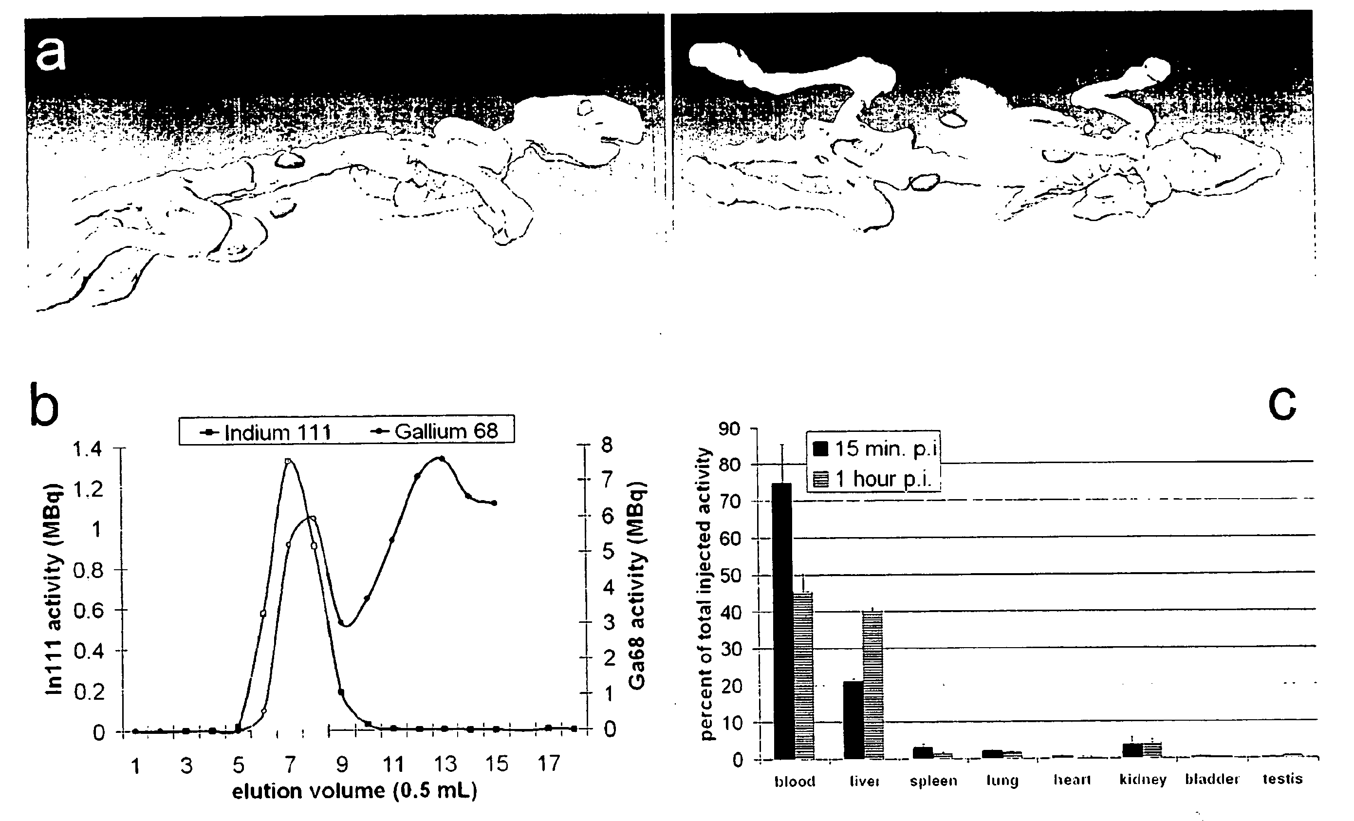
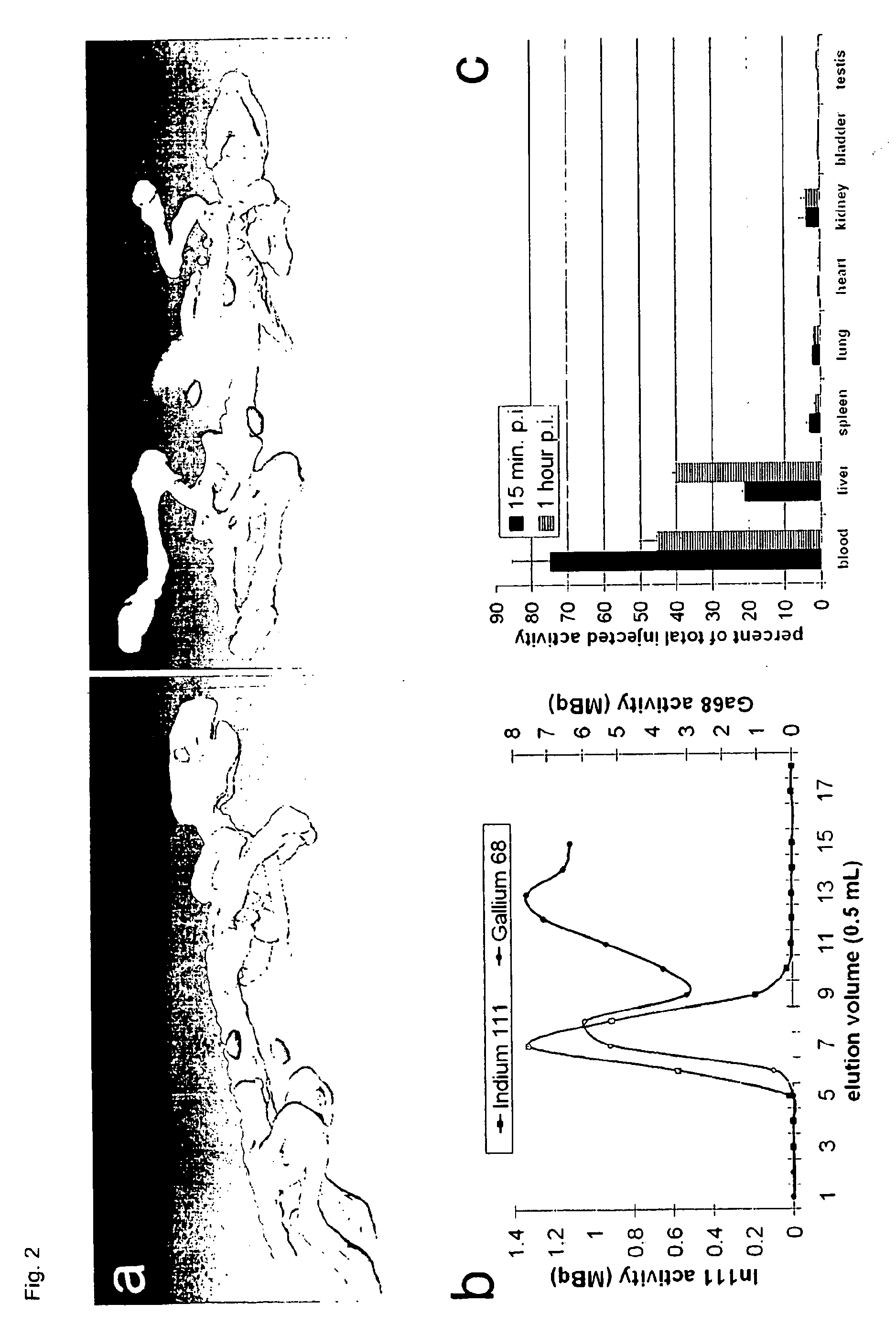
[0082]A characteristic of the three compartment polymer nanoparticles is the possibility to bind different tracers which can be used in different modalities of imaging and detection. To our knowledge, PET with labelled nanoparticles has only been reported with 64Cu for which the sources of supply remain limited [6,13]. In contrast, the polymer nanoparticles enable PET with the clinically well established radiotracer 68Ga. PET is a highly sensitive and non-invasive method allowing for a rapid whole body scan. On the other hand, MRI has the advantage of a higher temporal and spatial resolution and the acquisition of more precise anatomical information [33].

Problems solved by technology

Furthermore, half-life-time of the particle inside the body needs to be long enough to meet diagnostic and / or therapeutic purposes, but as short as possible to keep toxicological burden low.
In both cases, the descript particles need additional bioreactive or immunoreactive molecules to be cell specific and could not be designed for multimodal imaging due to limited structure-function capabilities.
However, the prior art fails to teach the synthesis of polymer particles for multimodal imaging with specificity for cells or cellular components without the use of additional biologically active materials.
To avoid biologically active material such as foreign antibodies is demanding but also of utmost importance in this regard, since the use of biologically active material in patents is risky.

Method used


Image

  • Multimodal imaging using a three compartment polymer nanoparticle with cell specificity
  • Multimodal imaging using a three compartment polymer nanoparticle with cell specificity
  • Multimodal imaging using a three compartment polymer nanoparticle with cell specificity

Examples


Embodiment Construction

[0007]Nanoparticle technology is a rapidly emerging field and advanced to clinical applications within the last few years [1-3]. High expectations have been raised for the development of novel high-resolution diagnostics and drug nanocarriers for more efficacious and personalized therapies [4]. The scientific ground was set through our increasing understanding of disease at the molecular level and recent developments in material sciences enabling the production of nanostructured and biocompatible materials.

[0008]One obvious advantage in using nanoparticles is to obtain a higher surface to mass ratio of the carrier system permitting relatively high local concentrations of the delivered substances [5]. Colloids also have particular physicochemical properties affecting their pharmacokinetics and in vivo compartmentalization [6]. Unlike soluble substances which generally disseminate from the site of injection and are rapidly cleared by the renal system nanoparticles are usually confined...


PUM


Abstract

The present invention pertains to a three compartment structured polymer nanoparticle (core-shell-corona) for multimodal imaging with specificity for cells or cellular components, thus enabling more advanced diagnostic approaches and targeted therapy on the cellular level without the use of additional biologically active materials.

Description

[0001]The present invention pertains to specifically designed polymer nanoparticles with image enhancing properties and cell specificity. These two basic functional properties of polymer particles became possible through controlled monomer addition during polymer particle growth to build a three compartment (core-shell-corona) structure. The outer layer of the particle, the corona, is designed to bind, e.g., CD42a positive cells (thrombocytes) or alternatively CD14 positive cells (monocytes). The intermediate and the inner layer, the hairy shell and the solid core, contain appropriate image enhancing compounds with the following differential localisation to allow multimodal imaging procedures: fluorophores and iron oxide crystals are located in the solid core; gadolinium derivatives, iodine salts and radioisotopes are located in the hairy shell. The resulting polymer nanoparticles may thus be used for imaging procedures including magnetic resonance imaging (MRI) or magnetic particle...

Claims


Application Information

Patent Timeline
IPC IPC(8): A61K51/00A61K49/00A61K49/06A61K49/04
CPCA61K49/0002A61K49/0041A61K49/0093A61K49/0404B82Y5/00A61K49/0485A61K49/1878A61K51/1255A61K49/0423
Inventor PISON, ULRICHPAULKE, BERND-REINERPIETSCHMANN, SILVIACARTIER, REGISKAUFNER, LUTZ
Owner TOPASS
Learn More