

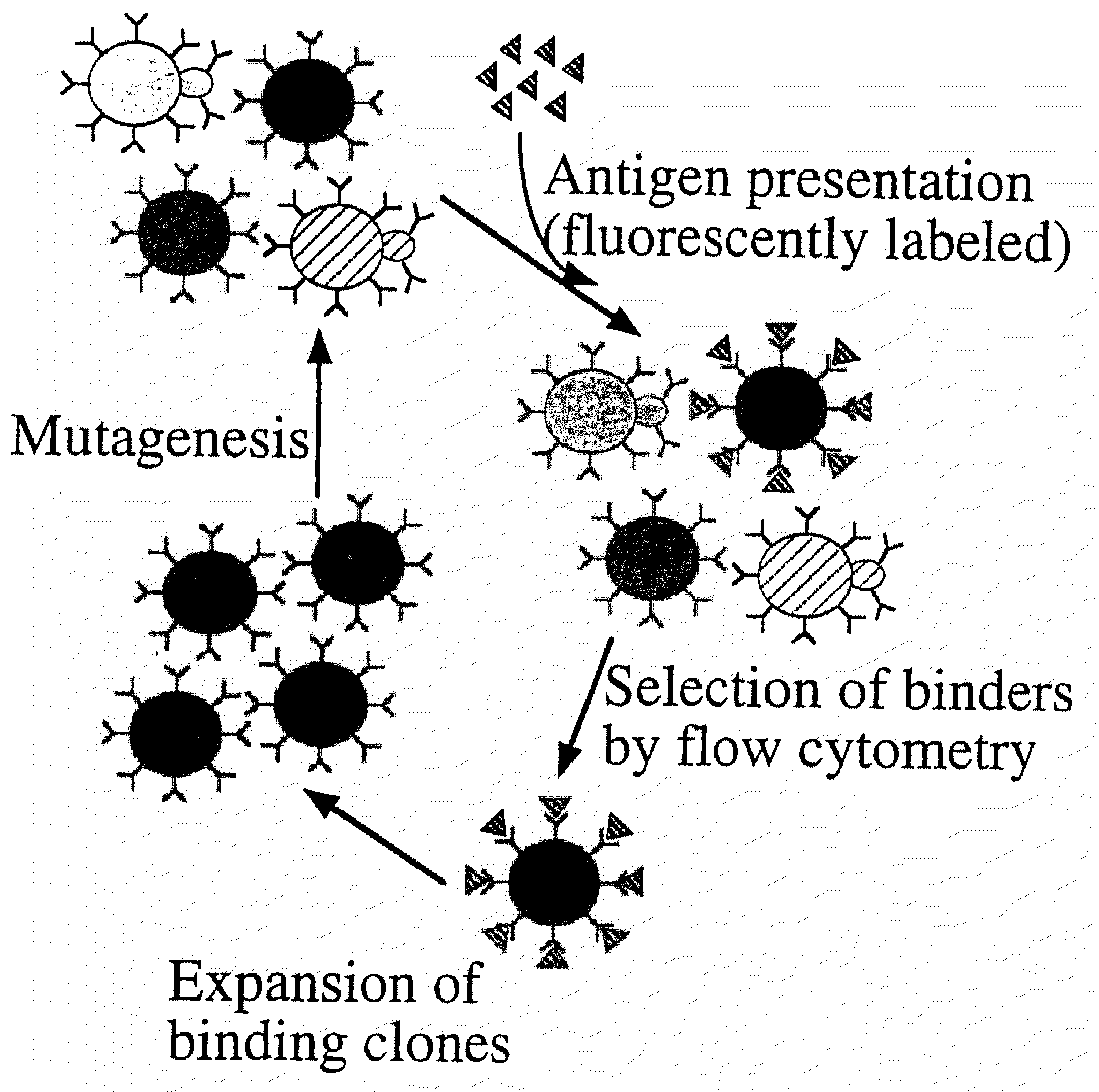
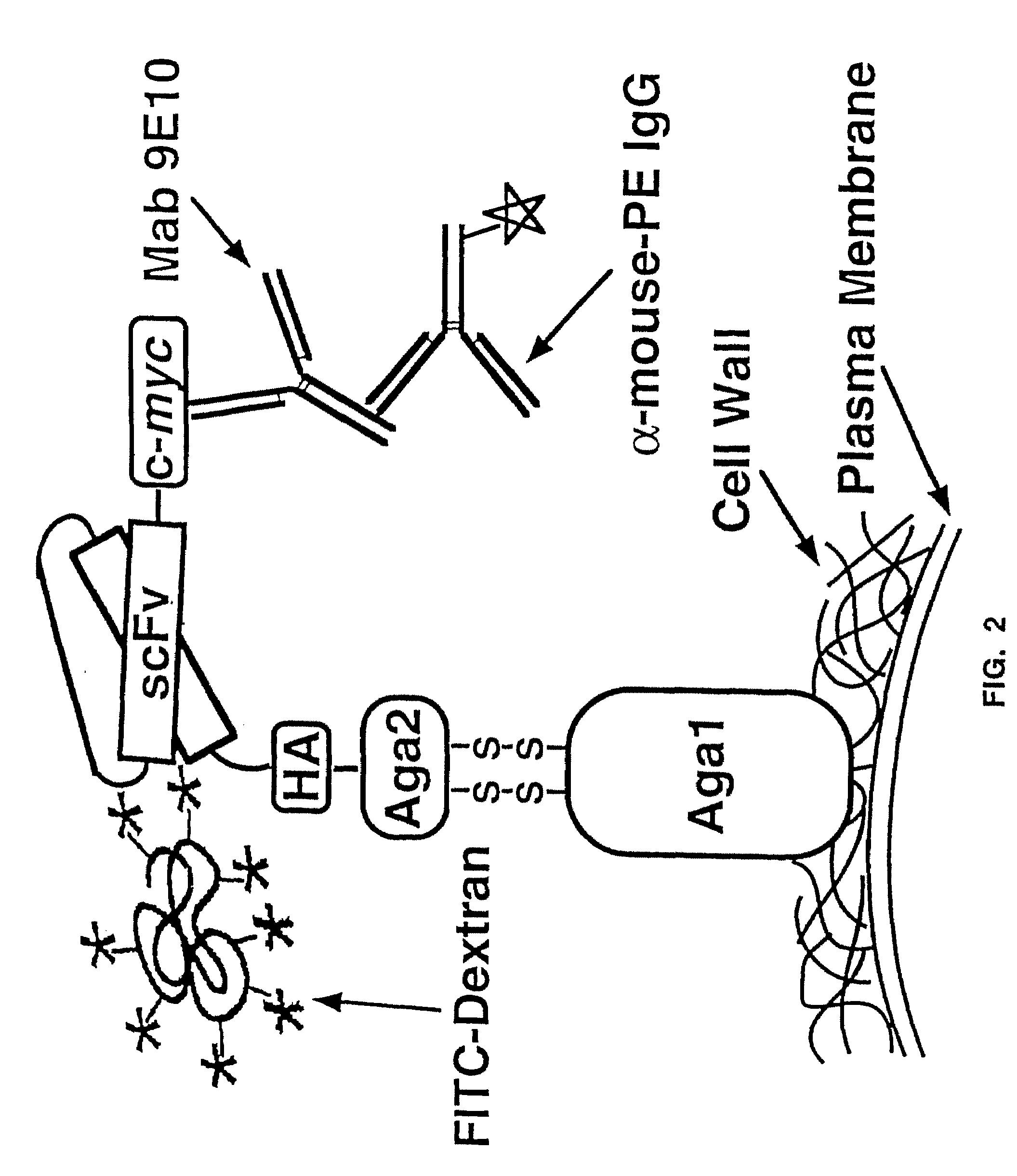
Yeast cell surface display of proteins and uses thereof
a technology of protein and cell surface, applied in the field of immunology and protein chemistry, can solve the problems of limited wide-spread success of antigen application, difficult panning of high affinity antibodies (ksub>d/sub>1 nm), and many artifactual difficulties of immobilizing antigens to solid surfaces, so as to enhance the level of cell surface expression increase the level of t cell receptors
- Summary
- Abstract
- Description
- Claims
- Application Information
AI Technical Summary
Benefits of technology
Problems solved by technology
Method used
Image

Examples
example 1
Media / Buffers
[0094]The following media / buffers were used herein:
E. coli
LB media (1×): Bacto tryptone (Difco, Detroit, Mich.): 10.0 g; Bacto yeast extract (Difco): 5.0 g; NaCl:10.0 g. Make up to 1 L, autoclave. For plates, add 15 μL Agar and autoclave.
[0095]Stock: 25 mg / ml of sodium salt of ampicillin in water. Filter sterilize and store in aliquots of 4 mls at −20° C. Working [ ]=35-50 μg / ml; 4 ml of aliquot in 1 L->100 μg / ml; 2 ml of aliquot in 1 L->50 μg / ml. Add to autoclaved LB only after it has cooled to ˜55° C.
SOC media 100 mL
[0096]2% Bacto tryptone: 2.0 g; 0.5% Yeast Extract (Difco):0.5 g; 10 mM NaCl: 0.2 ml 5 M; 10 mM MgCl9: 1.0 ml 1 M; 10 mM MgSO4: 1.0 ml 1 M; 20 mM Dextrose: 0.36 g. Autoclave or filter sterilize.
[0097]Synthetic Minimal+Casamino acids (SD-CAA) 500 mL
Dextrose(Glucose)10.00gYeast Nitrogen Base w / o Amino Acids (Difco)3.35gNa2HPO4•7H2O5.1gNaH2PO4•H2O4.28gCasamino Acids (Trp-, Ura-) (Difco)2.5g
[0098]Add dH2O to final volume. Filter sterilize and r...
example 2
Protocol
Replica Plating
[0103]1. Choose a material suitable for colony lifts, making sure it is washed, dried and sterile.
[0104]2. Mark the bottom of each fresh replica plate with an arrow to line up the plate. Also mark the starting plate.
[0105]3. Take the top off the starting plate, turn it upside down and line up the arrow on the bottom with the mark on the colony lift material. Lay the plate down onto the surface of the material and gently put pressure on the entire plate. Make sure the plate doesn't move around after it has touched the material. Remove the plate and replace the lid. Portions of the colonies chat transferred to the material can be seen.
[0106]4. Repeat this procedure with one of the fresh replica plates. Make sure the arrow lines up with the mark also to make an exact replica. Hold up to the light to see the tiny colonies that transferred.
[0107]5. Repeat the entire procedure for each replica plate to be made.
[0108]6. Incubate the replica plates at the appropriate ...
example 3
Protocol
Electrotransformation of Yeast
[0109]The cell preparation procedure was as follows: Step 1. Inoculate 50 ml of YPD with an overnight culture to an OD of 0.1. Step 2. Grow cells at 30° C. with vigorous shaking to an OD600 of 1.3 to 1.5 (approximately 6 hours). Step 3. Harvest in cold rotor at 3500 rpm for 5 minutes at 4° C. Discard supernatant. Step 4. Thoroughly wash the cells by resuspending in 50 ml cold sterile water. Centrifuge as above and discard supernatant. Step 5. Repeat step 4 with 25 ml cold water. Step 6. Resuspend in 2 ml of ice-cold sterile 1 M sorbitol. Centrifuge as above and discard supernatant. Step 7. Resuspend in 50 ml ice-cold 1 M sorbitol. Final volume of cells is about 150 ml (enough for 3 transformations).
Electrotransformation:
[0110]1. Place 0.2 cm cuvettes and white slide chamber on ice.
[0111]2. In an eppendorf tube, add 50 ml of yeast suspension and gently mix in <5 ml (0.1 mg) of plasmid DNA in TE. Make sure to add DNA to yeast already in eppendorf....
PUM
| Property | Measurement | Unit |
|---|---|---|
| OD | aaaaa | aaaaa |
| volume | aaaaa | aaaaa |
| time constant | aaaaa | aaaaa |
Abstract
Description
Claims
Application Information