

SlpA as a tool for recombinant protein and enzyme technology
- Summary
- Abstract
- Description
- Claims
- Application Information
AI Technical Summary
Benefits of technology
Problems solved by technology
Method used
Image

Examples
example 1
Cloning and Purification of SlpA and SlyD Fusion Polypeptides
Cloning of Expression Cassettes
[0108]On the basis of the pET24a expression plasmid of Novagen (Madison, Wis., USA) an expression cassette encoding the SlyD or SlpA fusion polypeptides was obtained. The sequence of the g,p41 ectodomain was retrieved from the SwissProt database. A synthetic gene encoding gp41 (aa 536-681) with a glycine-rich linker region fused in frame to the N-terminus was purchased from Medigenomix (Martinsried, Germany). BamHI and VhoI restriction sites were at the 5′ and the 3′ ends of the coding region, respectively. A further synthetic gene encoding two SlpA units (residues 1-146 and 2-149 according to SEQ ID NO: 1, SwissProt accession no. P0AEM0) connected via a glycine-rich linker region and encompassing part of a further linker region at the C-terminus were likewise purchased from Medigenomix. NdeI and BamHI restriction sites were at the 5′ and 3′ ends of this cassette, respectively. The genes and ...
example 2
Spectroscopic Measurements
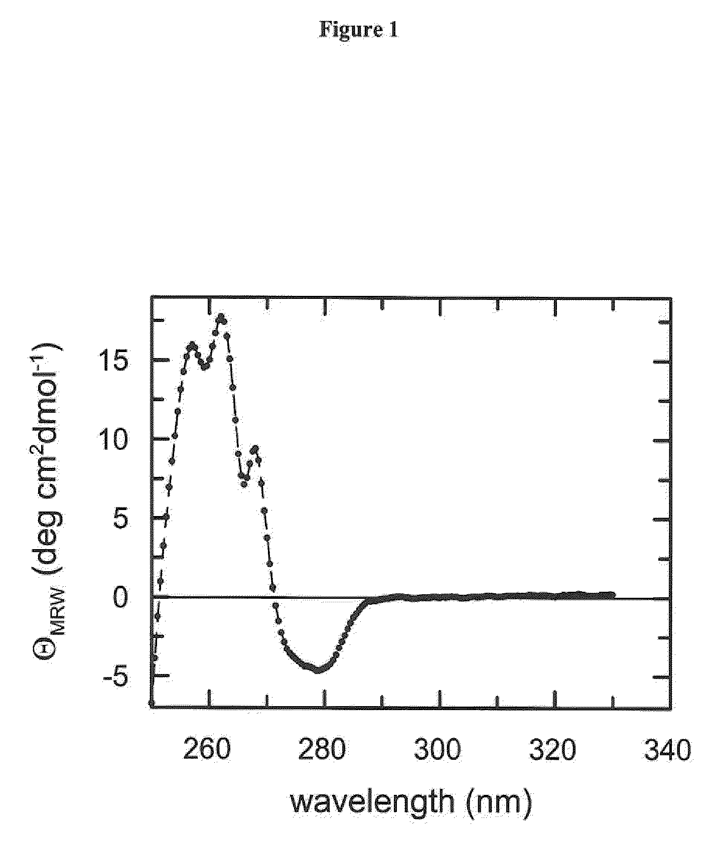
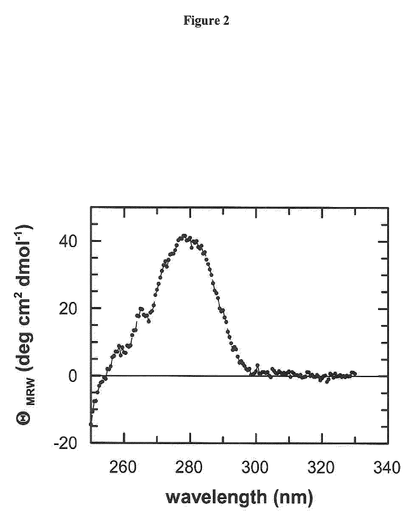
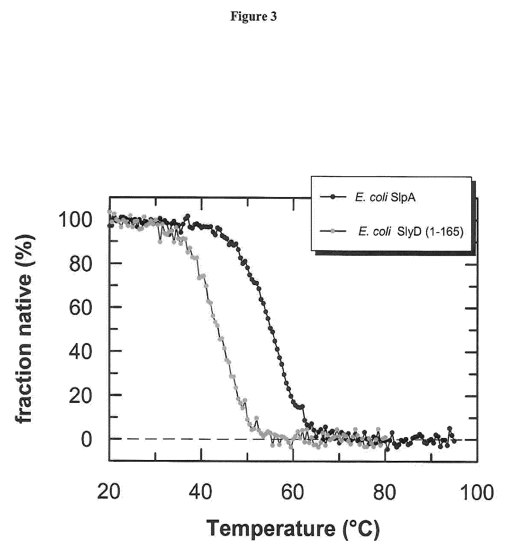
[0114]Circular dichroism spectroscopy (CD) is the method of choice to assess both the secondary and the tertiary structure in proteins. Ellipticity in the aromatic region (260-320 nm) reports on tertiary contacts within a protein (i.e., the globular structure of a regularly folded protein), whereas ellipticity in the amide region (190-250 nm) reflects regular repetitive elements in the protein backbone, i.e., the secondary structure.
[0115]Protein concentration measurements were performed with an Uvikon XL double-beam spectrophotometer. The molar extinction coefficients (E280) were determined by using the procedure described by Pace (1995), Protein Sci. 4, 2411-2423.
[0116]Near-UV CD spectra were recorded with a Jasco-720 spectropolarimeter with a thermostated cell holder and converted to mean residue ellipticity. The buffer was 50-150 mM potassium phosphate pH 7.5, 100 mM KCl, 1 mM EDTA. The path length was 0.5 cm or 1.0 cm, the protein concentration was 20-...
example 3
Coupling of Biotin and Ruthenium Moieties to the Fusion Proteins
[0118]The lysine E-amino groups of the fusion polypeptides were modified at protein concentrations of 10-20 mg / ml with N-hydroxy-succinimide activated biotin and ruthenium labels, respectively. The label / protein ratio varied from 2:1 to 5:1 (mol:mol), depending on the respective fusion protein. The reaction buffer was 150 mM potassium phosphate pH 8.0, 100 mM KCl, 1 mM EDTA. The reaction was carried out at room temperature for 15 min and stopped by adding buffered L-lysine to a final concentration of 10 mM.
[0119]To avoid hydrolytic inactivation of the labels, the respective stock solutions were prepared in dried DMSO (seccosolv quality, Merck, Germany). DMSO concentrations up to 15% in the reaction buffer were well tolerated by all fusion proteins studied. After the coupling reaction, unreacted free label was removed by passing the crude protein conjugate over a gel filtration column (SUPERDEX 200 HiLoad).
PUM
| Property | Measurement | Unit |
|---|---|---|
| Solubility (mass) | aaaaa | aaaaa |
Abstract
Description
Claims
Application Information