Methods and Compositions for Treating Mammalian Nerve Tissue Injuries

a nerve tissue and nerve tissue technology, applied in the field of methods for treating injured mammalian nerve tissue, can solve the problems of functional deficits, functional deficits, and blockage of nerve impulse traffic, and achieve the effect of rapid recovery of recorded nerve impulses and loss of effectiveness

Inactive Publication Date: 2010-01-21
PURDUE RES FOUND INC
View PDF20 Cites 7 Cited by
  • Summary
  • Abstract
  • Description
  • Claims
  • Application Information

AI Technical Summary

Benefits of technology

[0015]This is a dramatic and unexpected finding. A single dose of a biomembrane fusion agent such as PEG in aqueous solution administered beneath the back skin (subcutaneous injection) will reverse many functional deficits in severe or traumatic spinal cord injuries in guinea pigs when the dose is administered up to six (6) to eight (8) hours post injury. The PEG migrates to and selectively attaches to the site of a mammalian nerve tissue injury and functions there as a biomembrane fusion agent.
[0016]Tests show that the application or administration of a biomembrane fusion agent such as PEG to severe spinal cord crush / contusion injuries in situ produces functional recovery of an identified spinal cord mediated behavior in test mammals as well as a rapid recovery of recorded nerve impulses ascending the spinal cord through the original lesion. These physiological and behavioral recoveries following severe spinal cord injury in the test mammals are not temporary but rather stable, even improving with the passage of time. Moreover, the application of a biomembrane fusion agent such as PEG can be delayed for at least 8 hours after spinal cord injury without a loss in its effectiveness.
[0018]The present invention also contemplates the administration of an effective amount of a biomembrane fusion agent in the form of a hydrophilic polymer such as a polyalkylene glycol or in the form of a biocompatible surfactant such as a nonionic amphipathic polymer to the site of a nerve tissue injury by contacting the nerve tissue with an effective amount of the biomembrane fusion agent directly applied in a bath to the nerve tissue. Where the biomembrane fusion agent is a polyalkylene glycol, it can preferably and particularly take the form of C1 to C10 polyalkylene glycol such as polymethylene glycol, polyethylene glycol, polypropylene glycol, polybutylene glycol, polypentylene glycol, polyhexylene glycol, polyheptylene glycol, polyoctylene glycol, polynonylene glycol, and polydecylene glycol, including branched and structural isomers thereof. The biomembrane fusion agent may more generally take the form of any mixture of acceptable individual agents, such as mixtures of two or more polyalkylene glycols, including branched and structural isomers thereof, mixtures of polyalkylene glycols with block copolymers of polyalkylene glycols, and mixtures of block copolymers of polyalkylene glycols. The use of polyethylene glycol, polypropylene glycol and polyethylene glycol polypropylene glycol block copolymers (e.g., poloxamer 188) are particularly preferred for use in the present invention, with polyethylene glycol being most preferred. In some applications, administration is facilitated by using a biomembrane fusion agent having a reduced viscosity, e.g., reduced relative to room-temperature viscosity by heating. Polyethylene glycol polypropylene glycol block copolymers (e.g., poloxamer) appear to have an acceptably low viscosity. However, it is clear that a suitably low viscosity may be attained by selecting a low-molecular-weight molecule as the biomembrane fusion agent and injecting the agent after heating the agent to a permissibly elevated temperature.
[0026]3) Introduction of biomembrane fusion agents through the blood supply enormously facilitates the time in which these agents could be delivered clinically. The fusion agents can be delivered as a component of IV fluids that are standardly begun even at the accident site minutes to hours after injury.

Problems solved by technology

Mechanical damage to the nervous system of mammals results in sometimes irreversible functional deficits.
Most functional deficits associated with trauma to both the Peripheral Nervous System (PNS) or Central Nervous System (CNS) result from damage to the nerve fiber or axon, blocking the flow of nerve impulse traffic along the nerve fiber.
In either case, functional deficits occur because of the break in nerve impulse conduction.
The level of the injury is critical to clinical fascicular repair however, as the rate of regeneration (about 1 mm / day) may not be sufficient to avoid loss of target tissues dependent on its innervation (such as motor units in striated muscle).
However, severe, local, mechanical damage to any type of nerve fiber membrane may still initiate a process leading to axotomy and the irretrievable loss of distal segments.
This loss of sensory and motor communication across the injury site can produce a permanent paralysis and loss of sensation in regions below the level of the spinal injury.
Furthermore, it is clear the most damaging effects of progressive “secondary injury” [Young, W.
This in turn compromises both the structural integrity of this region of the nerve fiber and its ability to conduct impulses along the cable.

Method used


Image

  • Methods and Compositions for Treating Mammalian Nerve Tissue Injuries
  • Methods and Compositions for Treating Mammalian Nerve Tissue Injuries
  • Methods and Compositions for Treating Mammalian Nerve Tissue Injuries

Examples


example 1

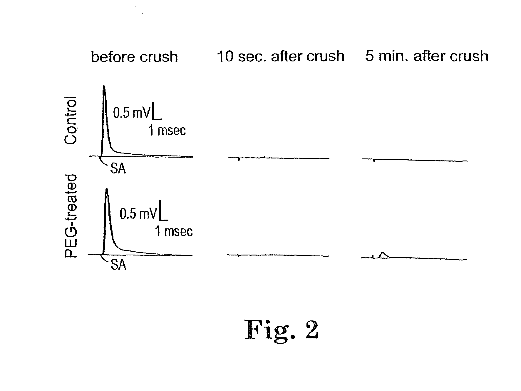
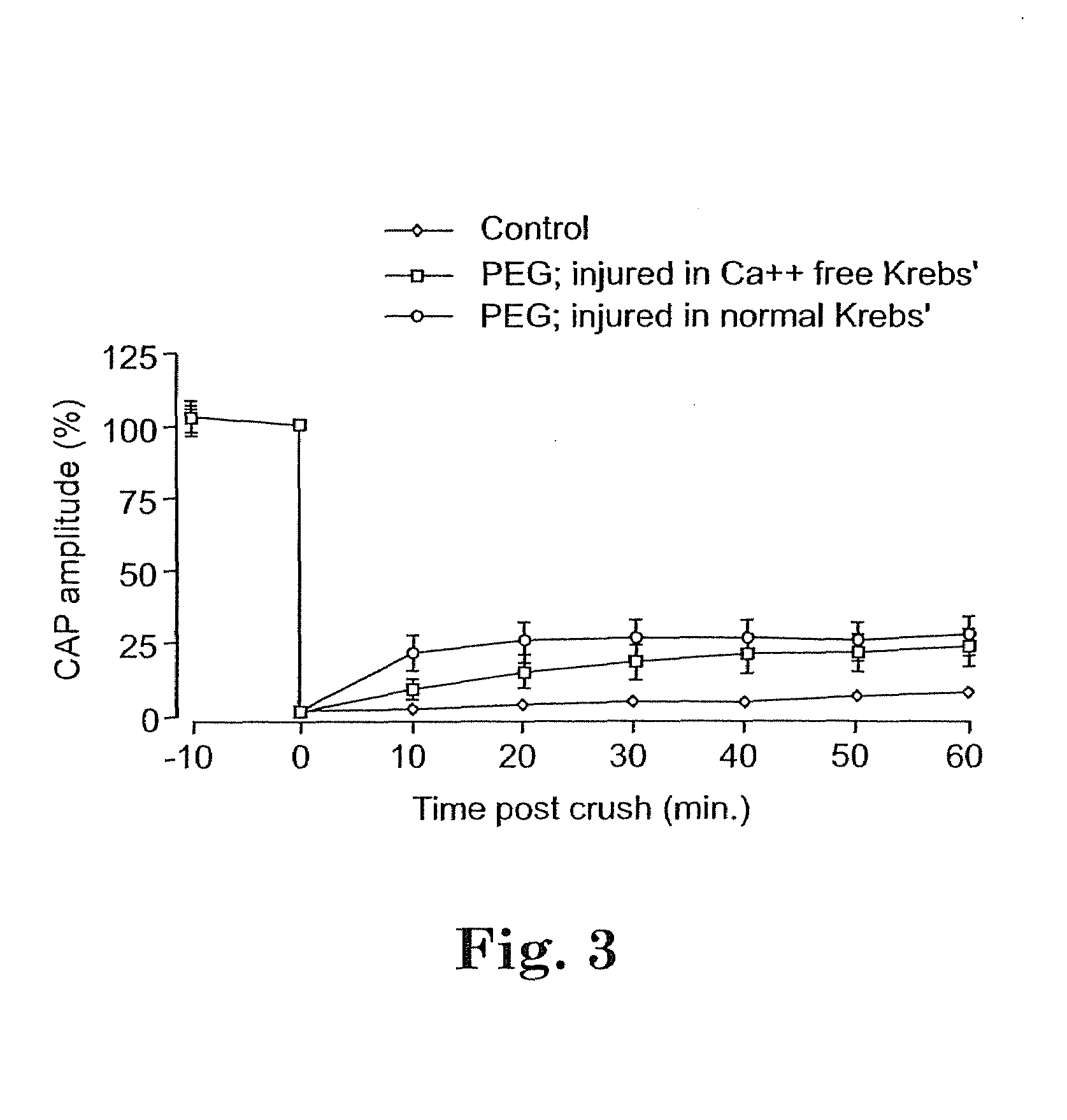
Acute In Vitro Response of Crushed Spinal Cord to PEG

[0104]This example demonstrates that compound action potentials are restored in a compressed spinal cord in vitro after it is treated with PEG.

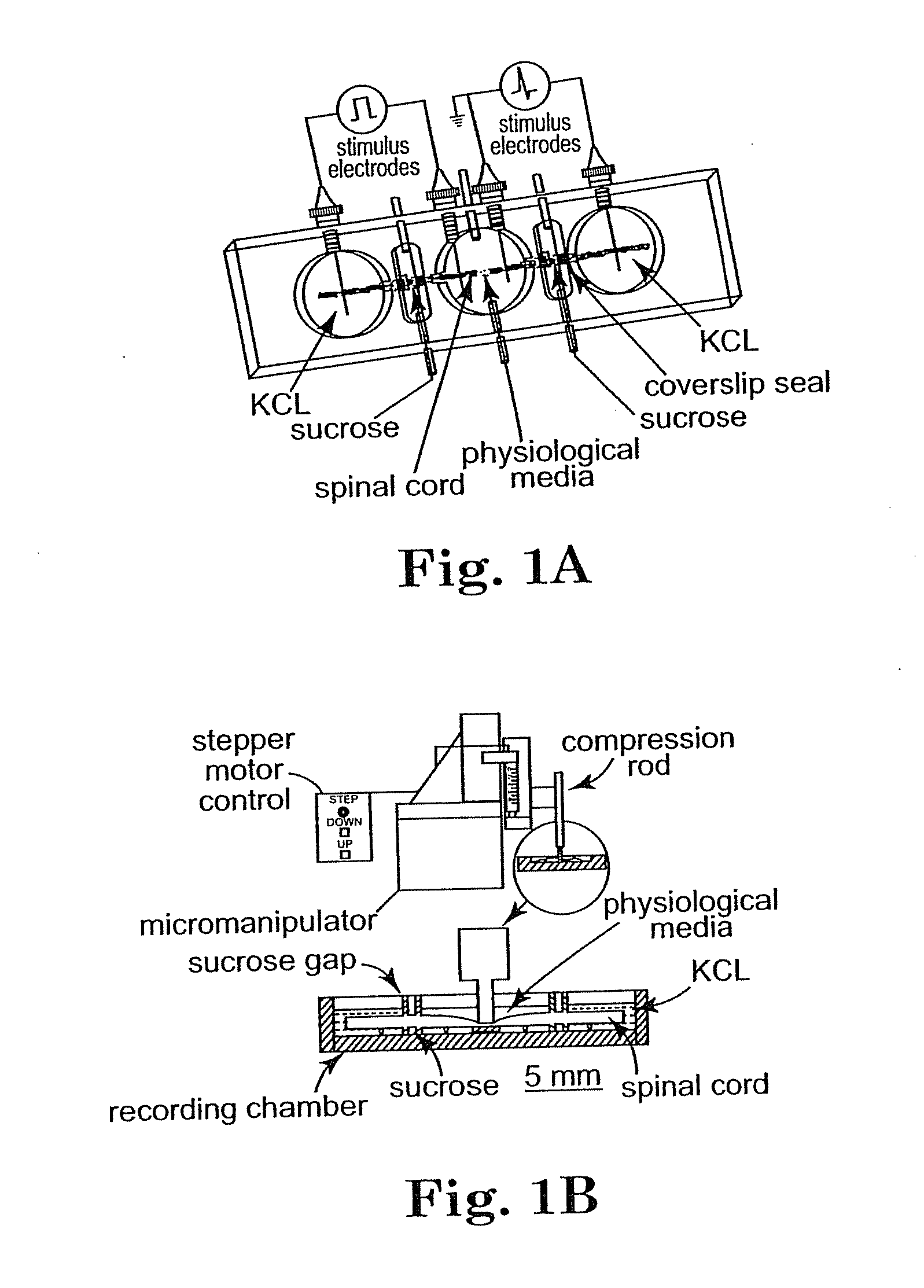
In Vitro Isolation of the Spinal Cord

[0105]Adult female guinea pigs of 350-500 gram body weight were used for these studies. The spinal cord was isolated from deeply anesthetized animals [(60 mg / kg ketamine hydrochloride, 0.6 mg / kg aoepromazine maleate, and 10 mg / kg xylazine, intramuscularly (i.m.)]. Following anesthesia, the animal was perfused transcardially with cold (1 50 C) Krebs' solution (NaCl, 124 mM; KCI, 2 mM; KH2PO4, 1.2 mM; MgSO4, 1.3 mM; CaCl2, 11.2 mM; dextrose, 10 mM; NaHC03, 26 mM; sodium ascorbate, 10 mM; equilibrated with 95% 02, and 5% C02). The vertebral column was rapidly removed using bone forceps and scissors by previously described techniques [Shi, R. and Blight, A. R. (1996) J. of Neurophysiblogy, 76(3):1572-1579; Shi, R. and Blight, A. R. (1997) Neuroscience 77(2):...

example 2

Potassium Channel Blockade as an Adjunct to PEG-Mediated Recovery of Conduction

[0125]This example shows that treatment of injured spinal cords in vitro with both a potassium channel blocker and a biomembrane fusion agent allows synergistic recovery of CAPs.

[0126]It is a common feature of injured cells to loose intracellular potassium to the extracellular milieu through compromised membrane. In axons, this may be sufficient to suppress action potential conduction. Thus, it was attempted to determine if blockage of fast potassium channels with 4-AP would affect the properties of conduction immediately following PEG repair.

[0127]Spinal cords were crushed, isolated and treated with PEG as described in Example 1. Analysis was also performed in the double sucrose recording chamber as described in Example 1.

[0128]FIG. 6A shows the enhancement of the CAP in crushed (but untreated with PEG) spinal cord by 4-AP. In this individual record, the initial recovered CAP at 1 hour post injury is sho...

example 3

Effect of PEG on Restoration of Caps in Severed Spinal Cord Axons

[0137]This example demonstrates that severed spinal cord axons may be fused with PEG, thus allowing restored conduction of CAPs through the lesion site.

[0138]In Vitro Isolation of Spinal Cord

[0139]The spinal cord of adult female guineas pigs was isolated according to the protocol of Example 1. After the cord was isolated, it was halved by midline sagittal division. The ventral white matter was separated from gray matter with a scalpel blade against a soft plastic block. Cords were maintained in continuously oxygenated Krebs' solution for at least an hour before mounting in the recording chamber. This was to ensure the recovery from dissection before each experiment was begun.

Double Sucrose Gap Recording Technique

[0140]The technique was followed according to the protocol in Example 1. The central bath was connected to instrument ground. The entire chamber was mounted on a Peltier temperature control system, which also m...


PUM

PropertyMeasurementUnit
molecular weightaaaaaaaaaa
weightaaaaaaaaaa
weightaaaaaaaaaa

Abstract

To achieve, an in vivo repair of injured mammalian nerve tissue, an effective amount of a biomembrane fusion agent is administered to the injured nerve tissue. The application of the biomembrane fusion agent may be performed by directly contacting the agent with the nerve tissue at the site of the injury. Alternatively, the biomembrane fusion agent is delivered to the site of the injury through the blood supply after administration of the biomembrane fusion agent to the patient. The administration is preferably by parenteral administration including intravascular, intramuscular, subcutaneous, or intraperitoneal injection of an effective quantity of the biomembrane fusion agent so that an effective amount is delivered to the site of the nerve tissue injury.

Description

CROSS-REFERENCE TO RELATED APPLICATIONS[0001]This application claims the benefit of U.S. Provisional Patent Application No. 60 / 286,200 filed Apr. 24, 2001, which is hereby incorporated by reference in its entirety.[0002]This invention was made in part with government support under grant number DAMD1 7-94-J-4242 awarded by the Department of the Army and grant number BES9631560 awarded by the National Science Foundation. Further financial support for development of this invention was provided by NSF grant CCR 92-22467 and NIH grant RO1 NS39288-01. The Government has certain rights in the invention.FIELD OF THE INVENTION[0003]The present invention relates generally to methods for treating injured mammalian nerve tissue including but not limited to a spinal cord. Specifically, the invention relates to methods for treating injured nerve tissue through an in vivo application of a biomembrane fusion agent. Pharmaceutical compositions for treating an injured spinal cord are also described.B...

Claims


Application Information

Patent Timeline
Patent Type & Authority Applications(United States)
IPC IPC(8): A61K31/08A61P25/00A61F13/00A61K45/00A61K31/00A61K31/74A61K31/765A61K31/77A61K31/785A61P25/02
CPCA61K31/00A61K31/74A61K31/765A61K31/77A61K2300/00A61P25/00A61P25/02A61P35/00A61P43/00
Inventor SHI, RIYIBORGENS, RICHARD B.
Owner PURDUE RES FOUND INC
Why Eureka
Try Eureka