Fluorochromes for organelle tracing and multi-color imaging

Active Publication Date: 2010-03-11
ENZO LIFE SCI INC
View PDF59 Cites 29 Cited by
  • Summary
  • Abstract
  • Description
  • Claims
  • Application Information

AI Technical Summary

Benefits of technology

[0036]A further aspect provided by this invention is a multimeric compound comprising two or more of the compounds described above and in further detail below, such multimeric compounds being joined together through a linkag

Problems solved by technology

This fundamental approach increases efficiency, reduces costs, and results in shorter time to market for new drugs.
To date, however, concerted efforts in developing such organic fluorochromes, specifically tailored to working in concert with fluorescent proteins, has been limited in scope.
While suitable for analysis of cell surfaces and permeabilized cells, fluorescently-labeled antibodies have few practical applications for intracellular imaging in living cells, due to their inherent inability to penetrate to their targets, which has given rise to development of cell-permeable small molecule organic fluorochromes, certain ones of which naturally sequester inside-specific organelles, based upon biophysical or biochemical properties favoring that distribution.
A problem with the classical organic probes from histology is that many of them require cofactors or, by requiring fixation or staining, report only on the static condition of a dead cell.
The required additional steps may be time consuming and expensive and, in the case of fixing and staining, may lack biological relevance.
While various organic fluorochromes have been developed in the past for live cell analysis, typically they were not devised with optimization of performance in conjun

Method used


Image

  • Fluorochromes for organelle tracing and multi-color imaging
  • Fluorochromes for organelle tracing and multi-color imaging
  • Fluorochromes for organelle tracing and multi-color imaging

Examples


example 1

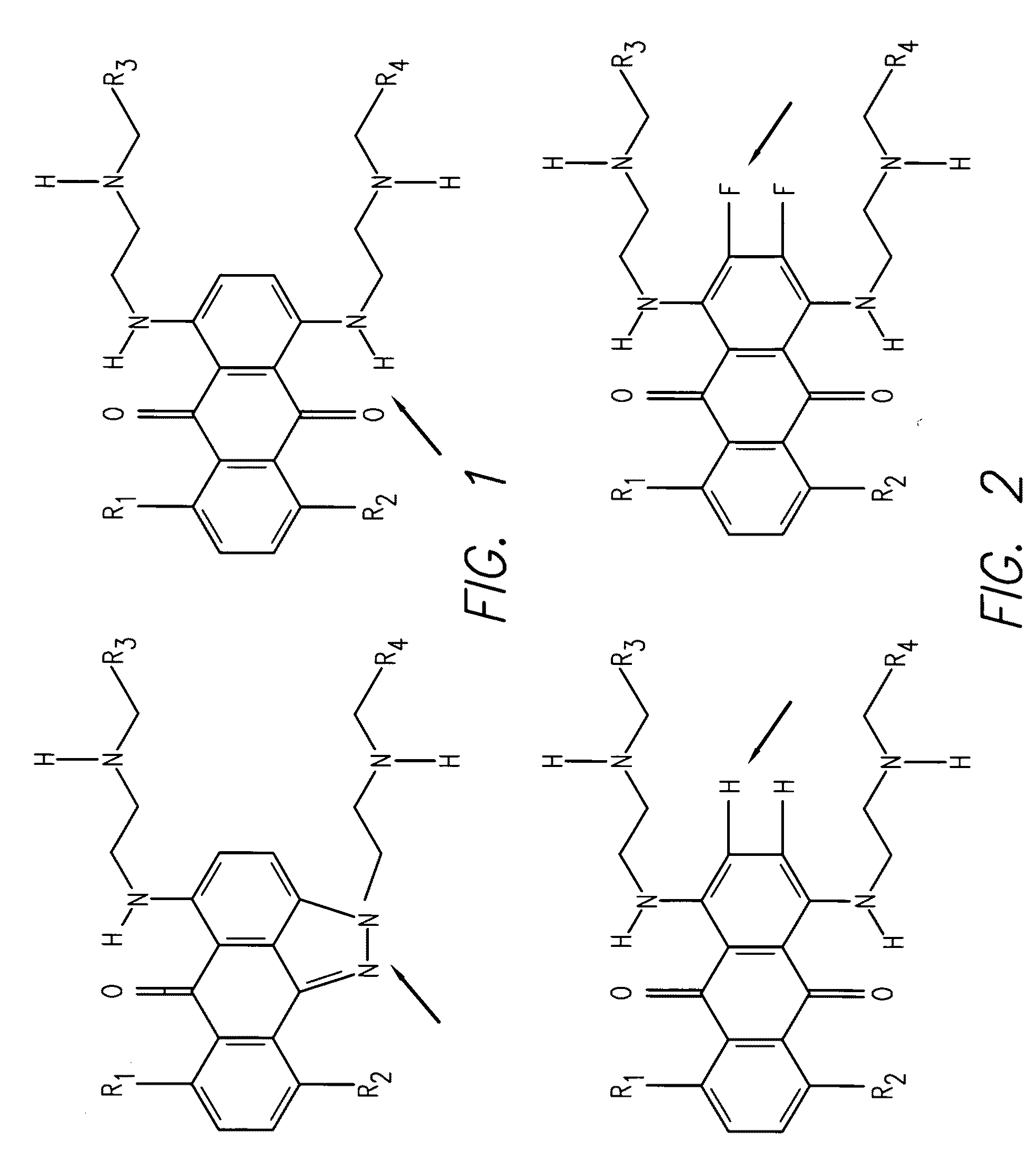
Synthesis of 1,4-bis(2-(dimethylamino)ethylamino)-2,3-difluoro-5,8-dihydroxyanthracene-9,10-dione

[0104](Compound 1)

[0105]A mixture of 1,2,3,4-tetrafluoro-5,8-dihydroxyanthraquinone (1.0 g, 3.2 mmol) and N,N-dimethylethylenediamine (3 mL) in CH2Cl2 (30 mL) was stirred at room temperature for 12 hours. After evaporation of the solvents, the residue was purified by silica gel chromatography using isocratic solvent system of EtOAc / MeOH / Et3N (10:10:1) yielding 830 mgs of Compound 1 as dark blue product. Abs (max, PBS pH 7.4)=568 nm; Em=675 nm. The structure of Compound 1 is given below:

example 2

Synthesis of trifluoro-anthraquinone ceramide (Compound 2) and difluoro-anthraquinone ceramide

[0106](Compound 3)

[0107]A mixture of 1,2,3,4-tetrafluoro-5,8-dihydroxyanthraquinone (62.4 mg, 10.2 mmol), D-sphingosine (123 mg, 0.4 mmol) in CH2Cl2 (8 mL) was stirred at room temperature for 12 h. After evaporation of the solvents, the residue was purified on silica gel chromatography eluted with EtOAc / MeOH / Et3N (10:10:1) to afford monoamine substituted Compound 2 (115 mg) and diamine substituted Compound 3 (34 mg). Abs (max, PBS pH 7.4)=533 nm; Em=625 nm for Compound 2 and Abs (max, PBS pH 7.4)=572 nm; Em=697 nm for Compound 3. The structures of these compounds are given below:

example 3

Synthesis of 1,2,3-trifluoro-5,8-dihydroxy-4-(2-(2-hydroxyethylamino)ethylamino)anthreacene-9,10-dione (Compound 4) and 2,3-difluoro-5,8-dihydroxy1,4-bis(2-(2-hydroxyethylamino)ethylamino)anthreacene-9,10-dione (Compound 5)

[0108]A mixture of 1,2,3,4-tetrafluoro-5,8-dihydroxyanthraquinone (1.0 g, 3.2 mmol) and 2-(2-aminoethylamino)ethanol (3.26 mL, 32 mmol) in CH2Cl2 (20 mL) was stirred at room temperature for 12 hours. After evaporation of the solvents, the residue was purified by silica gel chromatography using isocratic solvent system of EtOAc / MeOH / Et3N (10:10:1) yielding 200 mg of Compound 4 and 350 mg of Compound 5. Abs (max, PBS pH 7.4)=593 nm for Compound 4 and Abs (max, PBS pH 7.4)=574 nm for Compound 5. The structures of these compounds are given below:


PUM

PropertyMeasurementUnit
Dynamic viscosityaaaaaaaaaa
Compositionaaaaaaaaaa
Structureaaaaaaaaaa

Abstract

Provided are compounds, methods and kits for identifying in cells of interest organelles including nuclei and a wide variety of organelles other than nuclei (non-nuclear organelles), as well as cell regions or cell domains. These compounds and methods can be used with other conventional detection reagents for identifying the location or position or quantity of organelles and even for distinguishing between organelles in cells of interest.

Description

FIELD OF THE INVENTION[0001]The invention generally relates to fluorescent dyes suitable for applications involving wide-field fluorescence microscopy, flow cytometry, confocal microscopy, fluorimetry, high-content cell analysis, cell microarray analysis, high-content cell screening, laser-scanning cytometry and other imaging and detection modalities. The invention relates to fluorescent compounds useful for identifying organelles in live and dead cells, including nuclei and organelles other than nuclei (non-nuclear organelles), as well as subcellular organelles, cell domains and cell regions, whether within or on cells, or isolated from cells. In particular, the present invention relates to the identification of subcellular organelles, cell domains, cell regions, and the like, within living cells or extracellularly, with the identifying fluorescent compounds that are retained within or otherwise localize to the specified subcellular organelles, cell domains or cell regions. The flu...

Claims


Application Information

Patent Timeline
IPC IPC(8): C12Q1/68C07D241/04C07D231/54C07D241/02
CPCC07C225/34G01N33/5011C07C229/18C07C335/20C07C2103/24C07D213/74C07D295/135C07D487/06C07F9/5304C07F9/65038C09B1/005C09B1/51C09B1/512C09B1/5155C40B40/04G01N33/5005C07C225/36C07C2603/24
Inventor PATTON, WAYNE FORRESTPANDE, PRAVEENXIANG, YUEJUNLI, ZAIGUODONEGAN, JAMES J.RABBANI, ELAZAR
Owner ENZO LIFE SCI INC
Why Eureka
Try Eureka