Methods of treating neurological conditions with hematopoeitic growth factors

a growth factor and neurodegenerative disease technology, applied in the field of neurodegenerative diseases, can solve the problems of large socioeconomic burden, nmda antagonists, free radical scavengers and glutamate antagonists failing or showing severe side effects, etc., to enhance the survival of cells transplanted and enhance the survival of cells

Inactive Publication Date: 2010-05-06
AXARON BIOSCI
View PDF5 Cites 10 Cited by
  • Summary
  • Abstract
  • Description
  • Claims
  • Application Information

AI Technical Summary

Benefits of technology

[0042]Another object of the present invention is to provide a method of treating a neurological condition in a mammal by agonizing a GMCSF receptor, a GCSF receptor, IL-3 receptor, Il-5 receptor or combinations of these to treat the neurological condition. Another object of the present invention is to provide a method of enhancing the survival of a cell transplanted into a mammal, by introducing into the cell one or more polynucleotides which encode GCSF, GM-CSF, IL-3, IL-5, derivatives thereof, mimetics thereof and / or combinations thereof prior to transplanting the cell into the mammal, whereby the cell expresses the hematopoietic factor in an amount sufficient to enhance the survival of the cell relative to the cell survival prior to the introduction of the polynucleotides.

Problems solved by technology

Unfortunately, none of these prevailed to provide neuroprotective efficacy.
In particular, NMDA antagonists, free radical scavengers and glutamate antagonists failed or demonstrated severe side-effects.
It presents a large socioeconomic burden.
The cause of ischemic stroke is often embolic, or thrombotic.
So far, there is no effective treatment for the majority of stroke patients.
Cerebral ischemia may result from a variety of causes that impair cerebral blood flow (CBF) and lead to deprivation of both oxygen and glucose.
Traumatic brain injury (TBI), on the other hand, involves a primary mechanical impact that usually causes skull fracture and abruptly disrupts the brain parenchyma with shearing and tearing of blood vessels and brain tissue.
Therefore, the vast majority of the cardiac arrest victims annually in the United States is not treated successfully.
The major reason for the low survival rates after successful CPR, i.e., for postarrest in-hospital mortality, is persistent brain damage.
The personal, social, and economic consequences of brain damage following cardiac arrest are devastating.
Presently, it is not possible to decrease the primary damage to neurons that is caused by hypoxia during cardiac arrest by any post-arrest therapeutic measures.
Suicide remains one of the common and often unavoidable outcomes of depression.
Schizophrenia has a tremendous cost to society, estimated at $32.5 billion per year in the US.
However, neuroleptic treatment often does not reduce all of the symptoms of schizophrenia.
Moreover, antipsychotic treatment can have severe side effects, such as tardive dyskinesias.
While the brain does shrink in size, it does not do so uniformly.
Certain structures are more prone to shrinkage.
The slowing of mental processing may be caused by the deterioration of neurons, whether they are lost, shrink, or lose connections.
The shrinkage of both the frontal lobe and the hippocampus are thought to be responsible for memory difficulties.
Recent studies pointed out that axon damage occurs early in the disease and during lesion development.
The exact mechanisms and effector molecules of axonal degeneration, however, are not yet defined, and an axon-protective therapy has not yet been established.
Other neurological symptoms, such as dystonia, can be treated, but treatment is associated with a high risk of adverse events.
However, if left untreated, glaucoma can eventually lead to blindness.
Most of these diseases affect the brain and are fatal.
There has been limited success in only treating the symptoms of these diseases.
Severe SCI often causes paralysis (loss of control over voluntary movement and muscles of the body) and loss of sensation and reflex function below the point of injury, including autonomic activity such as breathing and other activities such as bowel and bladder control.
SCI patients are also prone to develop secondary medical problems, such as bladder infections, lung infections, and bed sores.
While recent advances in emergency care and rehabilitation allow many SCI patients to survive, methods for reducing the extent of injury and for restoring function are still limited.

Method used


Image

  • Methods of treating neurological conditions with hematopoeitic growth factors
  • Methods of treating neurological conditions with hematopoeitic growth factors
  • Methods of treating neurological conditions with hematopoeitic growth factors

Examples


example 1

Focal Cerebral Ischemia

Procedure for Inducing Focal Cerebral Ischemia (MCAO, Middle Cerebral Artery Occlusion)

[0311]Experimental protocols were approved by the local ethics committee. Twenty-four male Wistar rats (Charles River, Germany) weighing 280 to 320 g were randomly assigned to the following groups: A (Control group, n=12, ischemia for 90 min, treatment with 2 ml saline 0.9% for 90 min beginning 30 min after vessel occlusion); B (GCSF group, n=12, ischemia for 90 min, treatment with 60 μg / kg body weight of recombinant human GCSF, Neupogen®, Amgen, Europe B. V., Netherlands, soluted in 2 ml saline 0.9% for 90 min beginning 30 min after vessel occlusion. Alternatively, any GCSF or derivative or formulation of other source (another manufacturer (e.g. Lenogastrim™ by Roche or Granocyte™ by Chugai or Albugranin™ by HGS or Neulasta™ by Roche / Amgen) can be used here.

[0312]Animals then were anesthetized with an intraperitoneal injection of 100 mg / kg body weight ketamine hydrochloride...

example 2

Immunohistochemistry in the Context of Focal Cerebral Ischemia

Immunohistochemical Methods Used

[0318]For morphological analysis of STAT3 activation (FIG. 6) and GCSFR distribution in infarcted brains, and counts of neutrophilic granulocytes, a 2-mm-thick brain slice of GCSF-treated animals and controls was immersion fixed in 4% paraformaldehyde in 0.1 mol / l phosphate buffer for 24 hrs (n=5 per group). After paraffin-embedding, 1-μm-thick sections were cut and used for H&E staining, Nissl staining and immunohistochemical analysis. Immunohistochemical studies were performed with antisera against myeloperoxidase (DAKO, Carpinteria, Calif., USA), glial fibrillary acidic protein (GFAP) (DAKO, Carpinteria, Calif., USA), GCSFR (Santa Cruz Biotechnology Inc., Santa Cruz, Calif., USA) and STAT3 (Santa Cruz Biotechnology Inc., Santa Cruz, Calif., USA). For antigen retrieval, sections provided for GCSFR and STAT3 immunohistochemistry were heated for 20 min in a 10 mM citrate buffer at 99° C. Se...

example 3

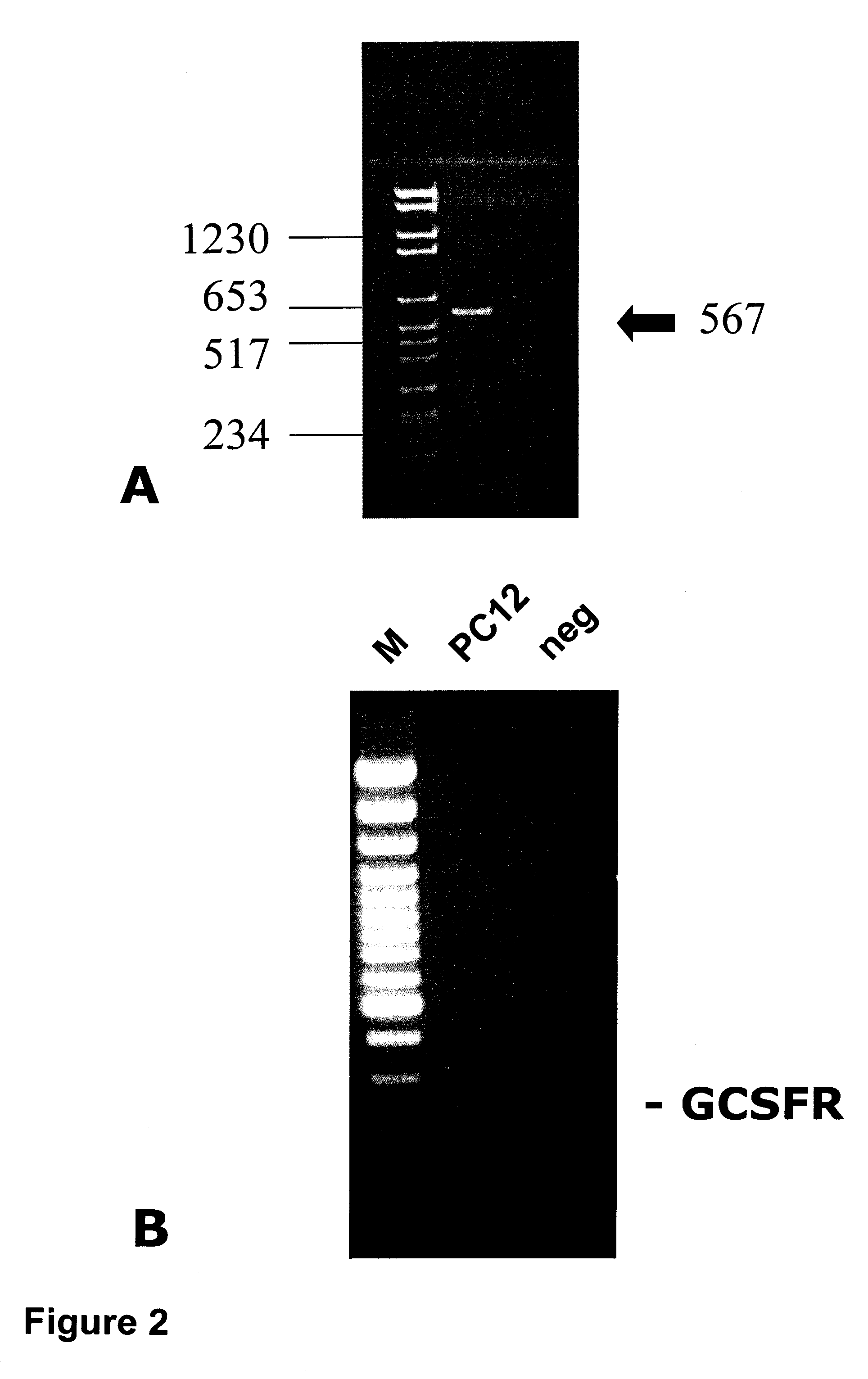
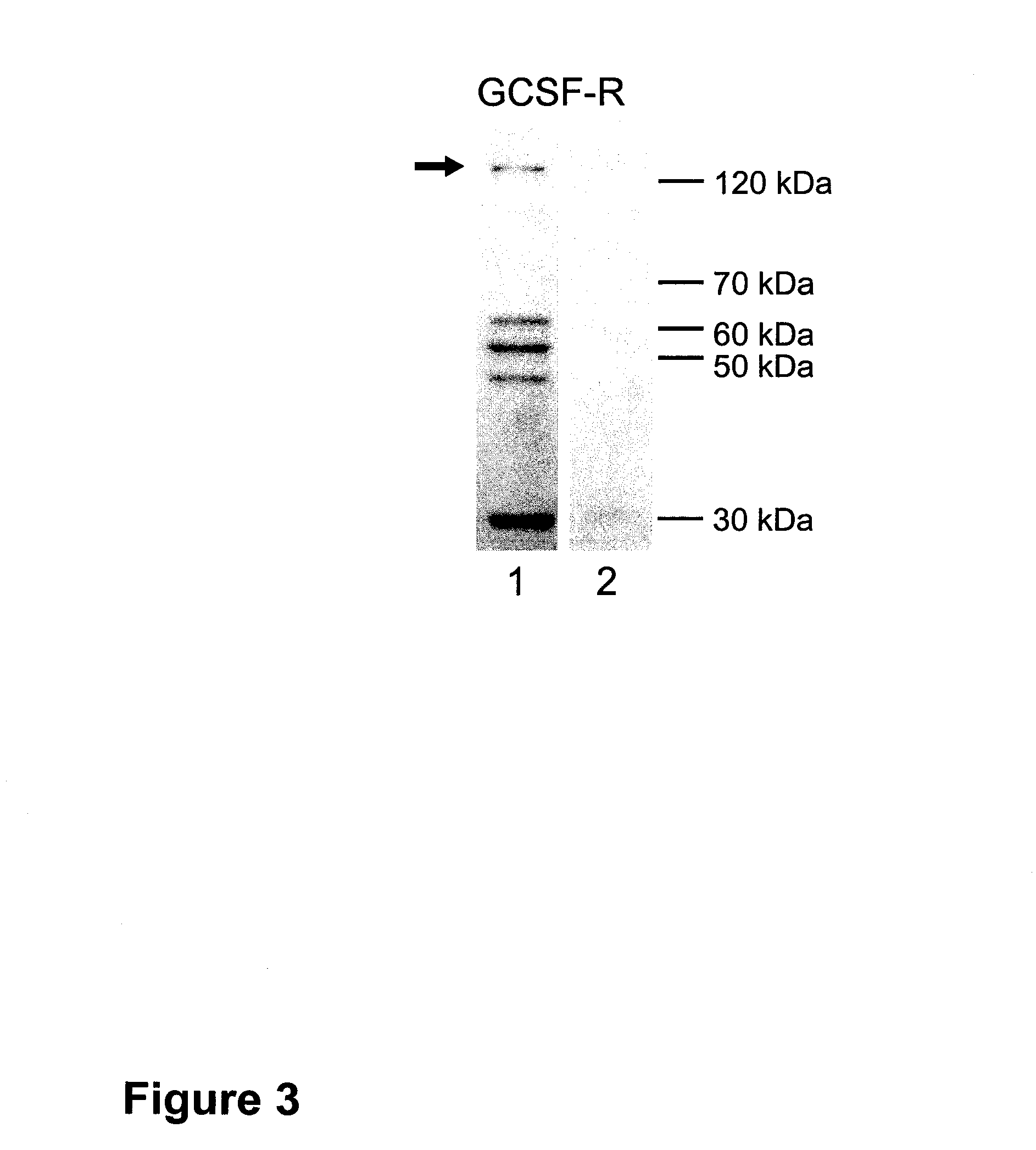
Western Blots and PCR in the Context of Focal Cerebral Ischemia

Western Blots (FIG. 3)

[0323]For immunoblotting, brain tissue (transient ischemia of 2 hours) was lysed in 20 volumes (w / v) of homogenization buffer (320 mM sucrose, 0.1 mM phenylmethylsulfonyl fluoride, and 2 μg / ml Pepstatin) at 4° C. Homogenates were centrifuged at 9,200 G for 15 min at 4° C. After resuspending pellets in 1 / 10 of the homogenization volume, aliquots for protein determination (Bio-Rad protein-assay, Munich, Germany) were separated and samples were rapidly frozen in nitric oxide and stored at −70° C. Per lane 15 μg protein were loaded on a 8% SDS polyacrylamide gel containing 4 M urea and electrophoresed under standard conditions. Proteins were electrophoretically transferred to Immobilon-P™ membranes (Millipore Corp., Eschborn, Germany) by semi-dry blotting. After blocking in 3% nonfat dry milk in TBST (20 mM Tris base, pH 7.6, 137 mM NaCl and 0.05% Tween-20) for 1 hour at room temperature (RT), membranes...


PUM

PropertyMeasurementUnit
concentrationsaaaaaaaaaa
concentrationaaaaaaaaaa

Abstract

The present invention relates to a method of treating a neurological condition in a mammal by administering at least one hematopoietic growth factor.

Description

CROSS-REFERENCE TO RELATED APPLICATIONS[0001]The present application is a continuation-in-part of PCT / IB03 / 006446 filed Dec. 31, 2003, pending, and is also a continuation-in-part of U.S. application Ser. No. 10 / 659,295 filed Sep. 11, 2003, pending, which is a continuation application of U.S. application Ser. No. 10 / 331,755 filed Dec. 31, 2002, abandoned.BACKGROUND OF THE INVENTION[0002]1. Field of the Invention[0003]The present invention relates to a method of treating a neurological condition in a mammal by administering at least one hematopoietic growth factor.[0004]2. Discussion of the Related Art[0005]Growth factors are proteins that are essentially involved in regulating survival, proliferation, maturation, and outgrowth of developing neuronal cells. For example, the expression of a large number of growth factors increases in response to various brain insults. Many factors display endogenous neuroprotective and neurotrophic effects (see Arvidsson A et al., Neuroscience 2001; 10...

Claims


Application Information

Patent Timeline
Patent Type & Authority Applications(United States)
IPC IPC(8): A61K9/127A61K38/18A61K38/20A61K38/16A61K31/7088A61K35/76A61K35/12C12N5/02A61K38/19A61K38/49A61P25/28G01N33/50G01N33/68
CPCA61K38/193A61K38/49G01N33/5008G01N33/5023G01N33/5041G01N33/5058G01N33/6872A61K2300/00A61P25/00A61P25/28A61K38/19
Inventor SCHAEBITZ, WOLF-RUEDIGERSCHNEIDER, ARMINKRUEGER, CAROLASOMMER, CLEMENSSCHWAB, STEFANKOLLMAR, RAINERMAURER, MARTINWEBER, DANIELAGASSLER, NIKOLAUS
Owner AXARON BIOSCI
Why Eureka
Try Eureka