
Cell-nanofiber composite and cell-nanofiber-hydrogel composite amalgam based engineered intervertebral disc

a technology of cell-nanofiber and composite resin, which is applied in the field of tissue engineered intervertebral discs, can solve the problems of adjacent segment disease, biomechanics of the disc, and further degeneration, and achieve the effect of increasing the interfiber distance and facilitating cell growth and differentiation

Inactive Publication Date: 2010-07-15
HEALTH & HUMAN SERVICES GOVERNMENT OF THE US SEC DEPT OF
View PDF10 Cites 42 Cited by
  • Summary
  • Abstract
  • Description
  • Claims
  • Application Information

AI Technical Summary

Benefits of technology

[0022]In yet another aspect, the invention provides a method of preparing a tissue engineered tissue comprising the steps of: preparing a nanofibrous biocompatible polymer support comprising a cavity; expanding the nanofibrous polymer support thereby increasing interfiber distance; contacting a suspension of cells with the support to form a polymer matrix having cells dispersed therein; injecting a hydrogel composition into the cavity; culturing the compressed cell-polymer matrix in a bioreactor with a culture medium under conditions to conducive cell growth and differentiation to tissue engineered tissue.

Problems solved by technology

However, for the subject requiring surgical management current interventions focus on fusion of the involved IVD levels, which eliminates pain but does not attempt to restore disc function (Shvartzman, L. et al.
Spinal fusion however is thought to significantly alter the biomechanics of the disc and lead to further degeneration, or adjacent segment disease.
These disc replacement technologies are designed to address flexion, extension, and lateral bending motions; however, they do little to address compressive forces and their longevity is limited due to their inability to biointegrate.
Current treatment for injuries that penetrate subchondral bone include subchondral drilling, periosteal tissue grafting, osteochondral allografting, chondrogenic cell and transplantation; but are limited due to suboptimal integration with host tissues.
However, few studies have investigated the potential of mesenchymal stem cells (MSCs) in IVD tissue engineering.

Method used


Image

  • Cell-nanofiber composite and cell-nanofiber-hydrogel composite amalgam based engineered intervertebral disc
  • Cell-nanofiber composite and cell-nanofiber-hydrogel composite amalgam based engineered intervertebral disc
  • Cell-nanofiber composite and cell-nanofiber-hydrogel composite amalgam based engineered intervertebral disc

Examples


example 1

Isolation and Culture of Bone Marrow-Derived hMSCs

[0108]With approval from the Institutional Review Board of Thomas Jefferson University, bone marrow-derived hMSCs were obtained from the femoral heads of subjects undergoing total hip arthroplasty, and processed as previously described (Noth U, et al. J Orthop Res 2002; 20:1060-9; Haynesworth S E, et al. Bone 1992; 13:81-8; and Wang M L, et al. J Orthop Res 2002; 20:1175-84). Briefly, whole bone marrow was curetted from the exposed cutting plane of the femoral neck, washed extensively in Dulbecco's Modified Eagle's medium (DMEM; BioWhittaker, Walkersville, Md.), separated from contaminating trabecular bone fragments and other tissues using a 20-gauge needle attached to a 10-cc syringe, and cultured in DMEM, 10% fetal bovine serum (FBS) from selected lots (Caterson E J, et al. Mol Biotechnol 2002; 20:245-56), and antibiotics (50 μg / mL streptomycin, 50 IU / mL of penicillin) at a density of 4×10⁵ cells / cm2. Forty-eight hours post-plating...

example 2

Fabrication of Electrospun Nanofibrous PLLA Scaffolds

[0109]Nanofibrous scaffolds were fabricated according to an electrospinning process described previously (Li W J, et al. J Biomed Mater Res 2003; 67A:1105-14). Briefly, PLLA polymer was dissolved in an organic solvent mixture (10:1) of chloroform and N,N, dimethylformamide (DMF) at a final concentration of 0.14.5 g / mL. The polymer solution was delivered through the electrospinning apparatus at a constant flow rate of 0.4 mL / h under an applied 0.8 kV / cm charge density, resulting in a 144 cm2 mat with an approximate thickness of 1 mm. To remove residual organic solvent, the non-woven polymer mat was placed within a vacuum chamber for 48 h, and subsequently stored in a dessicator. Prior to cell seeding, nanofibrous scaffolds were fashioned from the electrospun mat, sterilized by ultraviolet irradiation for 30 min per side in a laminar flow hood, and pre-wetted for 24 h in Hanks' Balanced Salt Solution.

example 3

Fabrication of Intervertebral Disc (IVD) Constructs

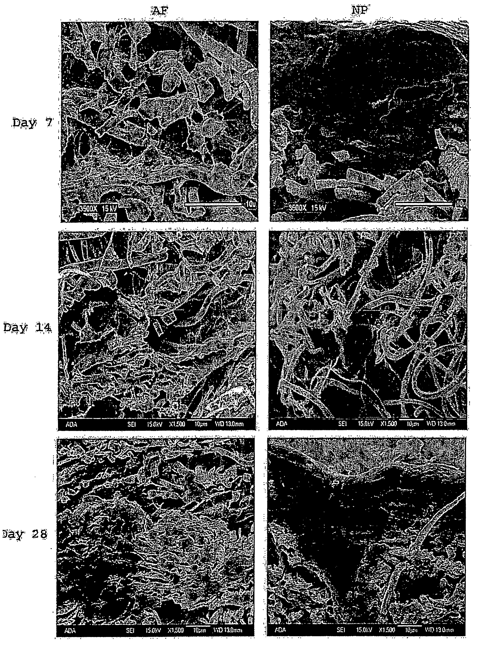
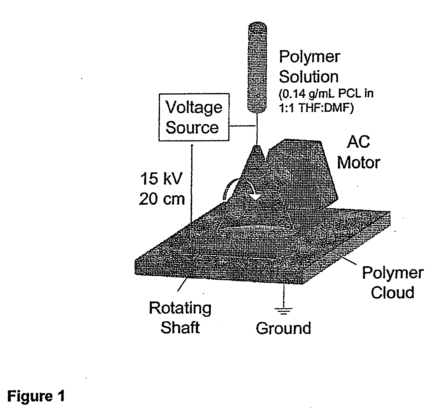
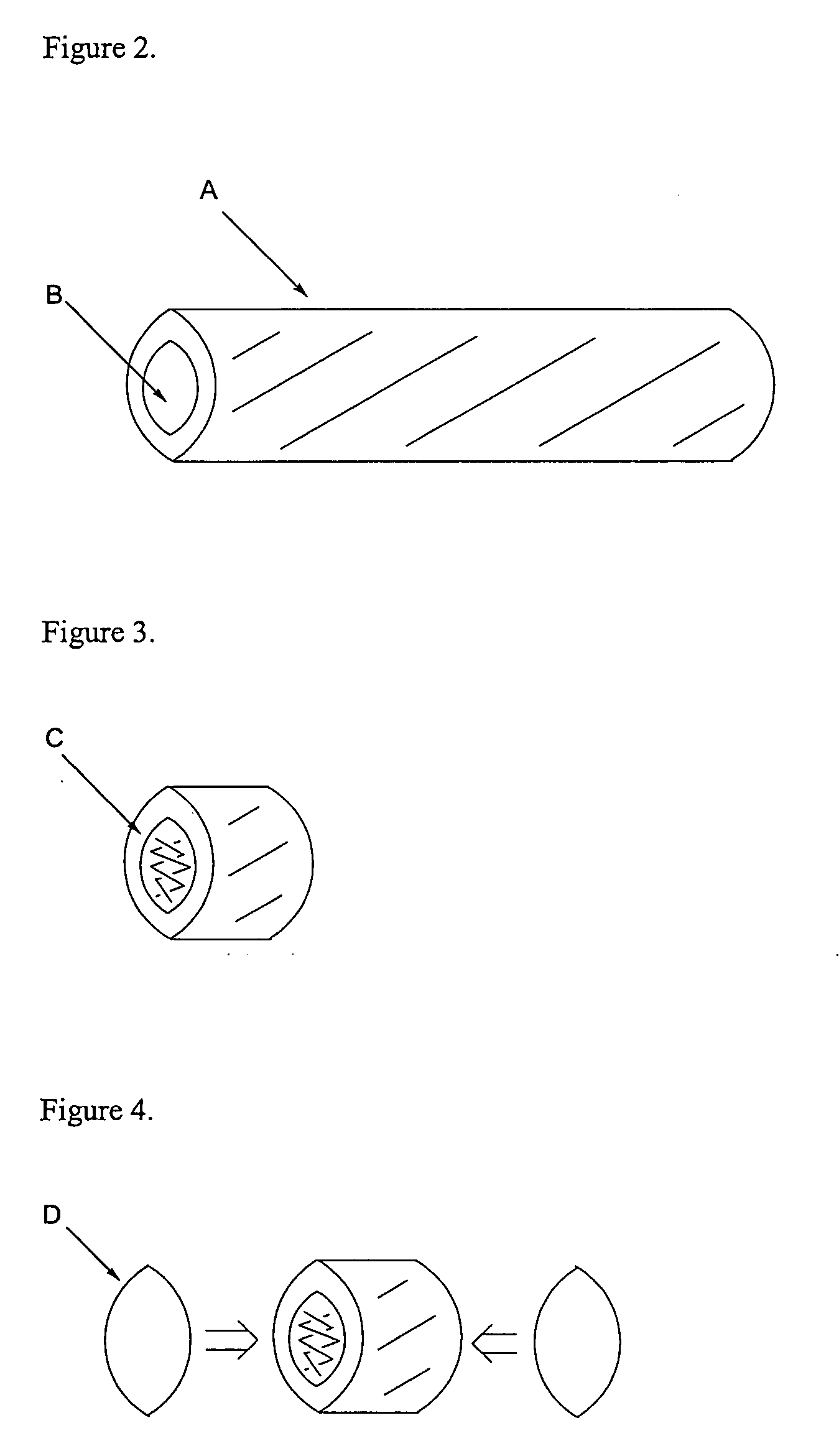
[0110]To make IVD constructs, PLLA nanofibers were electrospun onto a rotating rod (shaft) to produce homogeneous, non-woven or specifically oriented nanofibrous mats (FIG. 1), whose shape was dependent on the mechanical requirements for a construct. After pulling out the rod, a long hollow nanofibrous tube (FIG. 2) with the outer diameter of 1.1 cm and the inner diameter of 1.0 cm was produced. Nanofibrous rings with the height of 0.5 cm were obtained from cutting the nanofibrous tube into sections (FIG. 3). The open-to-outside ring was sealed with a circular nanofibrous mat with the diameter of 1.1 cm on each end of the ring after being inserted with fluffy nanofibers (FIG. 4). The inserted nanofibers with a loosened fiber structure serve the roles of mechanical reinforcement and biological enhancement.

[0111]A hydrogel such as hyaluron gel was mixed with nucleus pulposus cells isolated from human IVD, and was injected into the emp...


PUM

PropertyMeasurementUnit
porosityaaaaaaaaaa
porosityaaaaaaaaaa
size distributionaaaaaaaaaa

Abstract

The instant invention is directed to a tissue engineered intervertebral disc comprising at least one inner layer and an exterior layer, wherein: the exterior layer comprises a nanofibrous polymer support comprising one or more polymer nanofibers; the at least one inner layer comprises a hydrogel composition comprising at least one or more hydrogel materials and / or one or more polymer nanofibers; and a plurality of cells which are dispersed throughout the tissue engineered intervertebral disc. Additionally, the instant invention is directed to methods of making such intervertebral discs and methods of treating intervertebral disc damage.

Description

CROSS-REFERENCE TO RELATED APPLICATION[0001]This application claims the benefit of U.S. provisional application No. 60 / 847,839 filed Sep. 27, 2006 and U.S. provisional application No. 60 / 848,284 filed Sep. 28, 2006, both of which are fully incorporated herein by reference.STATEMENT OF RIGHTS TO INVENTIONS MADE UNDER FEDERALLY SPONSORED RESEARCH[0002]Research supporting this application was carried out by the United States of America as represented by the Secretary, Department of Health and Human Services.FIELD OF INVENTION[0003]The present invention relates to tissue engineered intervertebral discs comprising a nanofibrous polymer hydrogel amalgam having cells dispersed therein, methods of fabricating tissue engineered intervertebral discs by culturing a mixture of stem cells or intervertebral disc cells and a electrospun nanofibrous polymer hydrogel amalgam in a suitable bioreactor, and methods of treatment comprising implantation of tissue engineered intervertebral disc into a sub...

Claims


Application Information

Patent Timeline
Patent Type & Authority Applications(United States)
IPC IPC(8): A61F2/44C12N5/00
CPCA61F2/441A61F2002/30062A61L2430/38A61F2002/4445A61F2002/445A61F2002/4495A61F2210/0004A61L27/18A61L27/3852A61L27/3856A61L27/52A61L27/56A61L27/58C08L67/04A61L2400/12A61F2/442A61F2002/444C12N5/0068C12N2533/30
Inventor LI, WAN-JUNESTI, LEON J.TUAN, ROCKY S.
Owner HEALTH & HUMAN SERVICES GOVERNMENT OF THE US SEC DEPT OF
Learn More