Method for culturing stem cells

a stem cell and culturing technology, applied in the field of culturing stem cells, can solve the problems of inconvenient long-term hes cell culture, lack of simple methods for producing ebs of consistent and desired size from es cells, and inability to use 2-d sam monolayers for culturing primate es cells, etc., to achieve the effect of controlling the cell differentiation profile of ebs

Active Publication Date: 2010-08-05
WISCONSIN ALUMNI RES FOUND
View PDF9 Cites 30 Cited by
  • Summary
  • Abstract
  • Description
  • Claims
  • Application Information

AI Technical Summary

Benefits of technology

[0015]In a second aspect, the invention is summarized in that a method for forming EBs having a narrow size distribution includes the steps of harvesting substantially undifferentiated ES cells or iPS cells from the microwells and culturing the harvested cells under differentiating culture conditions until the culture contains differentiated cells. Because the undifferentiated pluripotent cells for use in the EB-forming method can be obtained from dimension-constrained microwells having uniform dimensions, supra, aggregates having a narrow size distribution can be harvested, thereby avoiding a shortcoming of existing EB-forming methods, namely that clumps of pluripotent cells from which EBs are now derived can vary widely in size, volume and cell number. The harvesting step can include harvesting e...

Problems solved by technology

Unfortunately, the art lacks simple methods for producing EBs of consistent and desired size from ES cells.
Unfortunately, 2-D SAM monolayers are of limited utility for culturing primate ES cells because of the cell's growth nature.
Likewise, initial efforts at 2-D micro-contact printing of Matrigel® on SAM surfaces indicated that this method was not suitable for long-term hES cell culture because of substrate instability and because growing colonies could span across unpatterned regions.
With few exceptions, current literature regarding patterned 2-D monolayers focuses primarily on cell attachment and replication to generate confluent monolayers in patterned regions, but does not investigate effects of th...

Method used


Image

  • Method for culturing stem cells
  • Method for culturing stem cells
  • Method for culturing stem cells

Examples


example 1

Microwells for Pluripotent Cell Culture and Embryoid Body Generation

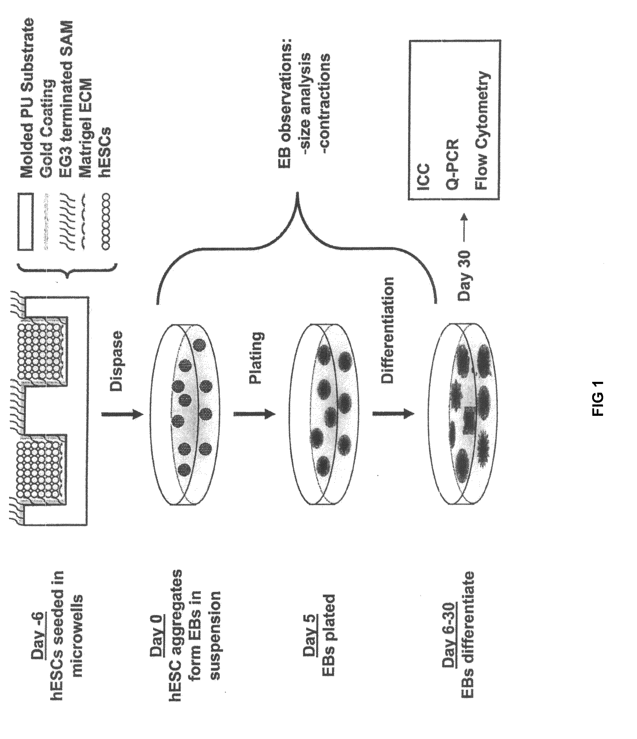
[0034]Reference is made to FIG. 1. Microscope slides having formed thereupon a homogeneous distribution of wells of identical size and shape were constructed in three steps using a polydimethylsiloxane (PDMS) stamp to shape a surface of a UV-crosslinkable polyurethane polymer substrate. First, silicon masters, each having desired microwell patterns formed into a surface thereof, were prepared using photolithography and plasma etching techniques similar to those described by Chen et. al. [22], incorporated herein by reference as if set forth in its entirety. The surfaces were passivated by fluorination with (tridecafluoro-1,1,2,2,-tetrahydrooctyl)-1-trichlorosilane vapor. Second, a mixture of PDMS elastomer pre-polymer with curing agent (10:1) (Sylgard 184 Silicon Elastomer; Dow Corning; Midland, Mich.) was poured over silicon masters to form PDMS stamps. The mixture was degassed under vacuum and incubated overnight ...

example 2

Directed Differentiation of hES cells From Microwell Cultures

[0053]hES cells were cultured either in microwells having depths of 50 μm-120 μm and lateral dimensions of 50 μm-500 μm or in TCPS plates as described above. Briefly, samples were incubated for 30 minutes at 37° C. to allow hES cells to settle into the microwells before adding 1.5 ml / well CMF+ to the wells of a 6-well plate. The medium was changed daily thereafter and the cells typically reached confluence within a week. TCPS EBs and microwell-derived EBS were then formed as described above.

[0054]Keratinocytes differentiation was achieved by initially forming EBs from microwell-derived hES cell aggregates as described above. EBs were grown in suspension for fourteen days in UMF− medium and then attached to gelatin-coated plates in Defined Keratinocyte Serum Free Medium (DSFM) (Invitrogen; Carlsbad, Calif.). Cells were cultured 2-3 weeks prior to analysis by immunocytochemistry and flow cytometry, as described for undiffere...

example 3

Microwells for hES cells Culture and Cryopreservation

[0057]hES cells were cultured in microwells having dimensions of 50 μm deep with 50 μm to 400 μm lateral dimensions as described above. Briefly, samples were incubated for 30 minutes at 37° C. to allow hES cells to settle into the microwells before adding 1.5 ml / well CMF+ to the wells of a 6-well plate. The medium was changed daily thereafter and the cells typically reached confluence within a week. Alternatively, hES cells were cultured in TCPS dishes, as described above.

[0058]After 7 days of culture, the hES cells in microwells, suspensions and TCPS plates were placed in freezers at −80° C. for up to 4 weeks. Following cryopreservation, cells frozen in suspension were thawed by immersion of the cryovial in a 37° C. waterbath with agitation. Cells were immediately diluted in 10 ml CMF (i.e. hES cell medium conditioned on mouse embryonic fibroblasts without bFGF) and centrifuged. hES cells were then diluted in 2 ml CMF and plated ...


PUM


Abstract

A three-dimensional microwell system that supports long term pluripotent cell culture and formation of homogeneous embryoid bodies (EBs) is described. Microwell-cultured pluripotent cells remain viable and undifferentiated for several weeks in culture and maintain undifferentiated replication when passaged to Matrigel®-coated, tissue culture-treated polystyrene dishes. Microwell-cultured pluripotent cells maintain pluripotency, differentiating to each of the three embryonic germ layers. Pluripotent cell aggregates released from microwells can be passaged for undifferentiated replication or differentiated to monodisperse EBs. The ability to constrain pluripotent cell growth in three dimensions advantageously provides for more efficient, reproducible culture of undifferentiated cells, high-throughput screening, and the ability to direct pluripotent cell differentiation by generating monodisperse EBs of a desired size and shape. Cardiomyocyte-rich EBs are obtained from pluripotent cells cultured in microwells of defined size and shape.

Description

CROSS-REFERENCE TO RELATED APPLICATIONS[0001]This application claims the benefit of U.S. Provisional Patent Application No. 61 / 112,368, filed Nov. 7, 2008, incorporated herein by reference as if set forth in its entirety.STATEMENT REGARDING FEDERALLY SPONSORED RESEARCH OR DEVELOPMENT[0002]This invention was made with United States government support awarded by the following agency: NSF DMR-0079983, NIH / NIBIB R01 EB007534, NIH / NHLBI R01 HL08846150, and NSF EFRI-0735903. The United States government has certain rights in this invention.BACKGROUND[0003]Primate embryonic stem (ES) cells and the recently-described induced pluripotent cells (iPS) (collectively, “pluripotent cells”) can proliferate without limit and can differentiate into each of the three embryonic germ layers [1-3]. To facilitate self-renewal, primate (including human) pluripotent cells are typically co-cultured with mouse embryonic fibroblast (MEF) feeder cells, or cultured in MEF-conditioned medium (MEF-CM) on a Matrig...

Claims


Application Information

Patent Timeline
IPC IPC(8): C12N5/071C12N5/02C12N5/00C40B40/14
CPCC07K16/18C12N2533/90C12N5/0603
Inventor KAMP, TIMOTHY J.ZHANG, JIANHUAMOHR, JEFFREY C.DEPABLO, JUAN J.PALECEK, SEAN P.
Owner WISCONSIN ALUMNI RES FOUND
Why Eureka
Try Eureka