Imaging of Activated Vascular Endothelium Using Immunomagnetic MRI Contrast Agents

Inactive Publication Date: 2010-11-25
JANSSEN DIAGNOSTICS LLC
View PDF13 Cites 4 Cited by
  • Summary
  • Abstract
  • Description
  • Claims
  • Application Information

AI Technical Summary

Benefits of technology

[0026]The present invention provides methods and compositions for improved medical diagnostic imaging. A novel contrasting agent is disclosed for use in MRI. The agent consists of conjugated monoclonal antibodies (mAb) directed against the murine isoform of an endothelial cell activation marker, such as, but not limited to, the murine isoform of anti-ICAM (CD54 endothelial cell activation marker). Typically, targeted MRI contrast agents provide enhanced relaxivity, improved signal-to-noise, targeting ability, and resistance to agglomeration. Methods of making such MRI contrast agents afford better control over particle size, and methods of using such MRI contrast agents typically afford enhanced blood clearance rates and distribution. CD54-FF is used as an MRI contrast agent in targeting vascular endothelial cells comprising a BSA coated iron oxide particle conjugated to a mono-thiolated anti-CD54. The quenched complex is stored in D1H20

Problems solved by technology

In general for contrast agents to be effective, they must interfere with the wavelength of electromagnetic radiation used in the imaging technique, alter the physical properties of tissue to yield an altered signal, or provide the source of radiation itself.
While extremely long half-lives (i.e., days or weeks) are unnecessary in clinical imaging situations and possibly dangerous (due to the increased chance for toxicity and metabolic breakdown into more toxic molecules), short half-lives are also not desirable.
If the image enhancement lasts for too short of time, it is difficult to acquire a high-quality image of the patient.
In addition, rapid clearance of a targeted agent will reduce the amount of the agent available to bind to the target site and thus reduce the “brightness” of the target site on the image.
2 mm), however, it offers poor sensitivity when compared with other imaging techniques.
However, dextran coatings have been claimed to be unstable at the alkaline conditions of the particle synthesis, and their chemical composition has therefore been questioned.
Additionally, dextran-induced anaphylactic reactions present potential problems (U.S. Pat. No. 5,492,814).
In addition, this method provides little control over the degree of coating leading to particles containing multiple iron oxide nanoparticles within a single agent.
Nanoparticles obtained using conventional methods also have a low level of crystallinity, which significantly impacts the sensitivity of the contrast agent.
Moreover, nanoparticles tend to agglomerate due to their high surface energy, which is a significant problem encountered during synthesis and purification steps.
Such agglomeration increases the size of the particle, resulting in rapid blood clearance as well as reducing targeting efficiency, and may result in a reduction in relaxivity.
When large particles are employed, only a few targeting ligands may be attached before the particles become large enough to be cleared from the blood and failure of the agent to reach the intended target.
When coatings are globular, reactive sites intended for ligand attachment are generally hindered, thereby reducing conjugation efficiency.
In addition, once bound, ligands may reside in the interior of globular coatings, preventing easy access to the biomarkers.
Contrast agents, however, have inherent problems that limit targeting efficiency, such as low sensitivity, low signal-to-noise ratio, large particle sizes, rapid blood clearance, low efficiency of ligand attachment and the accessibility of ligands to the biomarkers' targets.

Method used


Image

  • Imaging of Activated Vascular Endothelium Using Immunomagnetic MRI Contrast Agents
  • Imaging of Activated Vascular Endothelium Using Immunomagnetic MRI Contrast Agents
  • Imaging of Activated Vascular Endothelium Using Immunomagnetic MRI Contrast Agents

Examples


Embodiment Construction

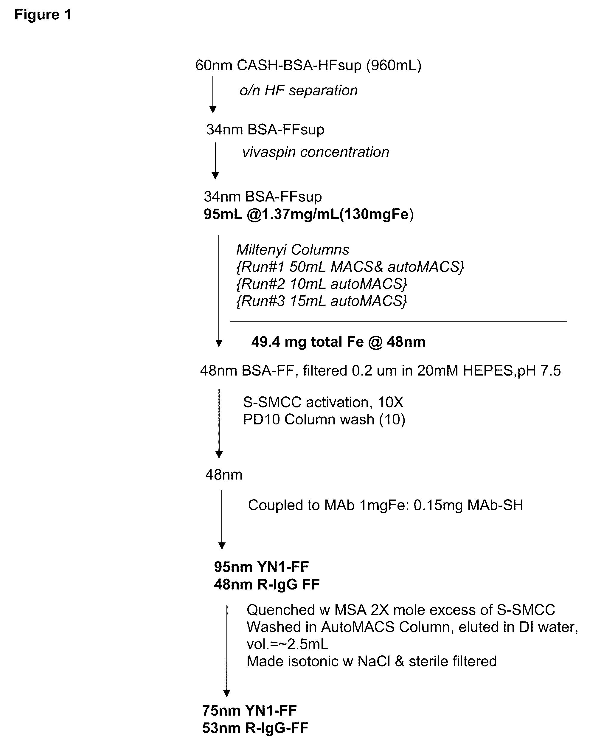
[0035]The present invention utilizes a coated, magnetic particle comprising a nanoparticle core of magnetic material, and a base coating material on the magnetic core (U.S. Pat. No. 6,365,362). These magnetic particles are characterized by extremely low non-specific binding. The magnetic core material of the particles described may comprise at least one transition metal oxide and a suitable base coating material comprises a protein. Proteins suitable for coating magnetic particles include but are not limited to bovine serum albumin and casein. The additional coating material may be the original coating proteins or one member of a specific binding pair which is coupled to the base material on the magnetic core. Exemplary specific binding pairs include biotin-streptavidin, antigen-antibody, receptor-hormone, receptor-ligand, agonist-antagonist, lectin-carbohydrate, Protein A-antibody Fc, and avidin-biotin. The member of the specific binding pair may be coupled to the base coating mate...


PUM

PropertyMeasurementUnit
Diameteraaaaaaaaaa
Paramagnetismaaaaaaaaaa

Abstract

Immunomagnetic nanoparticles are used as a contrast agent for enhancing medical diagnostic imaging such as magnetic resonance imaging (MRI). The present invention is directed to methods of making targeted MRI contrast agents from immunomagnetic particles, and to methods of using such MRI contrast agents. Typically, such targeted MRI contrast agents provide enhanced relaxivity, improved signal-to-noise, targeting ability, and resistance to agglomeration. Methods of making such MRI contrast agents typically afford better control over particle size, and methods of using such MRI contrast agents typically can afford enhanced blood clearance rates and distribution. The ability to use the contrast agents im MRI provides a tool in the diagnosis and treatment of several disease states.

Description

CROSS REFERENCE TO RELATED APPLICATIONS[0001]This application is a non-provisional application, which is incorporated by reference herein and claims priority, in part, of U.S. Provisional Application No. 60 / 856,127, filed 2 Nov. 2006.FIELD OF THE INVENTION[0002]This invention relates generally to in vivo diagnostic imaging with the use of nanoparticles. More specifically, this invention relates to a diagnostic imaging technique in which a disease state may be imaged using a targeted contrast agent formed by functionalizing nanoparticles in a coating process that incorporates a targeted moiety. These contrast agents are suitable for magnetic resonance imaging used to assess, diagnose, and treat disease states such as, but not limited to, cancer, cardiovascular, cerebrovascular, peripheral vascular, auto immune and all inflammatory diseases.BACKGROUND OF THE INVENTION[0003]The present invention relates to immunomagnetic nanoparticles as contrast agent and their use in medical diagnost...

Claims


Application Information

Patent Timeline
IPC IPC(8): A61K49/14A61K49/16
CPCA61K49/16B82Y5/00A61K49/1875A61K49/1869A61B5/055
Inventor DOYLE, GERALD V.
Owner JANSSEN DIAGNOSTICS LLC
Why Eureka
Try Eureka