Mitigation of pressure ulcers using electrical stimulation

a technology of electrical stimulation and pressure ulcers, applied in the field of treatment, can solve the problems of pressure ulcers that are not easily curable, prolong the damage of tissue, and high stress levels at the bone-muscle interface, so as to reduce damage, increase oxygenation to the loaded tissue, and restore blood flow

Inactive Publication Date: 2011-05-12
BIOMOTION
View PDF8 Cites 10 Cited by
  • Summary
  • Abstract
  • Description
  • Claims
  • Application Information

AI Technical Summary

Benefits of technology

[0025]Electrical transmission means may be at least two electrodes positioned on the skin of a person to provide electrical stimulus sufficient to effect contraction of the muscle underlying the skin. Electrical stimulation causes active contraction of the person's own muscle, thereby dynamically deforming and reshaping the muscle to reduce damage caused by mechanical compression of the muscle and restoring blood flow and increasing oxygenation to the loaded tissue.
[0026]Reconfiguration of loaded muscle tissue during the electrically-induced activation period temporarily redistributes pressure away from the loaded tissue, thereby mimicking a form of muscle deformation that occurs in able-bodied individuals during postural weight shifting or “fidgeting”, without lifting the person's body from the seating interface.
[0027]Further, reconfiguration of the loaded muscle tissue during the electrically-induced activation period, followed by a predetermined sustained relaxation period, persistently restores blood flow and tissue oxygenation to the muscle, thereby reducing damage caused by long periods of ischemia and reperfusion. Sustained restoration of tissue oxygenation (i.e. for an order of minutes in duration) during the predetermined relaxation period occurs independently of muscle mass, prevents or reduces muscle fatigue from occurring during treatment, and occurs without the need to precondition the muscle prior to treatment.

Problems solved by technology

People in intensive care units, hospital wards, or undergoing long surgical procedures are also at risk of developing pressure ulcers.
Paradoxically, the restoration of blood flow, vital to preserving tissue viability, has also been identified to cause extended damage of the tissue.
In addition to the injury caused by biochemical changes occurring during tissue ischemia and ensuing reperfusion, high stress levels at the bone-muscle interface and the duration of their application have also been reported to be direct causes of tissue injury.
Furthermore, injury to the muscle result in the formation of scar tissue, thus creating more foci for increased stress and leading to injury of adjacent previously healthy tissue.
It is the combined effects of these processes that cause the edema, inflammation and necrosis that ultimately leads to the formation of a pressure ulcer.
Such ulcers, known as “inside-out ulcers”, result from muscle breakdown due to prolonged pressure causing sustained and damaging mechanical deformation of muscle and ischemic reduction in blood flow to the tissue.
Muscle is considered to be more susceptible than dermis to tissue degradation from mechanical loading and oxygen deprivation.
In present clinical practice, pressure ulcers are normally detected by visual inspection of the skin, which often belies existing extensive damage occurring in deeper tissue.
Therefore, DTI can be perilous, as it can develop and evolve undetected by the patient or care giver until a significant destruction of the tissue has already occurred.
However, effective administration of these pressure-relieving techniques is difficult, expensive and often dependent upon patient compliance.
Specialized mattresses and cushions are heavy, expensive and not widely utilized.
Further, these techniques merely provide passive tissue load reduction, thereby failing to actively engage the patient's own muscles.
As with specialized cushions or mattresses, however, one primary disadvantage of this technique is that any benefits gained during EMS treatment of the loaded muscle (e.g., increase in muscle mass) are abolished when the work-out sessions are discontinued.
When the fatigued muscle can no longer contract, the work-out must be discontinued and the beneficial effects of the treatment are lost.
As with building muscle mass, however, pre-conditioning can be a laborious task necessitating that electrical stimulation be applied to the muscle daily for several months immediately prior to commencing treatment.
One main disadvantage of this technique, however, is that individuals who suffer from sustained and appreciable muscle atrophy still require prolonged EMS stimulation or pre-conditioning for lifting treatment of the loaded muscle to be effective.
Such restraint of the patient can lead to complications associated with reduced stability of the wheelchair user and / or fracture of weak bones.
Despite the foregoing attempts to use unloading or EMS, no single treatment has succeeded in preventing pressure ulcers effectively.

Method used


Image

  • Mitigation of pressure ulcers using electrical stimulation
  • Mitigation of pressure ulcers using electrical stimulation
  • Mitigation of pressure ulcers using electrical stimulation

Examples


example no.1

Example No. 1

[0074]Intermittent electrical stimulation may be a useful medical intervention that allows immobilized individuals to remain seated or supine for prolonged periods of time, reducing the frequency of assisted repositioning, and, most importantly, reducing the development of DTI. Experiments have been conducted to investigate the effectiveness of applying intermittent electrical stimulation (IES) to reduce muscle injury due to the presence of persistent external pressure.

experiment 1

s of IES in the prevention of DTI in the Rat

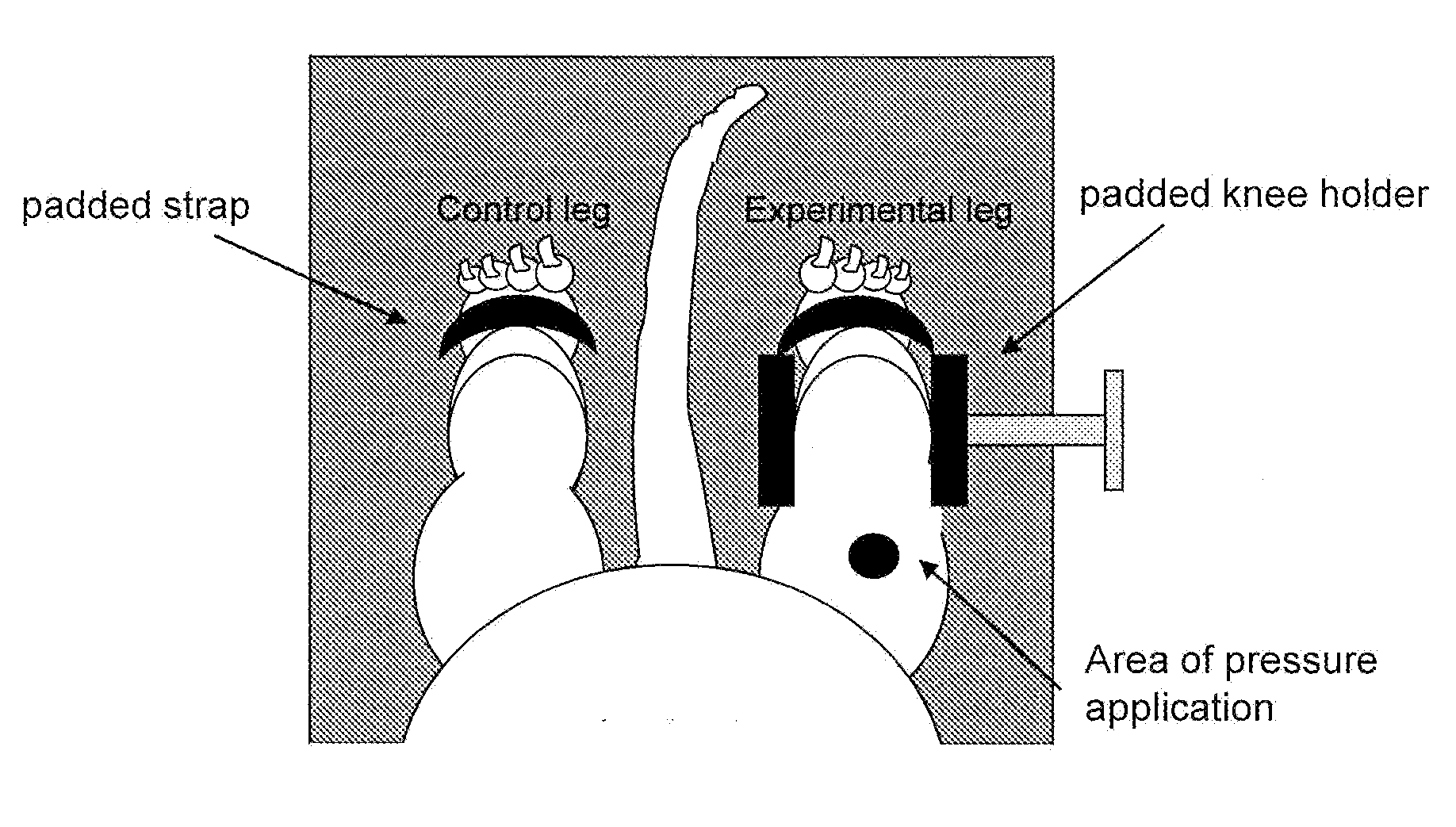
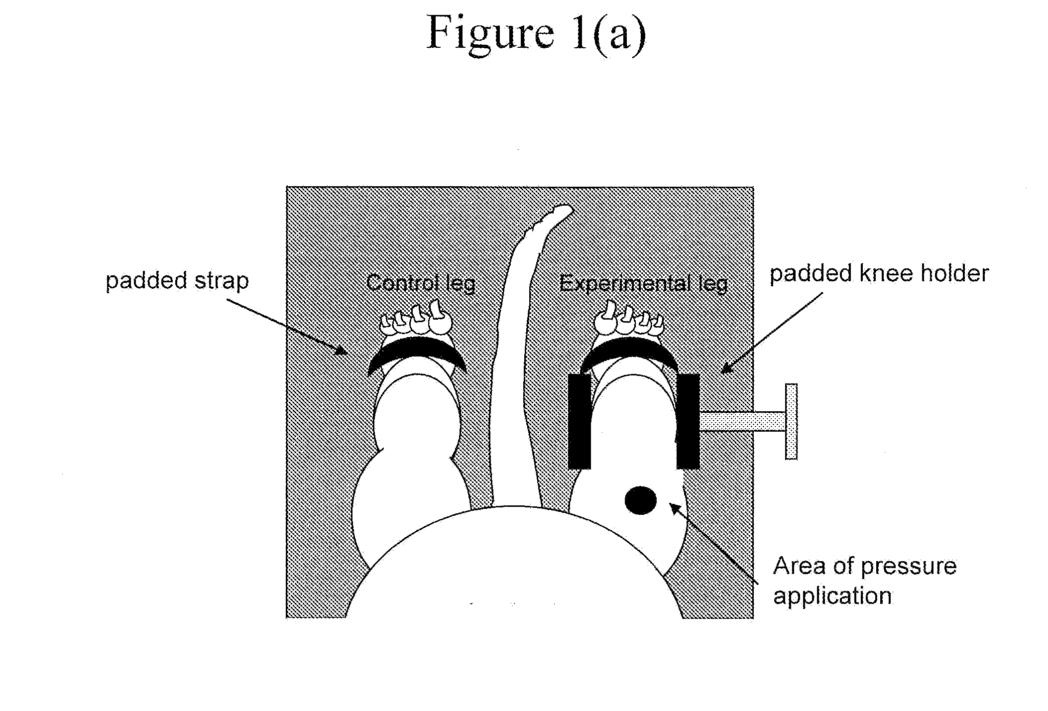
[0075]To investigate the effectiveness of IES in the prevention of DTI, a series of experiments were conducted in four groups of rats:[0076]A Control Group, which received 2 hours of external load applied to the quadriceps muscle of one hind limb,[0077]Experimental Group 1, which received the application of pressure and simultaneous application of a 10-s stimulus bout (biphasic, charge-balanced, constant current, 10-40 mA, 250 μs, 50 pulses / s) to the femoral nerve of the experimental leg every 10 minutes throughout the duration of pressure application (see FIG. 1c),[0078]Experimental Group 2, which received the same pressure as Group 1 and simultaneous electrical stimulation to the treated leg (10-s bouts) every 5 minutes, and[0079]Experimental Group 3, which received the application of IES at 5-minute intervals but without pressure application.

[0080]Eighteen adult female, Sprague-Dawley rats were anesthetized with isoflurane (2-3% isoflur...

experiment # 2

Experiment #2: Mechanisms of Action of IES in the Human

[0094]To obtain an insight into the mechanisms of action of IES, the effect of IES on tissue oxygenation was measured in two separate experiments with human volunteers. In the first experiment, tissue oxygenation measurements were obtained from an able-bodied volunteer by means of T2* MRI quantification in muscles in both unloaded and loaded conditions, respectively. In the second, changes in the surface (bed-buttocks interface) pressure profiles generated by the IES-elicited contractions were measured in an able-bodied volunteer.

Tissue Oxygenation in Able-Bodied Human

[0095]An initial experiment was conducted in an able-bodied volunteer (male, 22 yr) to assess changes in tissue oxygenation associated with contractions elicited by IES in an unloaded muscle. The experimental setup is illustrated in FIG. 7. Electrodes were placed on the body and electrical stimulation was delivered to induce muscle contractions. The change in the s...


PUM


Abstract

A method is provided for mitigating or preventing formation of pressure ulcers by transmitting an electrical stimulus sufficient to effect contraction of a loaded muscle, wherein the method comprises the steps of providing an electrical transmission for effecting contraction of the loaded muscle, transmitting sufficient electrical stimulation to the muscle to contract it for a predetermined period of time (the ON period), and ceasing transmission of the stimulus to the muscle for a predetermined period of muscle relaxation (the OFF period), whereby the predetermined period of relaxation is sufficient to cause sustained reoxygenation of the activated muscle.

Description

CONTINUATION IN PART[0001]This application is a continuation-in-part of U.S. patent application Ser. No. 12 / 362,725, filed Jan. 30, 2009, which claims priority of U.S. Provisional Patent Application No. 61 / 025,472, filed Feb. 1, 2008.FIELD OF THE INVENTION[0002]This invention relates to treatments designed to mitigate pressure ulcers and, in particular, the mitigation of pressure ulcers by means of electrical stimulation.BACKGROUND[0003]Pressure ulcers (also known as “bed sores” or “pressure sores”) are typically associated with individuals having compromised mobility or lack of sensation, such as the infirm, elderly and people suffering from stroke, spinal cord injury, bone and joint disease, vascular pathologies, tumours and diabetes. People in intensive care units, hospital wards, or undergoing long surgical procedures are also at risk of developing pressure ulcers.[0004]A pressure ulcer is a tissue abnormality or lesion resulting from pressure imposed upon soft tissue underlying...

Claims


Application Information

Patent Timeline
Patent Type & Authority Applications(United States)
IPC IPC(8): A61N1/36
CPCA61N1/36003A61N1/322
Inventor MUSHAHWAR, VIVIAN K.SOLIS-AGUILAR, LEANDRO RAFAEL
Owner BIOMOTION
Why Eureka
Try Eureka