Device for diagnosing tissue injury

a tissue injury and diagnostic device technology, applied in the field of tissue injury diagnostic devices, can solve the problems of lipid peroxidation and apotosis, difficult to make quantitative analysis of ros, and the detection current of superoxide anion radicals is reduced promptly, so as to achieve accurate diagnosis

Inactive Publication Date: 2011-05-19
YUASA MAKOTO +3
View PDF3 Cites 4 Cited by
  • Summary
  • Abstract
  • Description
  • Claims
  • Application Information

AI Technical Summary

Benefits of technology

[0030]According to the present invention, in vivo free radicals typified by superoxide anions in systemic organ or tissue injury caused by cerebral ischemia reperfusion

Problems solved by technology

Though ROS play various important roles in biophylaxis and homeostasis, production of excess ROS induced by oxidative stress induce radical toxicity, which leads to lipid peroxidation and apotosis.
However, it is considered very difficult to make quantitative analysis of ROS, because ROS are quickly eliminated by antioxidase and antioxidant in the living body.
In addition, it has also been confirmed that the detection current of superoxide anion radicals decreased promptly by administration of superoxide dismutase (SOD) and that

Method used


Image

  • Device for diagnosing tissue injury
  • Device for diagnosing tissue injury
  • Device for diagnosing tissue injury

Examples


example 1

[0094]In Vitro Validation of Radical Sensor in Rat Blood

[0095]The radical sensor 15 was verified in rat blood before it was applied to an in vivo model.

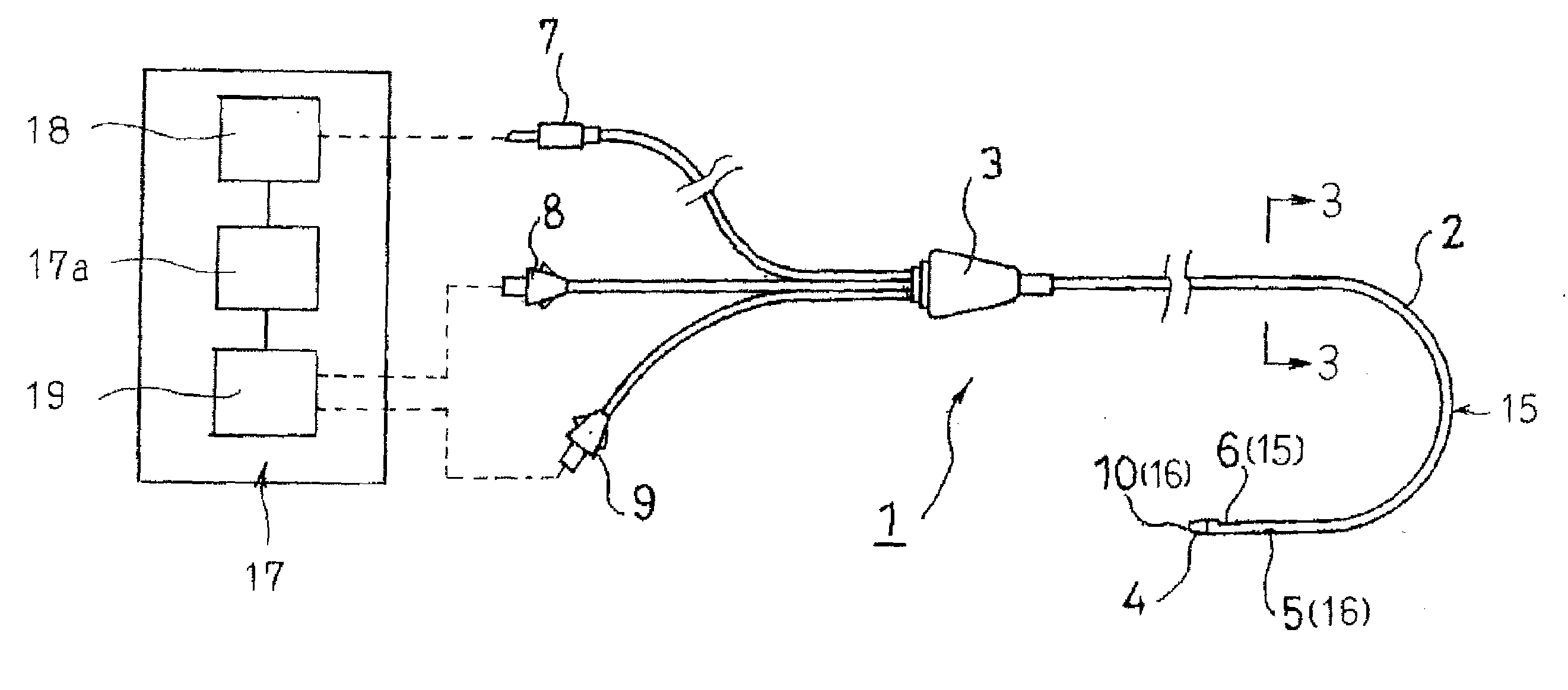
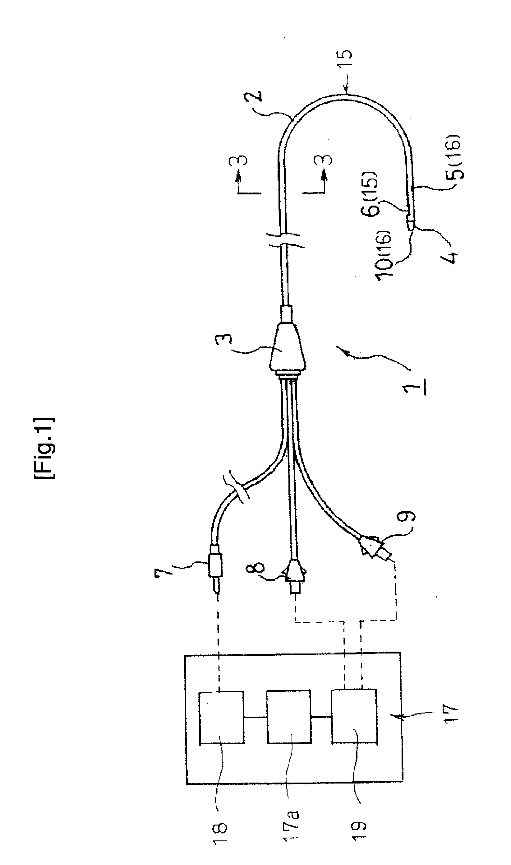
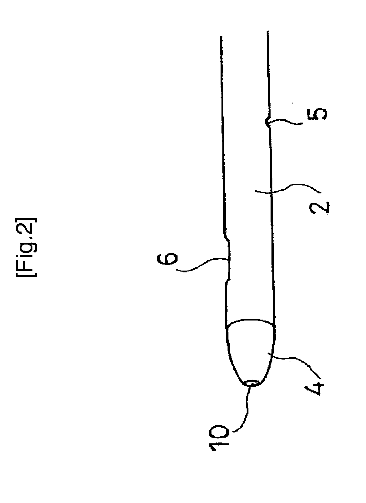
[0096]That is, the amount of superoxide anion radicals was measured by immersing the sensor electrode 6 formed at the tip end 4 of the catheter 1 of the device of the present invention 1 in rat blood collected from a rat (see FIG. 5).

[0097]1) Subject Rats

[0098]Twenty-eight male specific pathogen free Wister rats, weighing 250-300 g, were used. The rats were anesthetized by 4% isoflurane and 96% oxygen. After laparotomy, whole blood was sampled from the inferior vena cava with 500 U of heparin. Blood samples were used as soon as possible.

[0099]2) Preparation and Measurement of Rat Blood

[0100]Individual bloods were randomly assigned to one of three experimental groups: 30 mU / ml of xanthine (hereinafter referred to as “XAN”)+xanthine oxidase (hereinafter referred to as “XOD”) (n=7, low XOD group) (see (2) in FIG. 5), 60 mU / ml of XAN+XOD...

example 2

[0116]Measurement of Superoxide Concentration in an Acute Phase Ischemic Disease Model (Rats)

[0117]1) Subject Rats

[0118]Sixteen male Wister rats (260-280 g) were randomly assigned to 2 groups, i.e., the reperfusion group (n=8) and the reperfusion with SOD group (n=8). There is no statistical difference of body weight of rats between the 2 groups.

[0119]2) Configuration of the Radical Sensor 15

[0120]The radical sensor 15 installed in the device of the present invention is a catheter-typed one shown in FIG. 1. An electrodeposited film of a polymeric iron porphyrin derivative attached to a carbon electrode is placed in stainless steel tube as an auxiliary counter electrode.

[0121]3) Experimental Method

[0122]A dummy probe and a guide tube of microdialysis were embedded in the brain parenchyma of the rat on the previous day of the experiment under pentobarbital anesthesia (intraperitoneal administration, 50 mg / kg). The guide tube was fixed by dental resin at 3 mm outside and 3 mm anterior ...

example 3

[0137]Measurement of Superoxide Concentration in Acute Phase Sepsis Model (in Rats)

[0138]1) Subject Rats

[0139]Twenty-one male specific pathogen free Wister rats, weighing 250-300 g, were used in this example. The rats were randomly assigned to one of the 3 groups, i.e.; sham group: intravenous bolus and continuous administration of saline (n=7); LPS group: bolus lipopolysaccharide (hereinafter referred to as “LPS”) and continuous administration of saline (n=7); and bolus LPS and continuous administration of superoxide dismutase (SOD) (n=7).

[0140]2) Configuration of the Radical Sensor 15

[0141]The radical sensor 15 installed in the device of the present invention 1 is a catheter-typed one shown in FIG. 1. An electrodeposited film of polymeric iron porphyrin derivative attached to a carbon electrode is placed in stainless steel tube as an auxiliary counter electrode.

[0142]3) Experiment Method

[0143]The rats were anesthetized by 3% isoflurane and 97% oxygen. They were mechanically ventil...


PUM


Abstract

The device for diagnosing tissue injury of the invention has a catheter insertable into the body and a radical sensor provided in the catheter, and is characterized in that the radical sensor has a sensor electrode capable of measuring superoxide anion radicals provided at a tip end of the catheter, a lead wire connector for a sensor provided at a basal portion of the catheter, and a lead wire for a sensor for connecting the sensor electrode portion to the lead wire connector for a sensor. With the device, in vivo free radicals typified by superoxide anions in systemic organ or tissue injury caused by cerebral ischemia reperfusion injury, severe infection or sepsis can be promptly and quantitatively monitored, and whether the in vivo tissue conditions are good or not can be accurately diagnosed.

Description

TECHNICAL FIELD[0001]The present invention relates to a device for diagnosing tissue injury which diagnoses whether the in vivo tissue conditions are good or not, and particularly relates to a device for diagnosing tissue injury preferable for diagnosing whether the tissue conditions of a human body are good or not, and for diagnosing, for example, cerebral ischemia reperfusion injury, and systemic organ or tissue injury caused by severe infection or sepsis.BACKGROUND ART[0002]Cerebral ischemia reperfusion injury in stroke is a brain disorder caused by reactive oxygen species, reactive nitrogen species, free radicals or the like in the body (hereinafter collectively referred to as “in vivo free radicals”). In addition, severe infection, sepsis or the like is caused by in vivo free radicals.[0003]Among these in vivo free radicals, superoxide anion radical (O2−.), hydrogen peroxide, hydroxyl radical, singlet oxygen, lipid peroxide radical and the like are known as reactive oxygen spec...

Claims


Application Information

Patent Timeline
IPC IPC(8): A61B5/04
CPCA61B5/01A61B5/02152A61B5/14503A61B5/14546A61M2210/0693A61B5/412A61B5/6852A61M5/1723A61M2005/1726A61B5/1473A61B5/4076
Inventor YUASA, MAKOTOMAEKAWA, TSUYOSHIFUJITA, MOTOKIKIDO, SHIGERU
Owner YUASA MAKOTO
Why Eureka
Try Eureka