Tissue occluding agent

a tissue occluding agent and tissue technology, applied in the direction of drug compositions, extracellular fluid disorders, peptide/protein ingredients, etc., can solve the problems of time and effort, high death probability, and inability to remove hemorrhaged blood, etc., to achieve adequate tissue occluding effect, improve tissue adhesion, and improve the effect of curative

Active Publication Date: 2011-08-18
3 D MATRIX
View PDF7 Cites 40 Cited by
  • Summary
  • Abstract
  • Description
  • Claims
  • Application Information

AI Technical Summary

Benefits of technology

[0054]The self-assembling peptide as the major component in the tissue occluding agent of the invention can also serve as a scaffold for migrating cells in addition to its role as a occluding agent, thus allowing it to have a higher curative effect after surgery instead of simple occlusion. In addition, the tissue occluding agent of the invention has improved adhesion to tissue when excess body fluid has been removed from a body fluid leakage site (for example, when it ha

Problems solved by technology

Probability of death is high when hemorrhage cannot be immediately arrested.
2. Hemorrhage during surgery is a major concern, since hemorrhage can lead to systemic infection or organ dysfunction.
Hemorrhage not only interferes with the object of surgery, but removal of hemorrhaged blood is also a delaying factor in surgery.
3. Hemorrhage is also a problem with minimally invasive surgery (laparoscopic surgery and the like), and switching to incisive surgery may be necessary when hemorrhage cannot be sufficiently prevented.
The drawback to this method is that time and effort are required to maintain pressure, while the patient is also at risk of hematoma.
The drawback to these hemorrhage arresting methods is difficulty of management when the hemorrhaging is from numerous microvessels.
The drawback of such methods is that the surrounding tissue is subjected to thermal injury and the patient undergoes increased invasion, while the medical instruments used require expert skill (the method cannot be used outside of a medical institution).
Collagen fibers and fibrin paste are often clinically used as effective hemorrhage arresting materials, but their drawbacks include the fact that (1) gelatin and collagen fibers are animal collagen and fibrin paste is an animal-derived product obtained using a blood preparation and bovine thrombin, and therefore the risk of infection exists,

Method used


Image

  • Tissue occluding agent
  • Tissue occluding agent
  • Tissue occluding agent

Examples


example 1

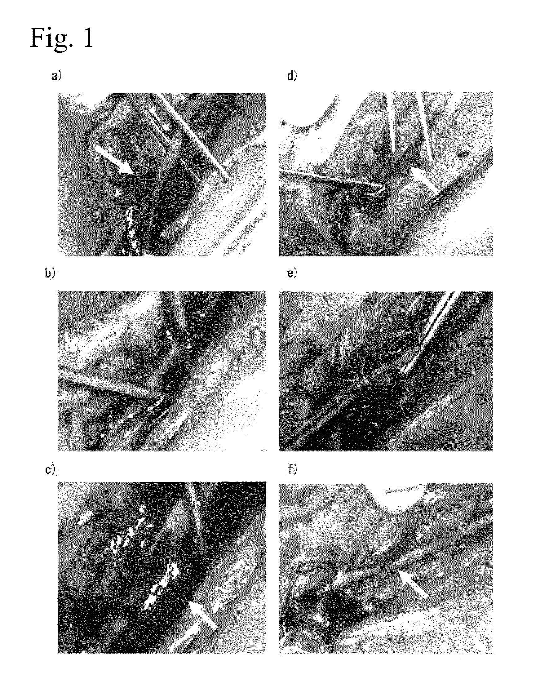
Hemostatic Effects of 1% Aqueous Peptide Solution and 3% Aqueous Peptide Solution in Rabbit Abdominal Aorta / Portal Vein Stem Injection Needle Perforation Model

[0094]A rabbit abdominal aorta and portal vein stem injection needle perforation model was prepared under vascular clamp, and the hemostatic effects of 1% aqueous peptide solution and 3% aqueous peptide solution were evaluated.

[0095]Aqueous Peptide Solutions

[0096]1.1% Aqueous peptide solution (peptide sequence: Ac-(RADA)4-NH2, CPC Scientific, Inc; concentration: wt / vol)

[0097]2.3% Aqueous peptide solution (peptide sequence: Ac-(RADA)4-NH2, CPC Scientific, Inc; concentration: wt / vol)

[0098]Animals

[0099]Japanese white rabbits (3.0-4.0 kg, Japan White, conventional, purchased from Funabashi Farm Co., Ltd.). The animals were bred with breeding pellets (JA Higashinihon Kumiai Shiryou) supplied in a breeding room controlled at a room temperature of 25° C., a humidity of 65% and an illumination time of 12 hours (7:00-19:00), and water ...

example 2

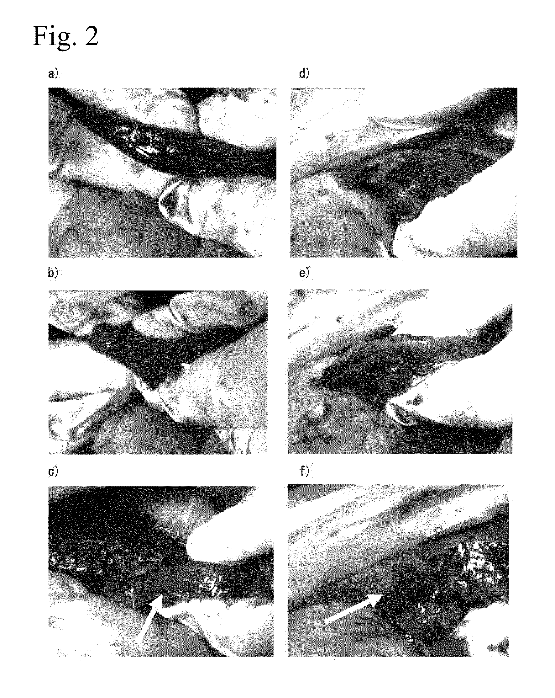
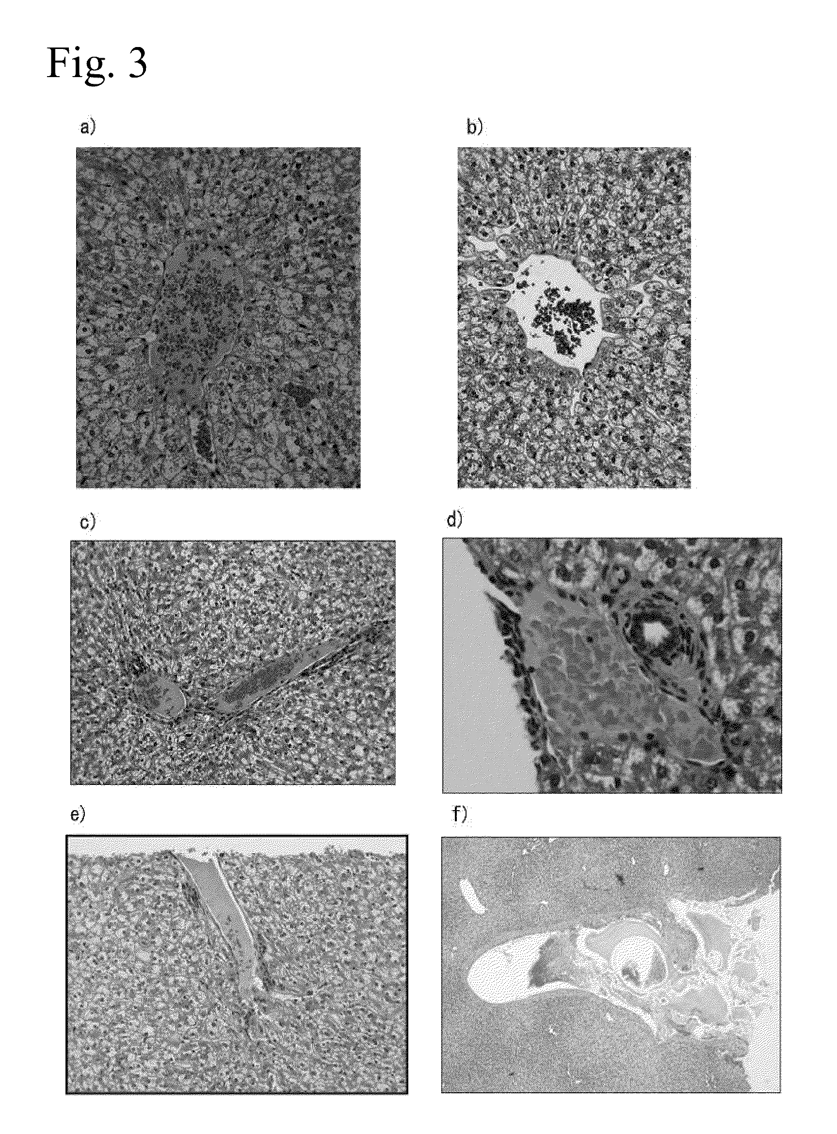
Hemostatic Effects of 1% Aqueous Peptide Solution and 3% Aqueous Peptide Solution in Rabbit Partial Liver Resection Model

[0105]A rabbit partial liver resection model was prepared and the hemostatic effects of 1% aqueous peptide solution and 3% aqueous peptide solution were evaluated.

[0106]Aqueous Peptide Solutions

[0107]1. 1% Aqueous peptide solution (peptide sequence: Ac-(RADA)4-NH2, CPC Scientific, Inc.)

[0108]2. 3% Aqueous peptide solution (peptide sequence: Ac-(RADA)4-NH2, CPC Scientific, Inc.)

[0109]Animals

[0110]Japanese white rabbits (3.0-4.0 kg, Japan White, conventional, purchased from Funabashi Farm Co., Ltd.). The animals were bred with breeding pellets (JA Higashinihon Kumiai Shiryou) supplied in a breeding room controlled to a room temperature of 25° C., a humidity of 65% and an illumination time of 12 hours (7:00-19:00), and water freely supplied with a water bottle. Fasting was from the morning of the test day, with water freely supplied.

[0111]The rabbits were subcutaneou...

example 3

Hemostatic Effect of Aqueous Peptide Solution in Abdominal Aorta Injection Needle Perforation Model of Rabbits with Suppressed Blood Clotting Function

[0117]The hemostatic effect of an aqueous peptide solution in rabbits administered an anticoagulant (heparin) was evaluated in an abdominal aorta injection needle perforation model.

[0118]Aqueous Peptide Solution

[0119]3% Aqueous peptide solution (peptide sequence: Ac-(RADA)4-NH2, CPC Scientific, Inc.)

[0120]Animals

[0121]Japanese white rabbits (3.0-4.0 kg, Japan White, conventional, purchased from Funabashi Farm Co., Ltd.). The animals were bred with breeding pellets (JA Higashinihon Kumiai Shiryou) supplied in a breeding room controlled to a room temperature of 25° C., a humidity of 65% and an illumination time of 12 hours (7:00-19:00), and water freely supplied with a water bottle. Fasting was from the morning of the test day, with water freely supplied.

[0122]The rabbits were subcutaneously administered (3 mg / kg) Celactar 2% injection (...


PUM

PropertyMeasurementUnit
Fractionaaaaaaaaaa
Fractionaaaaaaaaaa
Hydrophilicityaaaaaaaaaa

Abstract

There is provided a bioabsorbable peptide tissue occluding agent that can be applied to large mammals including humans, the peptide tissue occluding agent being obtained by artificial synthesis to avoid concerns of infection by viruses and the like.
The tissue occluding agent contains a peptide, wherein the peptide is an amphiphilic peptide having 8-200 amino acid residues with the hydrophilic amino acids and hydrophobic amino acids alternately bonded, and is a self-assembling peptide exhibiting a β-structure in aqueous solution in the presence of physiological pH and/or a cation.

Description

TECHNICAL FIELD[0001]The present invention relates to a tissue occluding agent comprising a self-assembling peptide hydrogel.BACKGROUND ART[0002]Tissue occlusion to prevent leakage of body fluids (blood, tissue fluids and the like) caused by tissue damage have major significance in clinical situations, including surgery. Effectively inhibiting body fluid leakage from damage sites is associated with improved life support during surgery and improved post-surgical quality of life (QOL).[0003]Hemostasis is considered clinically important for the following reasons.[0004]1. Blood loss is a major cause of death, with causes of blood loss including serious trauma, aneurysm, esophageal and gastric ulcers, and esophageal varix rupture. Probability of death is high when hemorrhage cannot be immediately arrested.[0005]2. Hemorrhage during surgery is a major concern, since hemorrhage can lead to systemic infection or organ dysfunction. Hemorrhage not only interferes with the object of surgery, b...

Claims


Application Information

Patent Timeline
IPC IPC(8): A61K38/00C07K7/08A61K38/08
CPCA61K38/16A61L31/047A61L31/148A61K31/7004C07K7/08A61K38/08A61K38/10C07K7/06A61K31/7016A61L31/145A61L2400/04A61K2300/00A61L2430/36A61P7/04A61L17/10A61K38/00A61L31/16
Inventor TAKAMURA, KENTAROGOJO, SATOSHIKOBAYASHI, SATORU
Owner 3 D MATRIX
Why Eureka
Try Eureka