
Production of Biologically Active Proteins

a technology of applied in the field of biologically active recombinant peptides and proteins, can solve the problems of poor host for the synthesis of correctly-folded mammalian proteins normally stabilized by disulfide bonds, lack of biological activity expected of that material, and poor efficiency of the oxi-redox system of bacteria for eukaryotic proteins, etc., to achieve the effect of increasing the accumulation rate and being readily availabl

Inactive Publication Date: 2012-01-26
ERA BIOTECH SA
View PDF4 Cites 6 Cited by
  • Summary
  • Abstract
  • Description
  • Claims
  • Application Information

AI Technical Summary

Benefits of technology

"The present invention provides a system and method for producing a fusion protein containing a protein body-inducing sequence (PBIS) and a biologically active polypeptide or protein of interest (target) in eukaryotic cells. The PBISs are able to induce the formation of recombinant fusion proteins that accumulate as recombinant protein body-like assemblies (RPBLAs) in the host cells. The RPBLAs are stable and can be isolated and purified from the host cells. The method involves the transformation of host cells with a nucleic acid sequence coding for the fusion protein and a PBIS. The biologically active polypeptide of the fusion protein exhibits at least 25% of the biological activity of the same polypeptide isolated from the second cell type in an assay of the activity of that polypeptide. The nucleic acid sequence used for transformation can also contain a linker or spacer amino acid sequence between the PBIS and the target polypeptide. The invention provides a novel method for producing a biologically active fusion protein in eukaryotic cells."

Problems solved by technology

In many instances, however, the protein or polypeptide that is ultimately produced can possess the correct primary amino acid residue sequence of the naturally-produced molecule, but lack the biological activity expected of that material.
For example, disulfide bond formation occurs spontaneously in the lumen of the endoplasmic reticulum (ER) of eukaryotic cells, but not in the reducing environment of the cytosol of prokaryotes, which makes bacterial cells such as Escherichia coli poor hosts for the synthesis of correctly-folded mammalian proteins that are normally stabilized by disulfide bonds.
However, even in this compartment the bacterial oxi-redox system is not very efficient for eukaryotic proteins.
Unfortunately, many such eukaryotic expression systems are inefficient with respect to protein product yield and cost of manufacture, even when proteins are secreted extracellularly.
The high costs frequently derived from low recombinant protein production levels and / or from complicated downstream protein isolation and purification procedures can invalidate a protein's commercial application.
This is an important issue because the efficiency of protein renaturation is highly limiting, particularly if the protein contains disulfide bonds (Clarc, Ed., April 2001 Curr. Opin. Biotechnol. 12(2):202-207).
As a consequence the yield of correctly refolded recombinant proteins from inclusion bodies is extremely low, and moreover the biological activities of such refolded proteins are typically much less than that of the native-formed proteins.
In addition, those authors constructed zein-GFP fusion proteins to determine the subcellular localization of zein proteins in yeast cells but did not observe formation of dense, concentrated structures characteristic of bona fide PBs.
(2002, Plant Cell 14: 655-672) concluded that yeast is a poor model for the study of zein interactions because zeins accumulated very poorly in transformed yeast.
Moreover, the presence of disulfide bonds in some natural PB-assembling protein domains, as for instance RX3, [Ludevid et al., 1984 Plant Mol. Biol. 3:227-234 and Kawagoe et al., 2005 Plant Cell April 17(4):1141-1153], which are probably involved in PB formation and stabilization, could represent an additional difficulty for production of a biologically active, native-folded protein in PBs.
However, the absence of immunomodulatory components having adjuvant properties associated with attenuated or killed vaccines often results in weaker immunogenicity for such vaccines.
Yet, this destructive environment generates peptides that are capable of binding to MHC class II molecules.
To a large degree, this diversity limits the use of molecular targeting techniques to cell surface markers and immune modulators such as cytokines, because for many species including wildlife, only minimal knowledge of these molecules is available.

Method used


Image

  • Production of Biologically Active Proteins
  • Production of Biologically Active Proteins
  • Production of Biologically Active Proteins

Examples


example 1

Accumulation of RX3-ECFP Derived Fusion Proteins in Dense Fractions of Transfected Mammal Cells

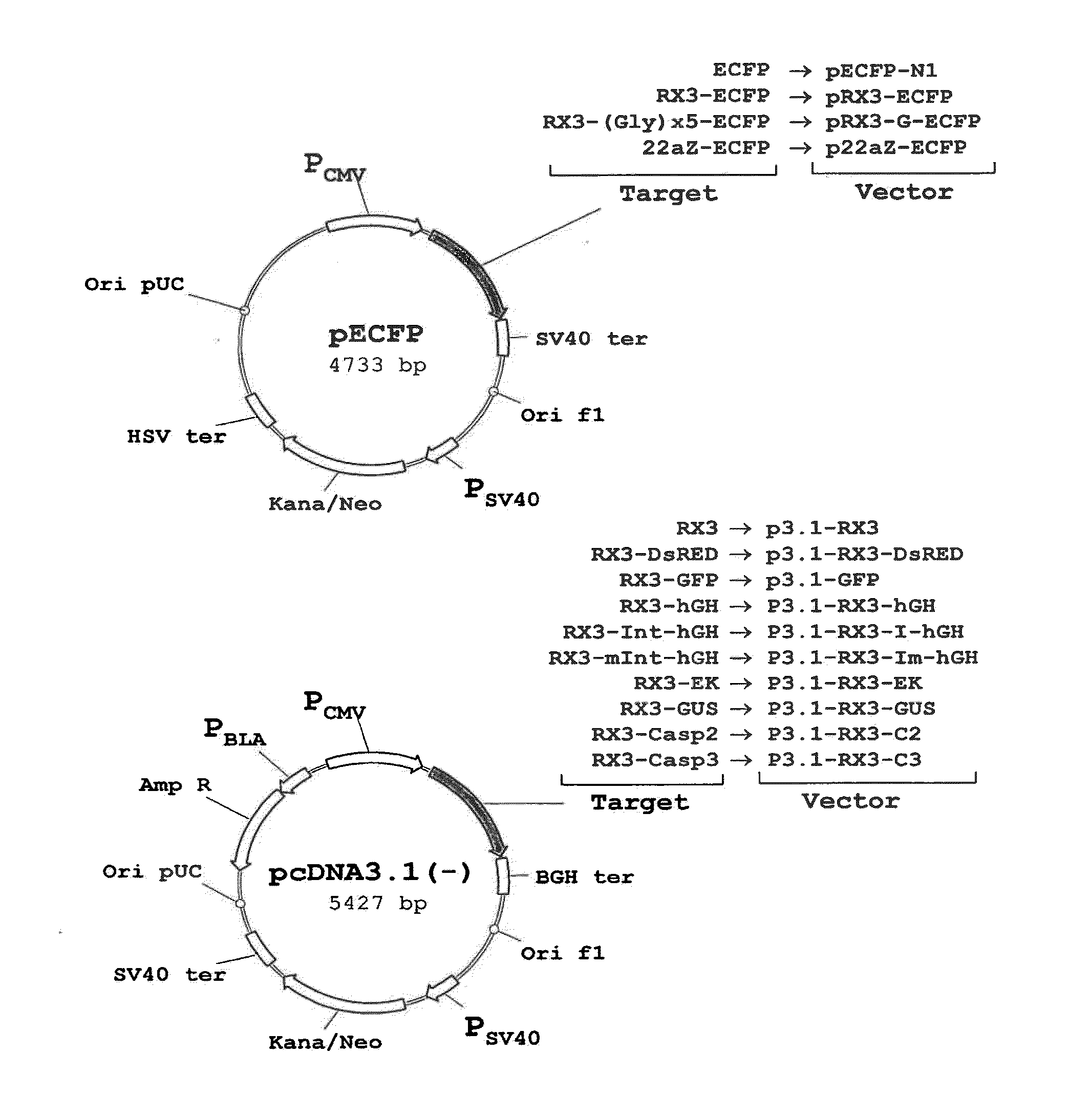
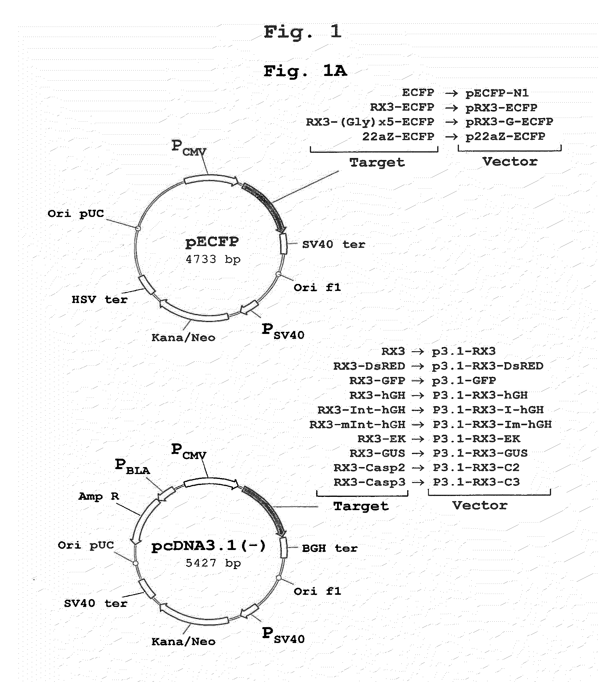
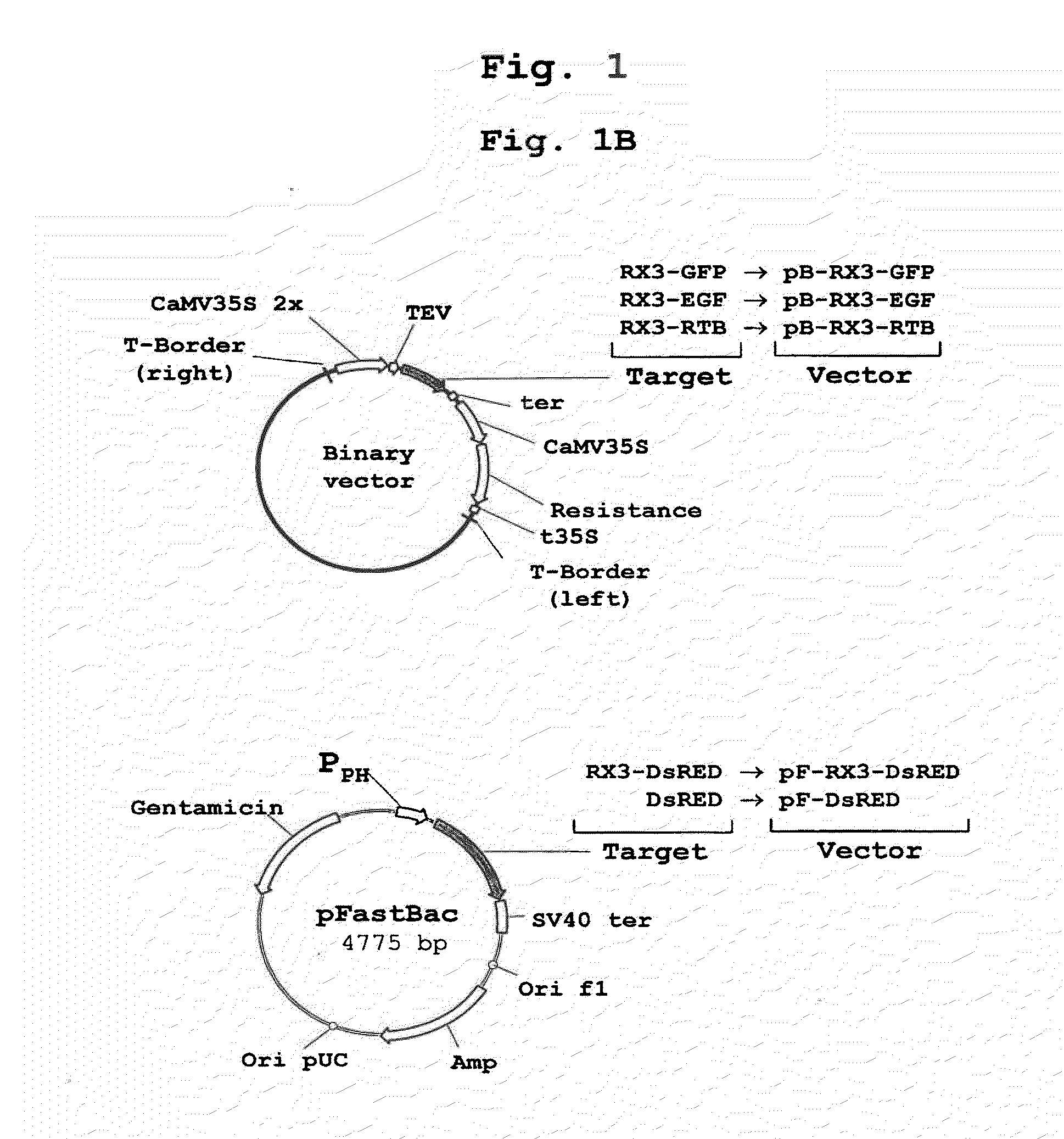
[0172]The polynucleotide sequence coding for the N-terminal gamma-zein coding sequence RX3 (WO2004003207) was fused directly, or through a linker consisting of five glycines, to the 5′ end of the sequence encoding ECFP, a cyan fluorescent variant of GFP. Those constructs (FIG. 1A) that code for the fusion proteins RX3-ECFP or RX3-Gx5-ECFP were introduced in cultured mammalian CHO cells by the Lipofectamine-based transfection method (Invitrogen). CHO cells transfected with plasmid pECFP-N1 (Clontech) containing the gene sequence of a non-targeted (cytosolic) ECFP were used as controls.

[0173]Transfected mammalian cell extracts were loaded on density step gradients and centrifuged. The accumulation of recombinant proteins in the different fractions was analyzed by immunoblot. The results shown in FIG. 2A indicate that RX3-ECFP and RX3-Gx5-ECFP sedimented in fractions F42, F56 and P correspond...

example 2

Accumulation of Active ECFP Fused to PBIS Domains in RPBLAs of Transfected Mammal Cells

[0175]To determine if the fusion proteins RX3-ECFP, RX3-Gx5-ECFP and 22aZ-ECFP are active inside the RPBLAs, confocal microscopic analysis was performed to visualize target protein fluorescence in transfected CHO cells (FIG. 1A). Cyan fluorescence was imaged by excitation at 458 nm with an argon ion laser with an emission window set at 470-530 nm. As shown in FIG. 3, the corresponding fusion proteins, RX3-ECFP (FIG. 3A) and RX3-Gx5-ECFP (FIG. 3B) and 22aZ-ECFP (FIG. 3D), were detected in proximity to the ER, indicating that the gamma-zein and the alpha-zein signal peptides are functional in mammalian cells where they mediates the translocation of the fusion protein into the ER.

[0176]In addition, the fusion proteins surprisingly also accumulated preferentially into large and dense spherical structures that strongly resembled both authentic PBs of cereal seed and RPBLAs in heterologous systems visua...

example 3

Subcellular Localization of Other Fluorescent Proteins Fused to RX3 in CHO Cells

[0178]The sub-cellular localization of RX3-DsRED and RX3-GFP fusion proteins in transiently transfected CHO cells was analyzed by confocal microscopy to analyze whether other fluorescent proteins than ECFP fused to RX3 are properly folded and bioactive inside RPBLAs. It is important to note that DsRED shares no homology to ECFP, which implies a completely different folding mechanism. Fluorescence images from the transfected cells were obtained by using a confocal laser scanning microscope (Leica TCS SP, Heidelberg, Germany) fitted with spectrophotometers for emission band wavelength selection. Green fluorescent images were collected by excitation at 488 nm with an Argon ion laser using an emission window set at 495-535 nm. Red fluorescent images were collected using 543 nm excitation with a HeNe laser and an emission window of 550-600 nm. Optical sections were 0.5 μm thick.

[0179]The expression of RX3-GFP...


PUM

PropertyMeasurementUnit
diameteraaaaaaaaaa
diametersaaaaaaaaaa
densityaaaaaaaaaa

Abstract

A fusion protein that is expressed in a recombinant protein body-like assembly (RPBLA) in host eukaryotic cells and organisms is disclosed. More particularly, a biologically active polypeptide fused to a protein sequence that mediates the induction of RPBLA formation is expressed and accumulated in host cells after transformation with an appropriate vector. The eukaryotic host cell does not produce protein bodies in the absence of the fusion protein. Methods for preparing and using the RPBLAs and the fusion protein are also disclosed, as are nucleic acid molecules that encode the fusion proteins.

Description

CROSS-REFERENCE TO RELATED APPLICATION[0001]This application claims benefit of provisional application Ser. No. 60 / 776,391 that was filed on Feb. 23, 2006.TECHNICAL FIELD[0002]The present invention contemplates the production of biologically active recombinant peptides and proteins, collectively referred to as polypeptides, in eukaryotic cells and organisms as host systems. More particularly, a biologically active polypeptide is fused to a protein body-inducing sequence (PBIS) that mediates the induction of recombinant protein body-like assemblies (RPBLA) to form a fusion protein that is stably expressed and accumulated in the host system as an RPBLA after transformation of the host cells with an appropriate vector.BACKGROUND ART[0003]The production of recombinant proteins for therapeutic, nutraceutical or industrial uses has enjoyed great success over the past several decades. Introduction of heterologous genes having desired nucleotide sequences into a variety of expression hosts ...

Claims


Application Information

Patent Timeline
Patent Type & Authority Applications(United States)
IPC IPC(8): A61K39/00A61P37/04C12N5/0783C12N5/0786C07K14/00C12N5/0781
CPCA61K39/00C07K14/415C07K14/425C07K14/485C07K14/61C12Y304/21009C12N9/6475C12N15/62C12N15/8257C12N2799/026C12P21/02C12N9/6424A61P37/04C12N5/10C12N15/79A61K9/0053A61K9/0019C07K14/43504
Inventor HEIFETZ, PETER BERNARDLLOMPART ROYO, BLANCAMARZ BAL LUNA, PABLOBASTIDA VIRGILI, MIRIAMLUDEVID M GICA, M DOLORESTORRENT QUETGLAS, MARGARITAO'CONNER, KEVIN JAMESBERGWERF, ROSER PALLISSELLOP TOUS, M IMMACULADA
Owner ERA BIOTECH SA
Learn More