
Anchorage devices comprising an active pharmaceutical ingredient

Active Publication Date: 2012-07-19
MEDTRONIC INC
View PDF7 Cites 31 Cited by
  • Summary
  • Abstract
  • Description
  • Claims
  • Application Information

AI Technical Summary

Benefits of technology

[0011]In some embodiments, the polymer coating can be capable of releasing one or more drugs into surrounding bodily tissue to reduce or prevent surgery-related complications associated with the implantable medical device (such as to the “pocket” surrounding the device). For example, an anesthetic agent in the polymer coating can be eluted into the surrounding bodily tissue, bodily fluid, or systemic fluid, to attenuate pain experienced at the implantation site. In another example, replacing the anesthetic agent with an anti-inflammatory agent can reduce the swelling and inflammation associated implantation of the mesh substrate and / or the implantable medical device. In yet another example, an antimicrobial agent can be provided at a rate of drug release sufficient to prevent or reduce colonization of the mesh substrate, the implantable medical device and / or the surgical implantation site by bacteria, for example, for at least the period following surgery necessary for initial healing of the surgical incision.
[0013]Without wishing to be bound by any particular theory, it is believed that by eluting an antimicrobial agent over time to an area surrounding or adjacent to a transdermal medical device, the incidence of microbial infections may be reduced, prevented, or mitigated, especially against those organisms described herein.
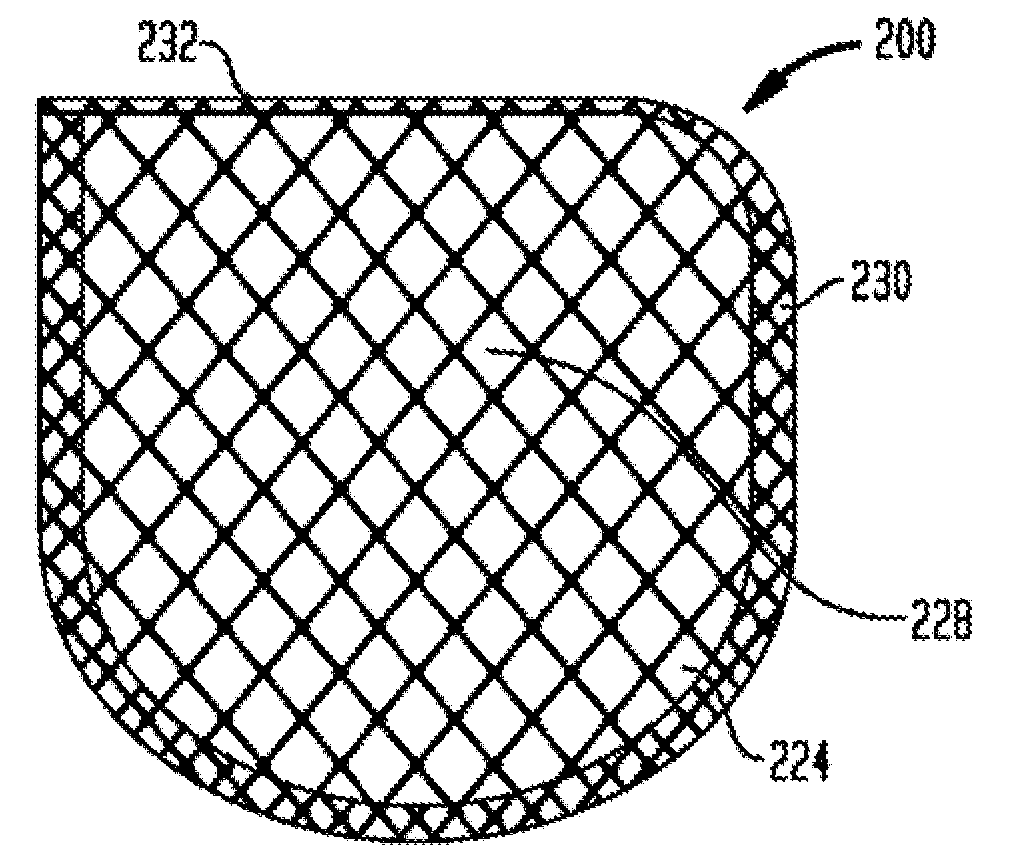
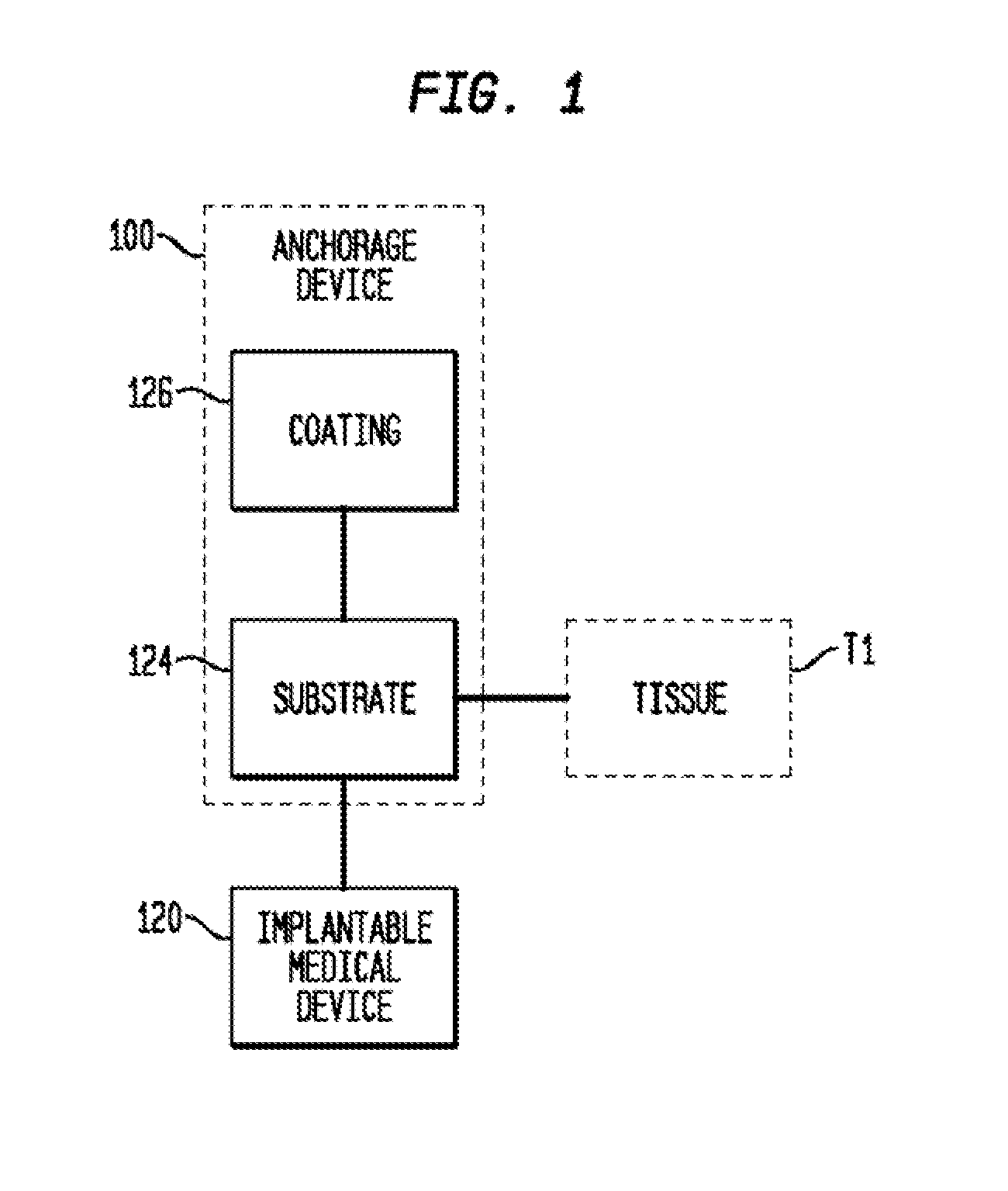
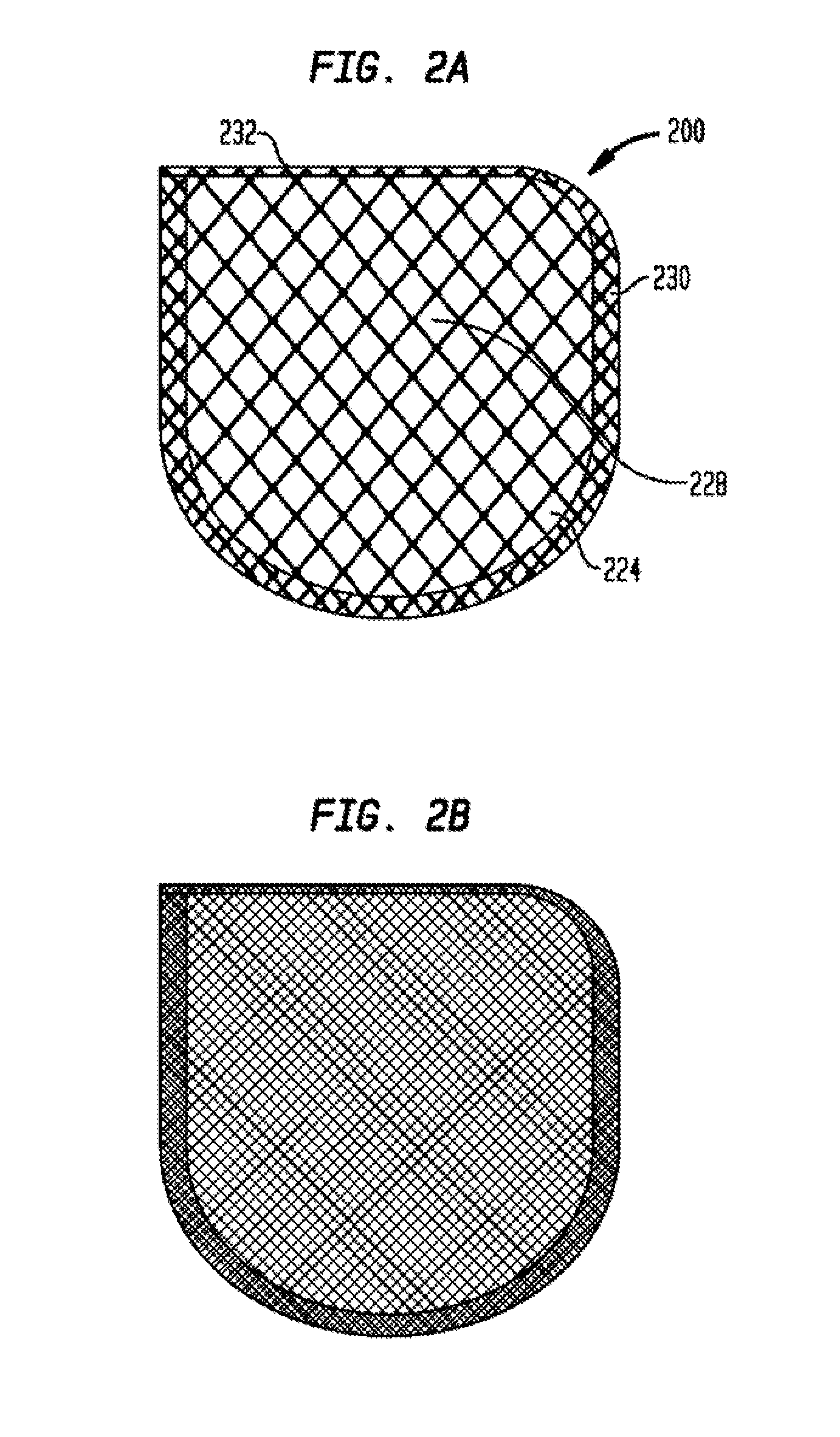
[0014]In another aspect of the present invention, devices and methods are described for use in supporting an implantable medical device, such as a cardiac defibrillator or a pacemaker, in a desired position at a treatment site within a body of a patient. In some embodiments, an anchorage device can include a mesh substrate that defines a pocket or envelope in which an implantable medical device can be at least partially disposed. In other embodiments, the mesh substrate can be secured to tissue to support the implantable medical device at the treatment site. In other embodiments, the mesh substrates of these embodiments have a smaller surface area, less tissue contact and therefore require less dissection to remove. In other embodiments, means of improving explantation include a device which is at least partially constructed of a mesh having very small or no pores at all, such that tissue will not grow into it, and that portion of the device will not require dissection of tissue to remove. In other embodiments, the implantable medical device can be configured to reduce the mass of the device such that it is not necessary to explant the device. In other embodiments, the means of reducing the mass of the device include using a mesh having a low areal density or a mesh with one or more apertures larger than the pore size of the mesh to reduce the mass of the substrate. In other embodiments, the entire device can be constructed of resorbable material. In other embodiments, a biodegradable polymer coating can be disposed on at least a portion of the mesh substrate. The polymer coating can include a drug that can be released at the treatment site.
[0015]In other embodiments, an anchorage device as described herein can be used to provide a physical barrier between various types of tissue or provide support and strength to a physical defect in soft tissue. In yet other embodiments, the anchorage devices described herein can be configured to reduce the amount of associated post-surgical complications that can occur with such implantable medical devices, such as, for example, post-implant infection, pain, excessive scar tissue formation and shrinkage of the prosthesis or mesh, excessive scar tissue formation, limited patient mobility, and / or chronic pain. In yet other embodiments, the size, shape, and / or mass of the anchorage device can be varied to reduce the amount of scar tissue formation and tissue in-growth. In yet further embodiments, an anchorage device can be configured with lighter weight meshes using smaller fibers, larger weaves, and / or larger pore sizes as well as meshes woven from both non-resorbable and resorbable materials.
[0017]In other embodiments, the shape and / or size of the anchorage device can be configured to reduce the surface area of the anchorage device in contact with tissue thus, it is believed, requiring less tissue dissection for removal while maintaining a desired amount of support for an implantable medical device and / or to tissue to which the anchorage device is to be secured.
[0018]As described herein, an anchorage device can include a variety of different configurations that provide for the anchorage device to be removed or explanted from a patient's body with reduced damage to surrounding tissue and / or reducing the portion of the anchorage device that remains within the patient's body after treatment has been completed. For example, various configurations of an anchorage device can have reduced mass to reduce the need to explant the anchoring device. Other examples include anchoring devices constructed at least partially of mesh with very small pores or entirely without pores to reduce tissue in-growth and / or scar tissue formation in that portion of the device

Problems solved by technology

In some known non-biodegradable anchoring devices, removal or explantation of the device can be difficult.
For example, the mass of the device can result in an undesirable amount of fibrotic in-growth of surrounding tissue to the anchoring device, which can make it difficult to remove the anchoring device without damaging the surrounding tissue.
In such a situation, the tissue in-growth can also result in an undesirable portion of the material of the anchoring device remaining in the patient's body after the treatment has been completed.
Some known non-biodegradable anchoring devices can be too stiff or have an undesirable mass which can also result in the device being difficult to explant.
Some known biodegradable anchoring devices may have insufficient strength for a particular use and / or may not provide the desired amount of support for a particular use.

Method used


Image

  • Anchorage devices comprising an active pharmaceutical ingredient
  • Anchorage devices comprising an active pharmaceutical ingredient
  • Anchorage devices comprising an active pharmaceutical ingredient

Examples


example 1

[0128]FIG. 9, and Table 3 below, shows the cumulative release of rifampin and minocycline from three formulations (Formulation A, Formulation B, and Formulation C). The mesh substrate used here is a fully resorbable terpolymer of glycolic acid 6-hydroxycaproic acid and 1-3 propanediol. The total weight of the drug would range from about 5 mg to about 50 mg for Rifampin and about 5 mg to about 20 mg for Minocycline HCl. This is to keep, it is believed, the maximum drug that can be released in 1 day to a maximum of 1 / 10 of the oral daily dose. This low dose is sufficient for the product to be efficacious, since the drug is delivered locally at the site of action. This results in high tissue concentrations, which are above the minimum inhibitory concentrations of common pathogens. In some embodiments, the amount of each of rifampin and minocyclin in the devices of FIG. 9 comprise from about 0.85 to about 1.20 micrograms / cm2.

TABLE 3Cumulative Release (%)FORMULATIONTime (h)AFORMULATION B...

example 2

[0129]Standard in vitro studies were conducted to demonstrate effectiveness against several pathogenic organisms.

Minimum Inhibitory Concentrations

[0130]Establishing the MIC of antimicrobials is a necessary step in the process of establishing effective use concentrations. Approved standards for this activity with antibiotics are published by NCCLS. These standards are primarily intended for use in clinical settings with patient isolates. However, they represent a consensus methodology for best practice in determining MICS that are reproducible and defensible. The principles upon which they are based provide a sound framework for determining the MIC for the test.

Materials:

[0131]The broth dilution method was used to measure quantitatively the in vitro activity of an antimicrobial agent against a given microbial isolate. To perform the test, a series of tubes were prepared with a broth to which various concentrations of the antimicrobial agent were added. The tubes were then inoculated ...

example 4

Tissue Concentration

[0151]In some embodiments, the antibiotic is minocycline and a tissue concentration of the minocycline is between about 0.65 μg / mL and 0.8 μg / mL after about 2 hours; where the tissue concentration of the minocycline is between about 2.55 μg / mL and about 2.75 μg / mL after about 6 hours; and where the tissue concentration of the minocycline is between about 1.2 μg / mL and about 1.9 μg / mL after about 24 hours. In other embodiments, the antibiotic is rifampin and a tissue concentration of the rifampin is between about 0.6 μg / mL and 1.4 μg / mL after about 2 hours; where the tissue concentration of the rifampin is between about 1.9 μg / mL and about 2.3 μg / mL after about 6 hours; and where the tissue concentration of the rifampin is between about 2.6 μg / mL and about 4.2 μg / mL after about 24 hours. (See, e.g., Table 7).


PUM

PropertyMeasurementUnit
Fractionaaaaaaaaaa
Fractionaaaaaaaaaa
Fractionaaaaaaaaaa

Abstract

An anchorage device comprising a mesh substrate coupled to an implantable medical device is disclosed, where the mesh substrate has a coating comprising a polymer, and the mesh further comprises at least one active pharmaceutical ingredient. The active pharmaceutical agent is designed to elute from the mesh over time. The mesh substrate can be configured to reduce the mass of the anchorage device such that tissue in-growth and / or scar tissue formation at the treatment site is reduced. In some embodiments, the mesh substrate can be formed with a mesh having a low areal density. In some embodiments, the mesh substrate can include one or more apertures or pores to reduce the mass of the substrate.

Description

CROSS-REFERENCE TO RELATED APPLICATIONS[0001]The present application claims the benefit of the filing date of U.S. Provisional Application No. 61 / 413,135, filed Nov. 12, 2010, entitled Devices And Methods For Anchoring A Medical Device Within A Body, which is hereby incorporated herein by reference.BACKGROUND OF THE INVENTION[0002]The invention relates generally to devices and methods for anchoring an implantable medical device within a body, where the anchorage device further comprises at least one API which is eluted over time.[0003]Some known anchorage devices used to secure an implantable medical device within a body of a patient can include a mesh structure that forms a pocket or pouch in which an implantable medical device, such as, for example, an implantable cardiac rhythmic management device (e.g., an implantable cardiac pulse generator or defibrillator) can be disposed. The anchoring structure and implantable medical device can be inserted into a desired location within th...

Claims


Application Information

Patent Timeline
IPC IPC(8): A61M37/00A61N1/39A61N1/362
CPCA61L31/10A61L31/16A61N1/059A61L31/08A61L2300/602
Inventor MCJAMES, WILLIAMDO, FRANKPULAPURA, SATISHGE, QING
Owner MEDTRONIC INC
Learn More