
Novel immobilizing fusion protein for effective and oriented immobilization of antibody on surfaces

a fusion protein and surface antibody technology, applied in the field of fusion proteins, can solve the problems of reduced antibody activity, antigen binding ability, and large difficulty in efficient attachment of antibody-binding proteins to solid surfaces, and achieve the effect of improving antibody binding ability and adhesion efficiency

Active Publication Date: 2012-09-20
POSTECH ACAD IND FOUND
View PDF8 Cites 16 Cited by
  • Summary
  • Abstract
  • Description
  • Claims
  • Application Information

AI Technical Summary

Benefits of technology

The present invention is about a new type of protein that combines two different proteins, called protein A and mussel adhesive protein. This invention also includes a polynucleotide that encodes this protein, a recombinant vector that carries this polynucleotide, and a transformed cell that has been altered to produce this protein. The invention also includes a method for making the protein and a biochip that can detect a specific antigen in a biological sample. The technical effects of this invention include improved adhesion, increased strength, and improved detection of target antigens.

Problems solved by technology

Because effective immobilization of antibody on solid support controls specificity and sensitivity of assay devices, immobilization of antibody with efficient manner is crucial and one of key issues for various fields including immunoassays and immunosensors.
However, these methods have been suffered from reduced antibody activity (that is, antigen binding ability) due to random orientation, denaturation, and chemical modification of antibodies (G. T. Hermanson, Bioconjugate Techniques, Academic press, California 1996.; H. Zhu, M. Synder, Curr. Opin. Chem. Biol.
However, efficient attachment of antibody-binding proteins on solid surfaces still remains as a major challenge.
However, the above fusion proteins have still limitation because immobilization is possible onto only corresponding reactive group-derived surfaces.
For protein immobilization, preparation of modified surface is time-consuming and sometimes difficult for non-experts.
However, the amounts of MAPs that can be extracted directly from mussels are extremely low and attempts to produce functional recombinant MAPs have been failed (D. Morgan, Scientist 1990, 4, 1; M. Kitamura, et al., J. Polym. Sci. Ser.

Method used


Image

  • Novel immobilizing fusion protein for effective and oriented immobilization of antibody on surfaces
  • Novel immobilizing fusion protein for effective and oriented immobilization of antibody on surfaces
  • Novel immobilizing fusion protein for effective and oriented immobilization of antibody on surfaces

Examples


example 1

Construction of Expression Vectors

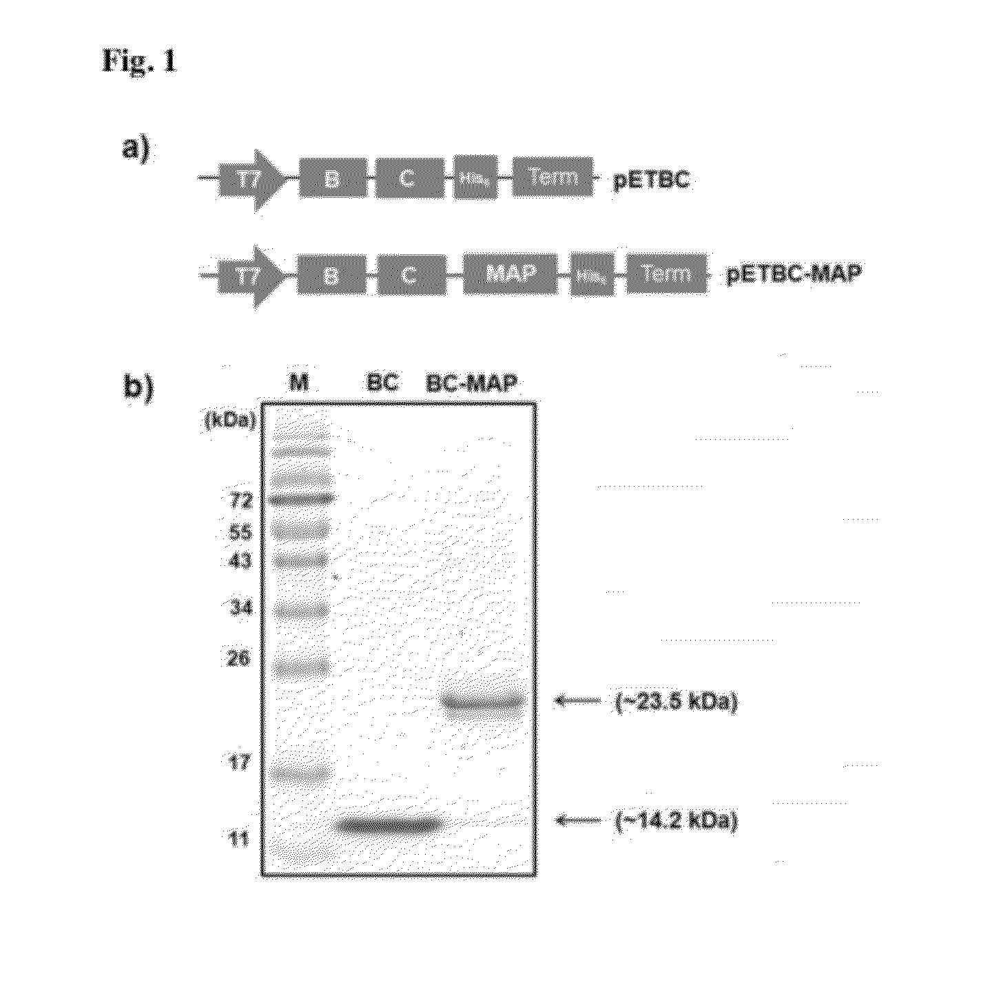
[0084]1.1: Construction of Expression Vector for BC-MAP Fusion Protein

[0085]The gene coding of MAP fp-5 was amplified by a polymerase chain reaction (PCR) from the previously constructed vector pMDG05. The forward primer (F1: 5′-GGGCCCATGGGGTGGCGGAGGGAGCTCTGAAGAATATAAAG-3′, SEQ ID No:1) was designed to contain a NcoI restriction enzyme site and the sequence encoding a GGGG amino acid linker. The backward primer (B1: 5′-CGCGCTCGAGGCTGCTGCCGCCATAATATTTTTTATAG-3′, SEQ ID No:2) was designed to contain a XhoI restriction enzyme site. The PCR product was digested with two restriction enzymes of NcoI and XhoI (Fermentas, Burlington, Ontario, Canada). The digested DNA fragment was ligated with a pET22b(+) vector (Novagen, San Diego, Calif., USA), which predigested with NcoI and XhoI, by using a ligation kit (TaKaRa, Shiga, Japan) to make the plasmid pET-MAP. The gene, encoding two domains (B and C domain) of protein A, was amplified by PCR from the genomic ...

example 2

Expression and Purification of BC-MAP and Sole BC Proteins

[0088]Each plasmid containing pET-BC-MAP or pET-BC was transformed into Escherichia coli BL21 (DE3). The transformants were grown to 0.8-0.9 OD600 at 37° C. in shake flasks containing of 400 mL of Luria-Bertani (LB) medium (0.5% yeast extract, 1% tryptophan, and 1% NaCl) with 50 μg mL−1 ampicillin. The expression was induced by adding isopropyl β-D-thiogalactopyranoside (IPTG) to a final concentration of 1 mM. The transformed cells were grown for an additional 10 h at 37° C. The cells were harvested by centrifugation at 4000 g for 10 min. The cell pellets were resuspended in lysis buffer (50 mM NaH2PO4, 300 mM NaCl, 10 mM imidazole, pH 8.0) and disrupted with a sonic dismembrator (Fisher scientific, USA) for 10 min at 50% power (3 sec pulse on and 2 sec pulse off). The soluble and insoluble fractions were then separated by centrifugation at 9000 g for 30 min at 4° C. The soluble fractions were loaded into a Ni-NTA column (Qia...

example 3

MALDI-TOF MS Analysis

[0089]Matrix-assisted laser desorption ionization mass spectrometry with time-of-flight (MALDI-TOF MS) analysis was performed on a 4700 Proteomics Analyzer (AB Sciex) in the positive ion linear mode. A-cyano-4-hydroxycinnamic acid in 50% acetonitrile and 0.1% trifluoroacetic acid was used as the matrix solution. Sample were diluted 1:20 with matrix solution, and 1 μL of the mixture was spotted onto the MALDI sample target plates and evaporated using a vacuum pump. Spectra were obtained in the mass range between 5,000 and 25,000 Da with ˜2000 laser shots. Internal calibration was performed using mix 3 of sequazyme peptide mass standard kit (AB Sciex).


PUM

PropertyMeasurementUnit
flow rateaaaaaaaaaa
flow rateaaaaaaaaaa
pHaaaaaaaaaa

Abstract

The present invention relates to a novel fusion protein comprising Staphylococcal protein A and mussel adhesive protein, a biochip comprising a solid substrate to which the fusion protein is attached, and a method for detecting a target antigen in a biological sample using the biochip. Furthermore, the present invention relates to a polynucleotide encoding the fusion protein, a recombinant vector comprising the polynucleotide, a transformed cell comprising the recombinant vector, and a method of preparing the fusion protein by transformed cell comprising the recombinant vector.

Description

BACKGROUND OF THE INVENTION[0001](a) Field of the Invention[0002]The present invention relates to a novel fusion protein comprising Staphylococcal protein A and mussel adhesive protein, a biochip comprising a solid substrate to which the fusion protein is attached, and a method for detecting a target antigen in a biological sample using the biochip. Furthermore, the present invention relates to a polynucleotide encoding the fusion protein, a recombinant vector comprising the polynucleotide, a transformed cell comprising the recombinant vector, and a method of preparing the fusion protein by transformed cell comprising the recombinant vector.[0003](b) Description of the Related Art[0004]Early detection of diseases is a key for their successful treatments despite of advancement of therapeutic tools. Immunoassay is one of the vital technologies for disease diagnosis due to high specificity and affinity of antibodies to target antigens. In most of immunoassays, antibody-antigen interact...

Claims


Application Information

Patent Timeline
Patent Type & Authority Applications(United States)
IPC IPC(8): G01N33/543C07H21/00C12N5/10C12P21/02C12N1/00C07K19/00C12N15/63
CPCC07K14/43504C07K2319/705G01N21/658C07K2319/21G01N33/531G01N33/54353
Inventor CHA, HYUNG JOONKIM, CHANG-SUP
Owner POSTECH ACAD IND FOUND
Learn More