Dimeric core-shell nanostructure labeled with raman active molecule localized at interparticle junction, use thereof, and method for preparing the same

a technology of active molecules and core shells, which is applied in the field of core shell nanoparticle dimers labeled with active molecules at interparticle junctions, can solve the problems of difficult handling of radioactive isotopes, short half-lives, and inability to store for a long period of time, and achieves enhanced raman scattering signals on the strong surface, high purity, and high purity

Inactive Publication Date: 2013-01-31
SEOUL NAT UNIV R&DB FOUND +1
View PDF1 Cites 12 Cited by
  • Summary
  • Abstract
  • Description
  • Claims
  • Application Information

AI Technical Summary

Benefits of technology

[0061]Because a Raman active molecule is localized at an interparticle junction and the distance between the Raman active molecule and the nano-core particle is precisely adjusted by the thickness of the silver or gold shell, the dimeric core-shell nanostructure of the present invention allows the production of strong surface enhanced Raman scattering (SERS) signals. Further, in spite of the localization of only one Raman active molecule, strong Raman signals can be detected using the dimeric core-shell nanostructure. In addition, the method for constructing the dimeric core-shell nanostructure guarantees the production of the dimer with high purity. Particularly, the nanostructure can be con

Problems solved by technology

However, radioactive isotopes are difficult to handle because the radiation they produce is harmful to the body.
Further, although their emission energy is high, some of the radioactive isotopes have short half-lives so that they cannot be stored for a long period of time or are not suitable for use in long-term experiments.
Particularly, as detection methods become increasingly simplified, radioactive substances face problems with detection limits and thus require long periods of time for detection.
However, unlike radioactive isotopes, fluorophores cannot substitute for elements of active ligands directly.
Another problem with fluorophores is the interference between different fluorophores because they re-emit a wide spectrum of light wavelengths while being excited over a highly narrow range of wavelengths.
Moreover, only a small number of fluorophores are available.
However, quantum dots suffer from the disadvantage of being highly toxic and being difficult to produce on a large scale.
In addition, the number of available quantum dots, although theoretically variable, is highly restricted in practice.
However, due to low signal intensity, the development of Raman spectroscopy has not yet reached the level where it can be used in practice in spite of research spanning a significant period of time.
However, there have been almost no advances in preparing single molecule SERS active substrates based on the salt-induced aggregation of silver (Ag) nanoparticles having Raman active molecules (e.g., Rhodamine 6G) since the first study.
Like this, randomly roughened surfaces provide a

Method used


Image

  • Dimeric core-shell nanostructure labeled with raman active molecule localized at interparticle junction, use thereof, and method for preparing the same
  • Dimeric core-shell nanostructure labeled with raman active molecule localized at interparticle junction, use thereof, and method for preparing the same
  • Dimeric core-shell nanostructure labeled with raman active molecule localized at interparticle junction, use thereof, and method for preparing the same

Examples


example 1

Preparation of Dimeric Au—Ag Core-Shell Nanostructure Labeled with Raman Active Molecule (Cy3) Localized at Interparticle Junction

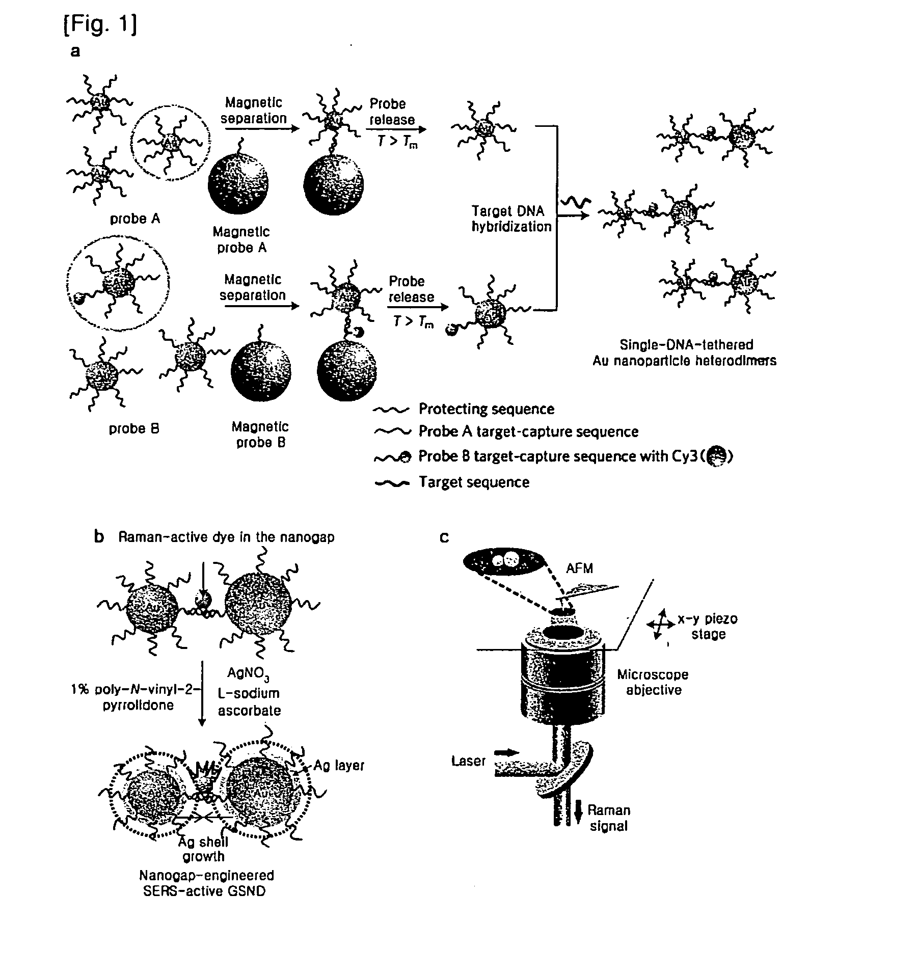
[0070]Based on a DNA-directed bridging method, the synthesis of a Raman active Au—Ag core-shell dimer was conducted using target oligonucleotide-tethered Au nanoparticles, with an Ag-shell being formed from a controlled amount of Ag precursor (FIG. 1).

[0071]Firstly, highly purified Au nanoparticle heterodimers were produced by precisely controlling the molar ratio between the protecting oligonucleotide and the target-capturing oligonucleotide, followed by an effective purification. Because the maximum distance (gap distance) between the Au nanoparticle (AuNP) surface and the Cy3 molecule still remained 7.5 nm, it needed to be decreased so as to give an amplified electromagnetic enhancement. A silver nano-shell was introduced since silver enhances SERS signals several times more than gold.

[0072]In detail, the DNA tethered Au nanoparticle dimers were coated...

example 2

UV-Visible Spectroscopy and HR-TEM Imaging Analysis

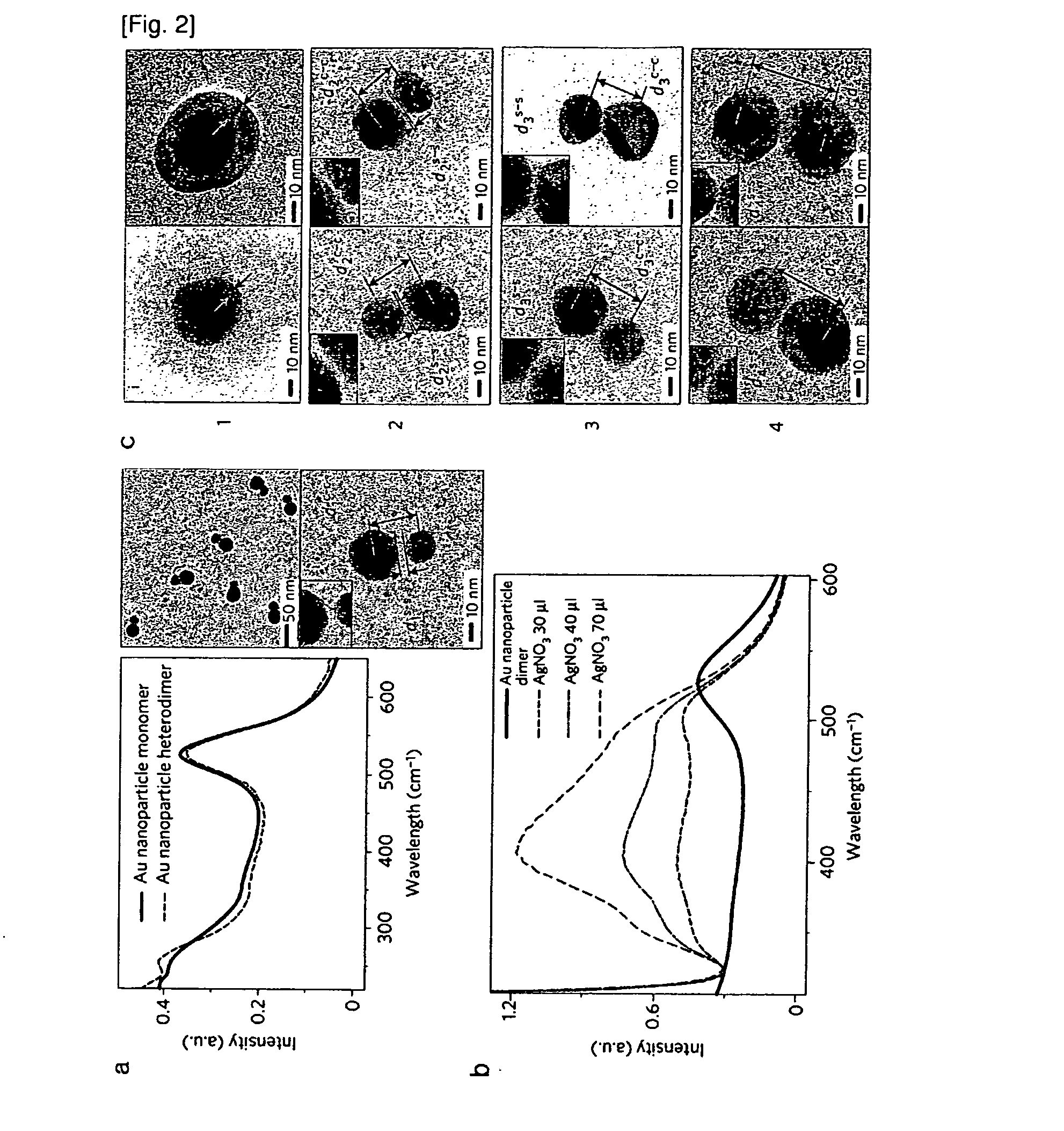
[0075]The formation of Au nanoparticle dimers (Cy3 used as a Raman Active molecule) was verified by UV-visible spectroscopy and high-resolution transmission electron microscope (HRTEM) images (FIG. 2). The UV-visible spectra show a very small red-shift after dimer formation, which is in agreement with the previously reported results by Oaul Alivisatos, et al. (Angew chem. 1999. 38(12), 1808). FIG. 2A is of typical HR-TEM images of the Au nanoparticle dimers. By a statistical analysis of at least 200 particles, it was found that 25% of the particles existed as a monomer and 65% of the particles as a dimer, and less than 10% as a multimer (trimer, tetramer and so on). The interparticle distance between the gold particles was found to be ca. 2-3 nm as measured by HR-TEM. In a solution (0.3M PBS), the interparticle distance was ˜15 nm which was expected to be far longer than that under dried conditions. Also, silver nanoparticles were i...

example 3

AFM (Atomic Force Micrograph) Analysis of Au—Ag Core-Shell Nanoparticles

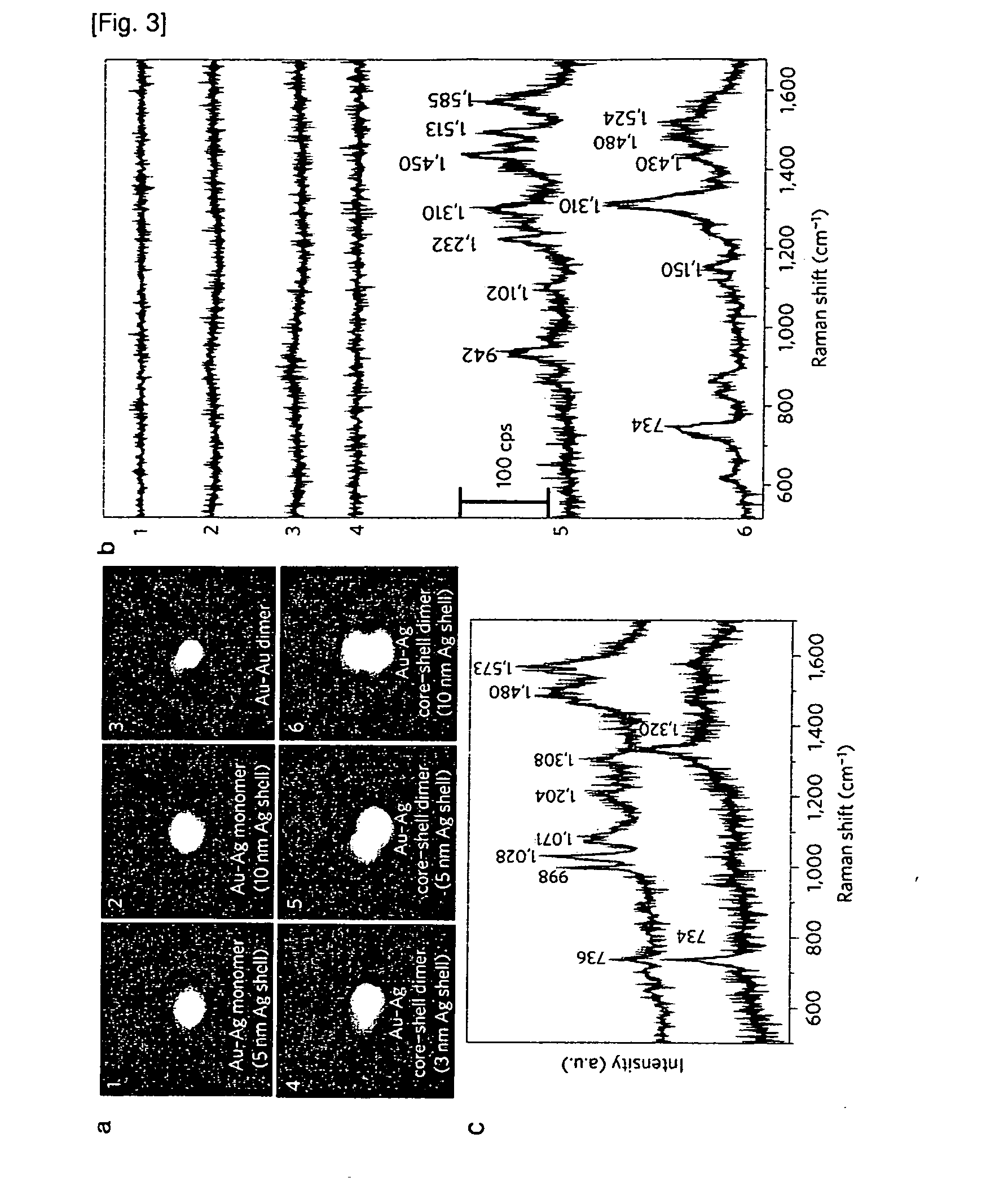
[0077]FIG. 3A shows magnified AFM images (1×1 μm) of the core-shell monomer and heterodimer nanostructures (using Cy3 as a Raman active molecule), which were coincident in shape and diameter with HR-TEM images. FIG. 3B shows the correlated SERS spectra from the corresponding single AFM-imaged particles in FIG. 3A. No Raman signals were detected for the monomeric Au—Ag core-shell nanoparticles with 5 nm (FIG. 3A-1) and 10 nm (FIG. 3A-2) silver shells, respectively because they had no hot spots and only one Cy3 molecule per particle. The Au nanoparticle heterodimers without Ag shells or with a gap distance less than 1 nm therebetween did not show any detectable SERS signal either. This is due to insufficient electromagnetic enhancement under 514.5 nm laser excitation conditions. When the Ag shell thickness was <3 nm (FIG. 3A-4), no Raman signals were detected even after using an elevated incident laser power (˜200...


PUM

PropertyMeasurementUnit
Thicknessaaaaaaaaaa
Thicknessaaaaaaaaaa
Diameteraaaaaaaaaa

Abstract

The present invention relates to a nanoparticle dimer in which Raman-active molecules are located at a binding portion of the nanoparticle dimer, and more particularly, to a core-shell nanoparticle dimer comprising: a gold or silver core having a surface to which oligonucleotides are bonded; and a gold or silver shell covering the core. In addition, the present invention relates to the core-shell nanoparticle dimer, to a method for preparing same, and to the use thereof.

Description

TECHNICAL FIELD[0001]The present invention relates to a core-shell nanoparticle dimer labeled with a Raman active molecule at an interparticle junction. More particularly, the present invention relates to a dimeric nanostructure comprising two nanoparticles, with a Raman active molecule localized at a junction therebetween, each nanoparticle consisting of a gold or silver core with oligonucleotides attached to the surface thereof, and a gold or silver shell sheathing the core. Also, the present invention is concerned with uses of the dimeric nanostructure and a method for preparing the dimeric nanostructure.BACKGROUND ART[0002]Highly sensitive, accurate detection of single molecules from biological or other samples is being extensively applied to many fields including medical diagnosis, pathology, toxicology, environmental sampling, chemical analysis, etc. Recently, to this end, the biology-chemistry field has widely utilized specifically labeled nanoparticles or chemical materials ...

Claims


Application Information

Patent Timeline
IPC IPC(8): G01N33/566C07K16/00C07H21/00C12N9/96C07K14/00B82Y15/00
CPCC12Q1/6834G01N33/54346G01N33/54373C12Q1/6816C12Q2565/628C12Q2563/155C12Q2563/107C12Q2565/632C12Q2563/143C12Q2537/125C12Q1/6827G01N33/5302
Inventor SUH, YUNG DOUGNAM, JWA MINLIM, DONG KWONJEON, KI SEOK
Owner SEOUL NAT UNIV R&DB FOUND
Why Eureka
Try Eureka