
Method for Determining Effectiveness of Medicine Containing Antibody as Component

a technology of medicine and antibody, which is applied in the field of determining the effectiveness of a medicine containing an antibody as a component, can solve the problems of insufficient method of ihc method using an anti-her2 antibody, limited quantitative analysis use of cultured cells alone, and insufficient quantitative analysis. achieve the effect of high sensitivity

Inactive Publication Date: 2013-09-05
TOHOKU UNIV
View PDF1 Cites 31 Cited by
  • Summary
  • Abstract
  • Description
  • Claims
  • Application Information

AI Technical Summary

Benefits of technology

This patent describes a new method for quantitatively measuring the expression level of the HER2 protein in breast cancer using a tissue staining method that is more sensitive than conventional methods. This new method can be used to diagnose and treat breast cancer more effectively by evaluating the therapeutic effect of trastuzumab and other medicines containing antibodies. Combining this method with other techniques can also allow for a more comprehensive evaluation of the gene copy number of HER2 and the amount of HER2 protein, which can lead to improved cancer treatment for breast cancer.

Problems solved by technology

Since the fluorescent particles of a quantum dot and the like show their power in stability of light emitted by themselves and quantitative analysis, they are expected as a novel tool to be used in immunofluorescence staining, but their use for the quantitative analysis is limited to cultured cells alone at the present.
Although the amount of long wavelength autofluorescence of biological tissues is smaller than the amount of short wavelength autofluorescence, the influence of the autofluorescence cannot be ignored in observation, and hence, the quantitative analysis is difficult.
Furthermore, since an antibody used for detecting HER2 protein in the diagnosis of breast cancer is different, in an antigenic determinant (epitope), from a therapeutic agent (an antibody) targeting HER2 protein, it is regarded that the current IHC method using an anti-HER2 antibody is insufficient as a method for selecting patients eligible for administration of trastuzumab in which HER2 including an antigenic determinant of trastuzumab is overexpressed.

Method used


Image

  • Method for Determining Effectiveness of Medicine Containing Antibody as Component
  • Method for Determining Effectiveness of Medicine Containing Antibody as Component
  • Method for Determining Effectiveness of Medicine Containing Antibody as Component

Examples


example 1

Method for Preparing Tissue Sample Immunostained by IHC-QDs Method

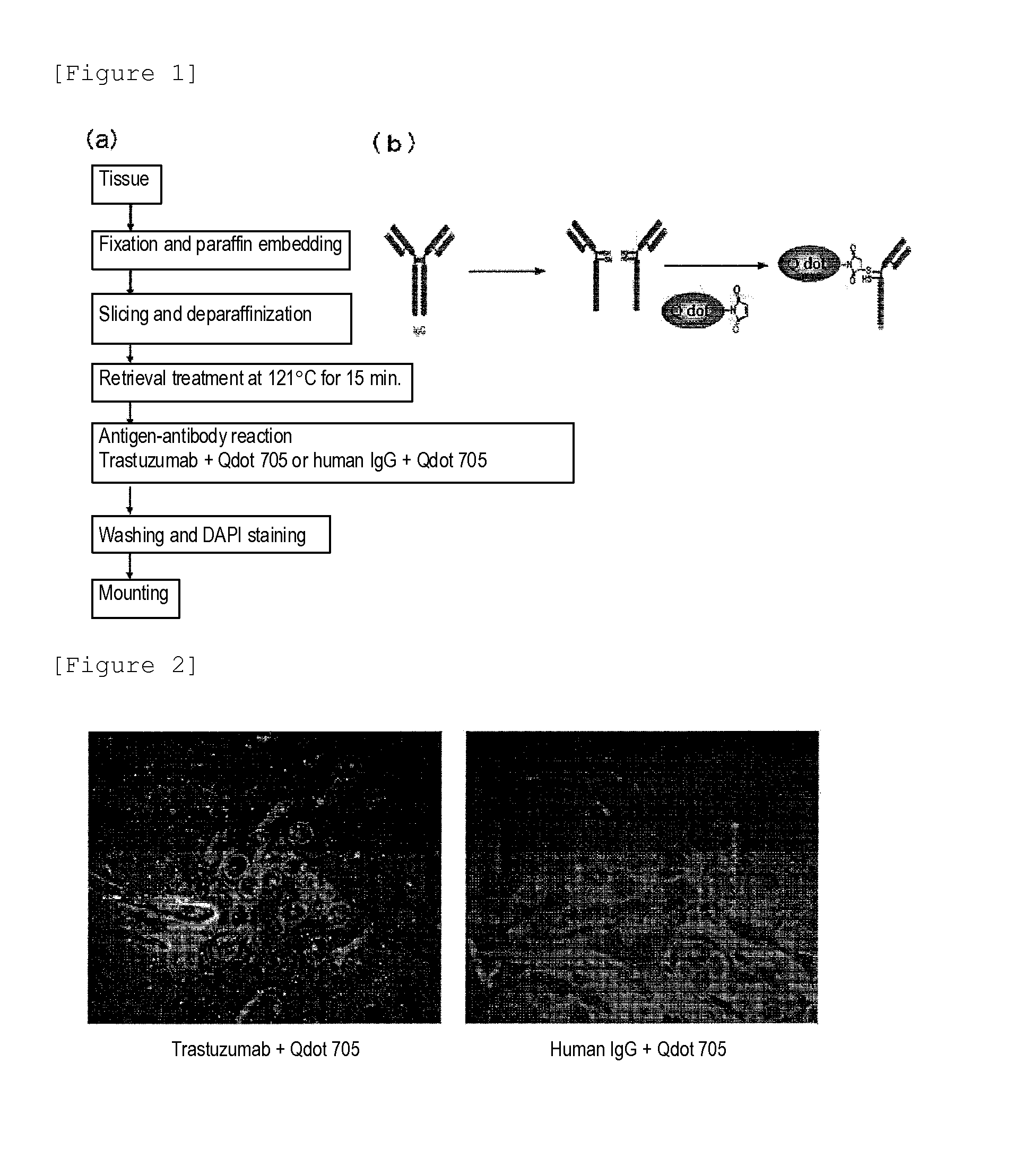
[0046]As human breast cancer pathological tissues, 6 cases evaluated based on the IHC-DAB method as Score 0, 6 cases evaluated as Score 1, 11 cases evaluated as Score 2 and 14 cases evaluated as Score 3, namely, 37 cases in total, were selected. These cases were selected carefully so that the background of the cases might be well-balanced as shown in Table 1. Tissue samples of tissues of the 37 cases were prepared by a method generally employed for pathological tissue diagnosis. Specifically, a breast tissue specimen of a cancer patient was fixed with formalin, followed by dehydration with alcohol and a xylene treatment, and the resultant was immersed in paraffin at a high temperature for paraffin embedding, and thus, a tissue sample was prepared (FIG. 1(a)). Subsequently, the tissue sample was cut into sections of 2 to 4 μm, followed by deparaffinization with xylene and an alcohol treatment, and the resultant was was...

example 2

Problem of Immunofluorescence Tissue Staining Method

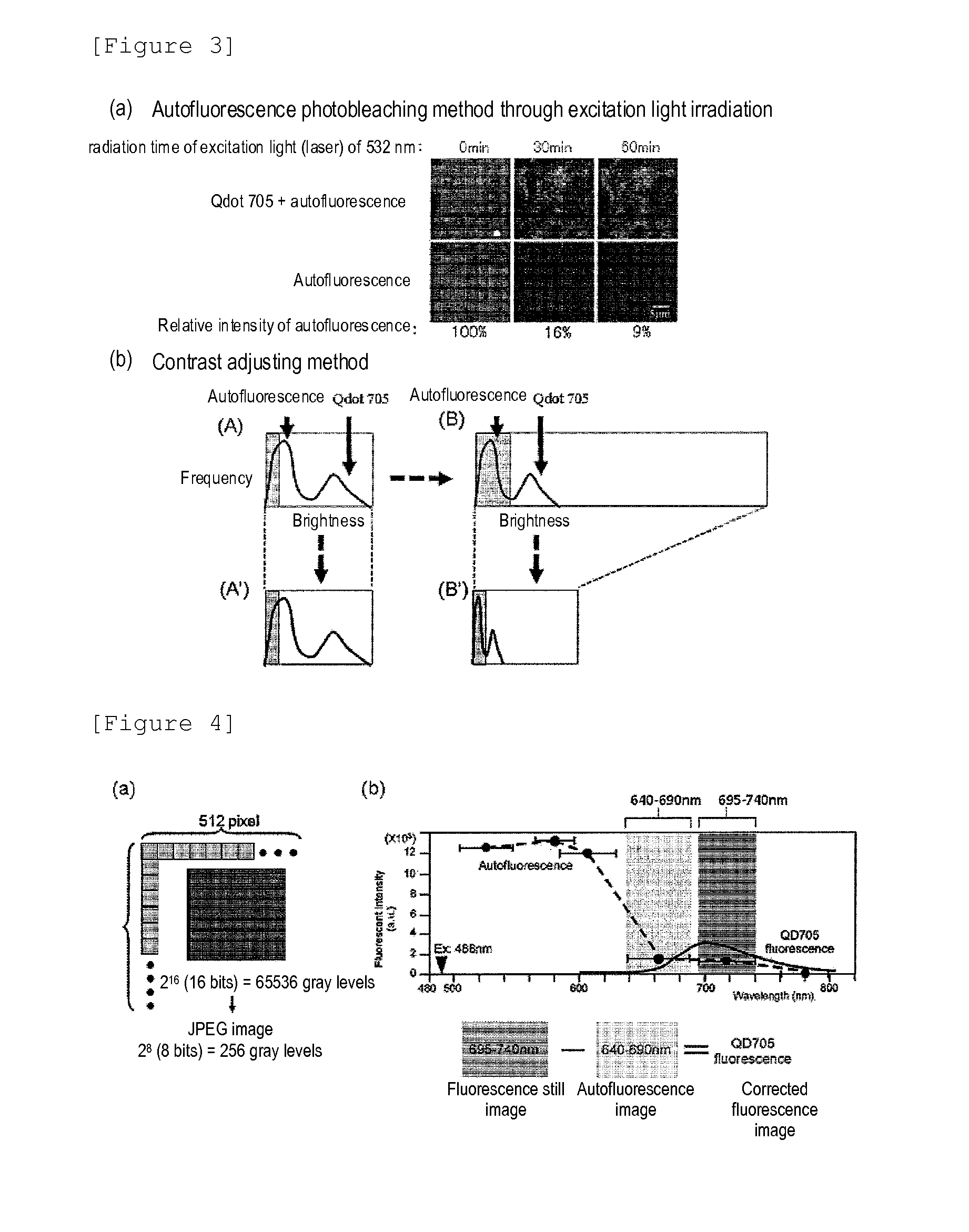
[0048]Each of the immunostained tissue samples prepared in Example 1 was irradiated with excitation light of 488 nm by using an apparatus obtained by combining a confocal unit (manufactured by Yokogawa Electric Corporation), a fluorescence microscope (manufactured by Olympus Corporation) and an electron-multiplier CCD (EM-CCD) camera (manufactured by Andor Technology), and thereafter, a fluorescence image (a fluorescence still image) of the quantum dot fluorescent particles (used for labeling trastuzumab) was obtained by using a band-pass filter having a pass range of a wavelength of 695 to 740 nm. FIG. 2 exemplarily illustrates a case evaluated as Score 3 by the IHC-DAB method, and thus, fluorescence derived from the quantum dot fluorescent particles was observed if trastuzumab+Qdot 705 was used while fluorescence derived from the quantum dot fluorescent particles was minimally observed if the control of human IgG+Qdot 705 was use...

example 3

Generation of Corrected Fluorescence Image Excluding Autofluorescence

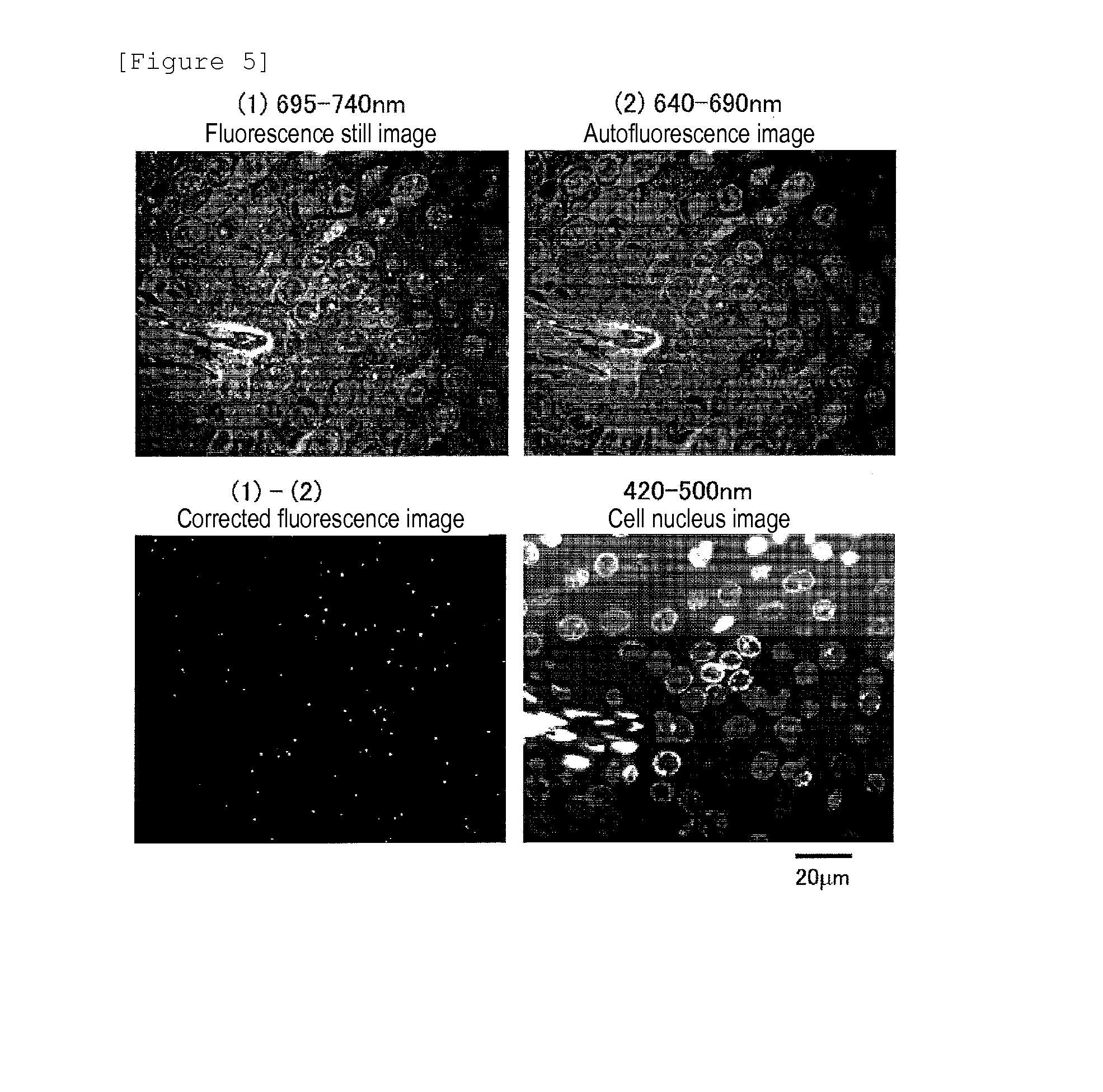
[0050]It is a fluorescence still image in which the autofluorescence of the background has fluorescent intensity of zero (0) that is necessary. If such an image is obtained, the total fluorescence included in the fluorescence still image can be calculated as the fluorescence derived from the quantum dot fluorescent particles. The contrast adjusting method described in Example 2 is conducted as division of the fluorescent intensity, and zero (0) cannot be obtained by division, and hence, an image processing method using subtraction, which can give zero (0), is necessary. Therefore, as an image processing method for removing autofluorescence from a fluorescent still image, the following method was devised: First, a breast cancer tissue sample immunostained with quantum dot fluorescent particles was irradiated with excitation light (laser) of an excitation wavelength of 488 nm, so as to obtain a fluorescence image of ...


PUM


Abstract

Protein recognized by an antibody used as an active ingredient of an antibody medicine such as trastuzumab or an antibody used for targeting a target site of an active ingredient is highly accurately quantitatively determined by employing a quantitative tissue staining method of biological tissues, thereby providing a method for determining therapeutic effectiveness of a medicine containing such an antibody as a component. The effectiveness of a medicine containing an antibody as a component is determined by employing a tissue staining method comprising the steps of: labeling the antibody in the medicine containing an antibody as a component with a fluorescent material and contacting the thus fluorescence-labeled antibody with a tissue sample; obtaining a fluorescence image by irradiating, with excitation light, a tissue site contacted with the antibody; obtaining an autofluorescence image in the same field of view and at the same focus as in the fluorescence image in a close region on a shorter wavelength side or a longer wavelength side of an acquisition wavelength region of fluorescence emitted by the fluorescent material; obtaining a corrected fluorescence image by performing image processing for removing fluorescence brightness of the autofluorescence image from fluorescence brightness of the fluorescence image; counting the number of cells in the tissue site contacted with the antibody; measuring average fluorescence brightness per fluorescent particle; and calculating the number of fluorescent particles per cell.

Description

TECHNICAL FIELD[0001]The present invention relates to a method for determining effectiveness of a medicine containing an antibody as a component by employing a tissue staining method in which highly accurate and quantitative analysis can be conducted with influence of autofluorescence effectively removed.BACKGROUND ART[0002]Cancer is a disease that is one of the two main causes of death of adults together with vascular diseases such as cardiac infarction and brain infarction. An incidence rate of, for example, breast cancer is lower in Japan than in Western countries but recently shows an increasing tendency, and overtook the incidence rate of stomach cancer and came top of the incidence rate among women in 1998. According to a recent report of statistics of Ministry of Health, Labour and Welfare in 2005, the annual number of breast cancer cases is over 50000. The number is increasing year by year also in the whole world, and according to a WHO report in 2008, breast cancer is a dis...

Claims


Application Information

Patent Timeline
IPC IPC(8): G01N21/64
CPCG01N21/6428G01N21/6456G01N33/6854G01N33/582G01N21/6486G01N2800/52G01N33/94
Inventor MIYASHITA, MINORUGONDA, KOHSUKEOHUCHI, NORIAKITAKEDA, MOTOHIRO
Owner TOHOKU UNIV
Learn More