Method for analyzing proteins contributing to autoimmune diseases, and method for testing for said diseases

a protein and autoimmune disease technology, applied in the field of analysis methods for proteins involved in autoimmune diseases, can solve the problems of inability to diagnose early-stage atherosclerosis by conventional image diagnosis, almost half of the genes on the genome remain unknown for their functions, and can not be detected by conventional image diagnosis. , to achieve the effect of high efficiency and high sensitivity

Active Publication Date: 2013-10-17
PUBLIC UNIV CORP YOKOHAMA CITY UNIV
View PDF2 Cites 11 Cited by
  • Summary
  • Abstract
  • Description
  • Claims
  • Application Information

AI Technical Summary

Benefits of technology

[0032]In the present invention, there is provided means for analyzing a protein involved in an autoimmune disease, in particular, arteriosclerosis with high sensitivity and high efficiency. There is also provided an examination method for arteriosclerosis, in particular, atherosclerosis, involving detecting an antibody titer of an autoantibody against the protein involved in arteriosclerosis obtained by the means.
[0034]“1. An examination method for arteriosclerosis, including detecting, from a sample derived from a patient, an antibody titer of an autoantibody against any one of the following groups of proteins:
[0035](1) a group of proteins involved in a cytokine, in which the group of proteins involved in a cytokine consists of any one or more of the following:
[0036]IL-5; STX1A; CSNK2A1; VAMP2; GSK3B; PRKCZ; PCNA; PIN1; STX4; HRB; HMGN1; HIST1H1C; TUBB; NPM1; VAMP8; VAPA; STX3; RABAC1; and CCND2; and
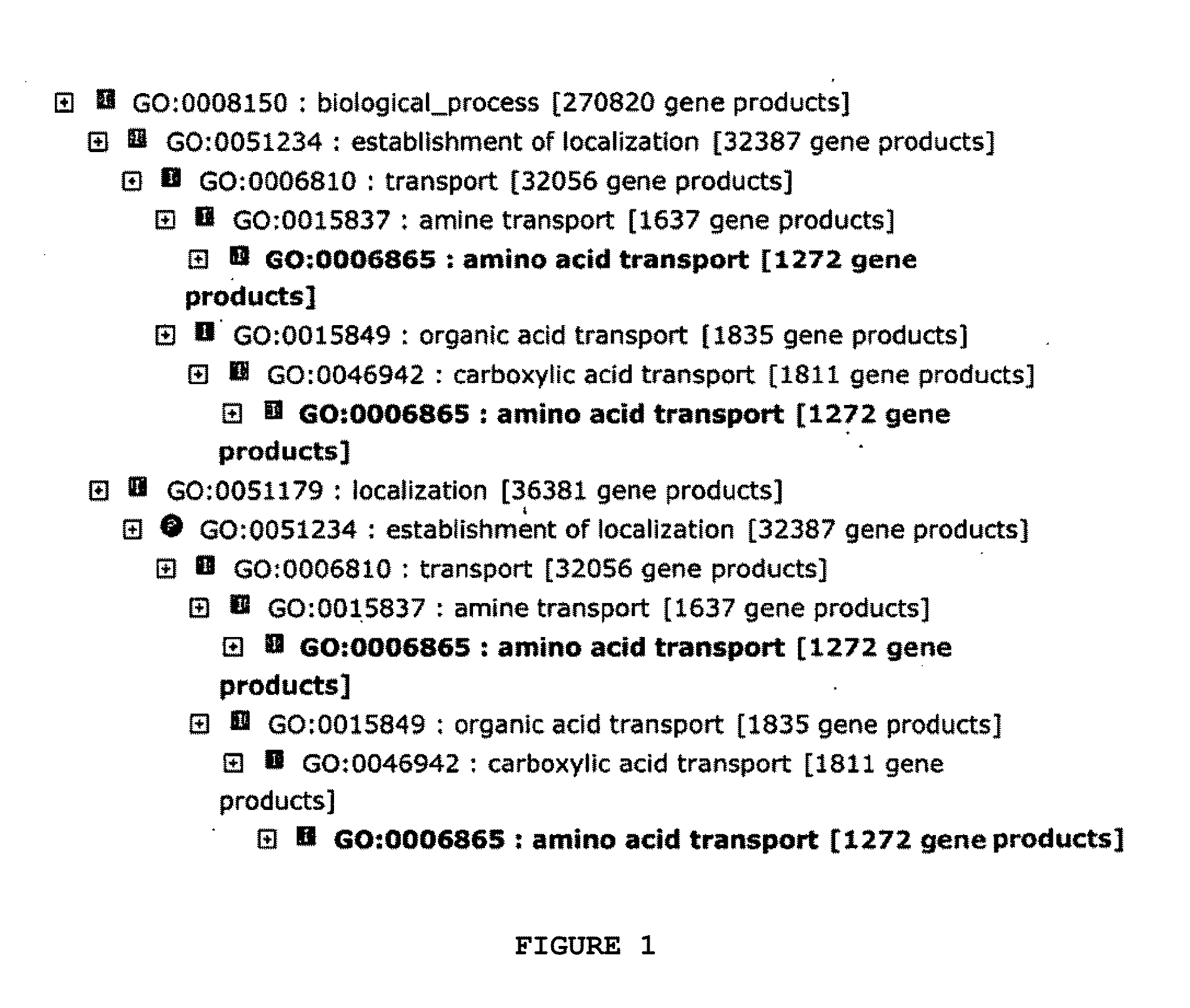
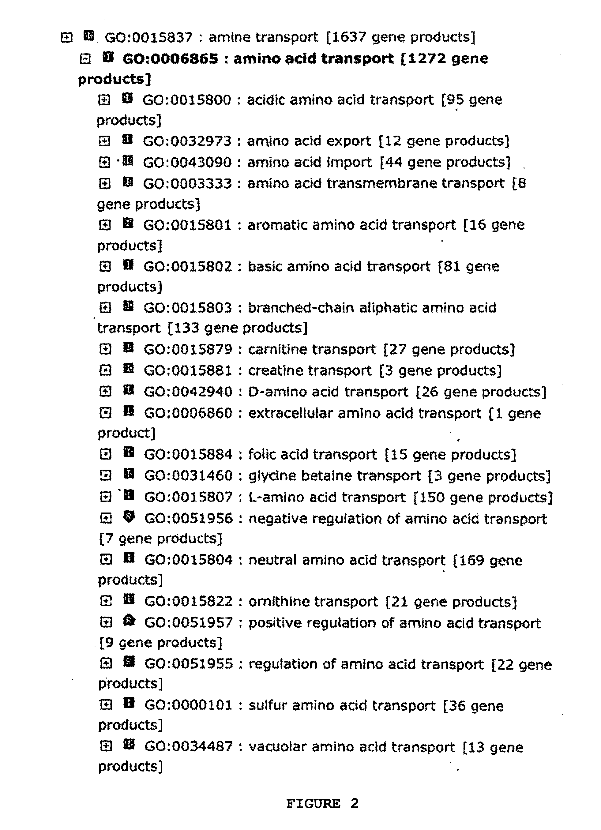
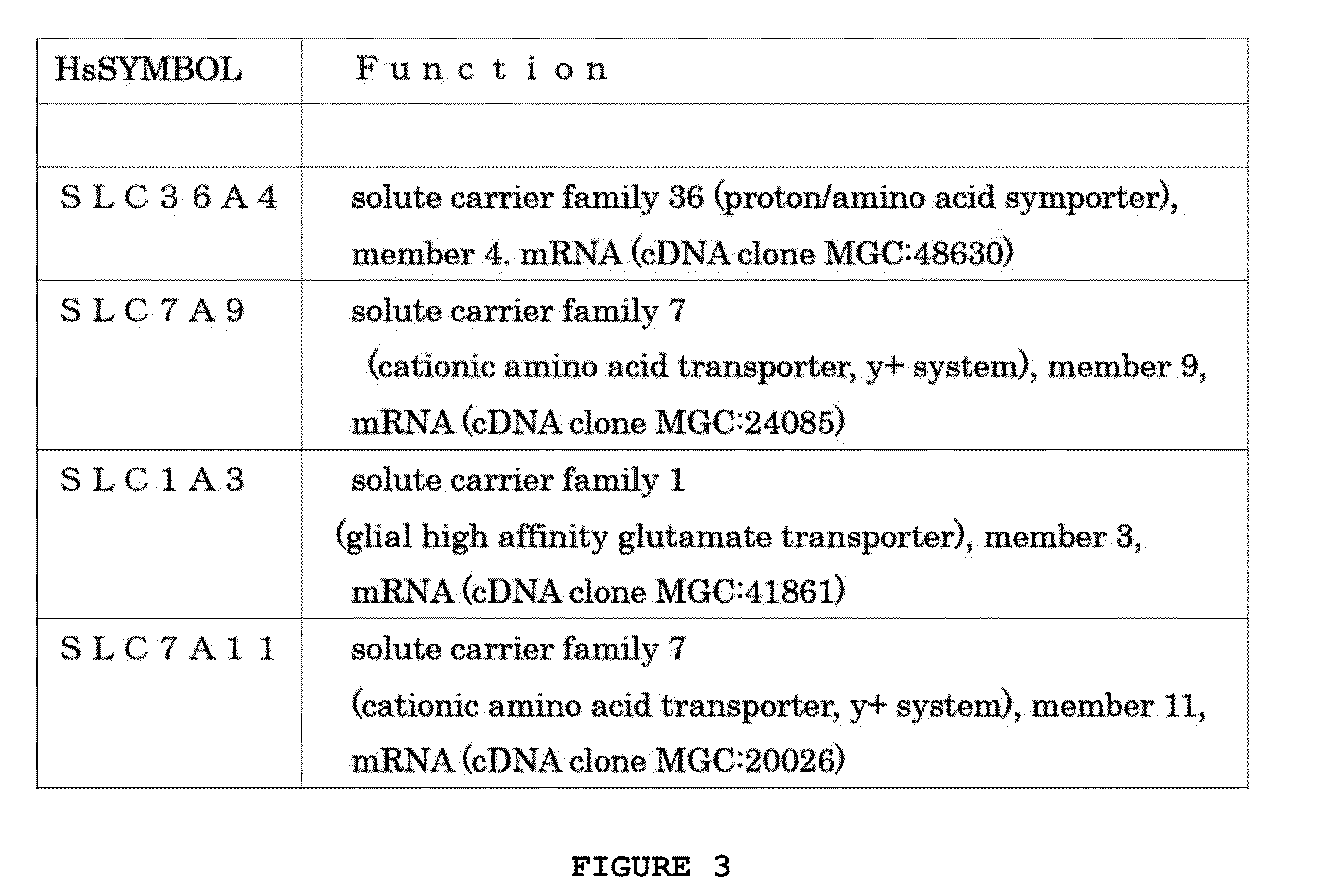
[0037](2) a group of proteins involved in an amino acid transporter, in which the group of proteins involved in an amino acid transporter consists of any one or more of the following:
[0039]2. An examination method according to the above-mentioned item 1, in which the group of proteins involved in a cytokine consists of any one or more of the following:

Problems solved by technology

However, almost half of genes on a genome remain unknown for their functions.
Atherosclerosis, a final presentation of a lifestyle-related disease such as hypertension, diabetes, or hyperlipidemia, causes myocardial infarction, cerebral infarction, peripheral artery disease, and the like, and thus is lethal or significantly impairs an activity of daily living (ADL) in some cases.
Under the present circumstances, however, early-stage atherosclerosis cannot be diagnosed by conventional image diagnosis, and moreover, there is no serum marker or the like useful for the diagnosis.
However, there are still many unknown things.
However, it cannot be considered that factors involved in an autoimmune disease are only the IL-1α autoantibodies.
However, it cannot be considered that the factors involved in an autoimmune disease are only the phosphorylcholine autoantibodies.

Method used


Image

  • Method for analyzing proteins contributing to autoimmune diseases, and method for testing for said diseases
  • Method for analyzing proteins contributing to autoimmune diseases, and method for testing for said diseases
  • Method for analyzing proteins contributing to autoimmune diseases, and method for testing for said diseases

Examples


example 1

Preparation of Sample Derived from Patient with Autoimmune Disease

[0234]In this example, sera obtained from patients with atherosclerosis (23 patients) were used as samples. In addition, the samples were divided into ones from mild patients, serious patients, mild and old patients, and serious and old patients to be analyzed.

[0235]It should be noted that sera derived from healthy subjects were each used as a control.

example 2

Preparation of Translation Template Encoding Biotinylated Mammal-Derived Protein

[0236]mRNA as a translation template was prepared as described below. Vectors as biotinylated protein transcription templates obtained by fusing a biotin tag to genes encoding various mammal-derived proteins (about 3,000 kinds) were prepared (pEU-biotinylated tag-various mammal-derived proteins). Based on each of the vectors, a PCR product including the Q sequence portion of a tobacco mosaic virus (TMV) was used as a template. The transcription template was added to a transcription reaction solution (final concentration: mM HEPES-KOH, pH 7.8, 16 mM magnesium acetate, 10 mM dithiothreitol, 2 mM spermidine, 2.5 mM 4NTPs (4 kinds of nucleotide triphosphates), 0.8 U / μL RNase inhibitor, 1.6 U / μL SP6 RNA polymerase), and the mixture was subjected to a reaction at 37° C. for 3 hours. The resultant RNA was extracted with phenol / chloroform, precipitated with ethanol, and then purified with Nick Column (manufactur...

example 3

Translation Reaction Step of Biotinylated Mammal-Derived Protein

[0237]A 96-well titer plate was used as a reaction vessel. First, 125.0 μL of a feed phase (62.5 μL of 2× Substrate Mixture, 1.25 μL of 50 μM biotin, 61.25 μL of MilliQ) were added to the titer plate. Next, a mixture obtained by adding each translation template {pelleted translation template (mRNA) dissolved with 25 μL of a reaction solution} of Example 2 above to 25.0 μL of a reaction phase (0.25 μL of 4 μg / μL creatine kinase, 6.5 μL of a wheat germ extract for cell-free protein synthesis (200 O.D.), 1.0 μL of a biotinylation enzyme (180 O.D.), 8.75 μL of 2× Substrate Mixture, 2.5 μL of 5 μM biotin, 3.5 μL of MilliQ) was added carefully and gently to the bottom of the titer plate. The resultant was left to stand still at 26° C. for 15 to 20 hours to perform a protein synthesis reaction.

[0238]The biotinylated protein after the completion of synthesis was used in the following examples without purification.


PUM

PropertyMeasurementUnit
pHaaaaaaaaaa
pHaaaaaaaaaa
antibody titeraaaaaaaaaa

Abstract

Provided are a detection method for a myriad of proteins involved in an autoimmune disease with high sensitivity and high efficiency, and an analysis method for data resulting from the detection method. In order to construct the detection method and analysis method, there is provided means for comprehensively analyzing the proteins involved in an autoimmune disease by bringing a mammal-derived protein expressed in a cell-free protein synthesis system into contact with a sample derived from a patient with an autoimmune disease to detect autoantibody production, and subjecting the detected data to statistical analysis processing, and further, gene ontology analysis and / or pathway analysis.

Description

TECHNICAL FIELD[0001]The present invention relates to an analysis method for a protein involved in an autoimmune disease, and an examination method for the disease.[0002]It should be noted that the present application claims priority from Japanese Patent Application No. 2010-256418, which is incorporated herein by reference.BACKGROUND ART[0003]After completion of human genome sequencing, while development of bioinformatics being promoted, analysis of genes has been made mainly based on their nucleic acid or amino acid sequences, such as search of amino acid sequences (domains) conserved across species or classification of ortholog genes based on the search. However, almost half of genes on a genome remain unknown for their functions.[0004]In actuality, even a majority of annotated genes as well as genes quite unknown for their functions found on the genome remain unknown for their biochemical functions.[0005]Therefore, in the post-genome era after the genome sequencing, in order to ...

Claims


Application Information

Patent Timeline
Patent Type & Authority Applications(United States)
IPC IPC(8): G01N33/68
CPCG01N33/6869G01N33/564G01N33/6845G01N2800/323
Inventor SAWASAKI, TATSUYAENDO, YAETAISHIGAMI, TOMOAKIAOKI, ICHIRO
Owner PUBLIC UNIV CORP YOKOHAMA CITY UNIV
Why Eureka
Try Eureka