Artificial Tissue Systems and Uses Thereof

a technology of artificial tissue and tissue, applied in the field of medical implants, can solve the problems of limiting the use of implants, limiting the application range, and currently designing implantable sensors to overcome their loss of function, so as to improve the function, improve the integration effect, and prolong the life of the devices

Active Publication Date: 2013-10-17
UNIV OF CONNECTICUT
View PDF2 Cites 2 Cited by
  • Summary
  • Abstract
  • Description
  • Claims
  • Application Information

AI Technical Summary

Benefits of technology

[0012]Briefly stated, the present invention in a preferred form includes an apparatus and a method for controlling the natural and artificial microenvironments surrounding an implanted device using an artificial tissue system (ATS). The ATS, among other things, induce better integration, improved function, and an extended lifespan of the devices at the site of implantation. The ATS includes cells, such as naturally occurring, engineered, and / or artificial cells and matrices such as a natural, engineered, artificial and / or hybrid matrix. Tissue response modifiers (TRM) and / or cell response modifiers (CRM) as also preferentially included. The specific composition of the ATS is based on the nature of the tissue in which also are preferred included the ATS-device combination is implanted and the nature of the implanted device, as well as the required function and lifespan of the implanted device. Additionally, the ATS, as well as ATS-device combinations can be utilized in vitro to aid in the design of improved artificial tissue systems, devices and ATS-device combinations for in vivo uses.

Problems solved by technology

Unfortunately the development of these implantable sensors has been hampered by the inability of currently designed implantable sensors to overcome their loss of function and short lifespan in vivo.
Frequently this loss of function is a result of the acute and chronic tissue reactions to the implanted sensors.
Ultimately, these chronic inflammatory reactions at sites of sensor implantation result in tissue destruction and fibrosis and complete loss of sensor function in vivo.
Unfortunately these approaches have not been successful, and the use of various coatings has seen limited success because of the body innate and acquired host defense systems (immunity) that can detect minute differences between normal tissue elements and foreign materials such as sensor coatings.
However, in the case of sensor coatings it has been found that 1) frequently only “analyte permeable coatings” can be used as sensor coatings, thus limiting the type of coating available for implantable sensors; 2) binding of sufficient quantities of bioactive agents such as peptides and proteins, can be difficult and often they do not remain active after being bound to the sensor coating; 3) the intense tissue reactions (proteins and cells) frequently “mask” or degrade the bioactive agents on the coatings and limit their effectiveness 4) because of the limited quantities of bioactive agents that can be incorporated into sensor coatings, the coating and therefore the sensor have a limited lifespan in vivo and must be replaced frequently.
Additionally the device and its byproducts can also damage both the tissue and the sensor and its coatings.
Additionally, the glucose oxidase used in the sensor continuously breaks down glucose into gluconic acid and hydrogen peroxide, both of which are tissue toxic as well as potentially “sensor toxic”.
The hydrogen peroxide is further broken down in reactive oxygen radicals, which are also toxic.
In the case of traditional drug delivery systems such as micro beads, they frequently do not incorporate (load) and or release bioactive agents in quantities and for durations that are useful for implantable sensors.
Since the drug delivery system used with implantable sensors are usually located near the sensor, “foreign body” tissue reactions to the drug delivery system often have negative “bystander” effects on the sensor and its function.
For example, the breakdown of the drug delivery systems such as micro beads result in the release of tissue toxic and sensor toxic byproducts that hinder sensor function in vivo.
Ultimately the combination of inflammation, fibrosis and loss of blood vessels also decrease tissue levels of both glucose and oxygen, both essential to glucose sensor function in vivo, with a resulting loss of sensor function and lifespan in vivo.
Unfortunately, because of the pain and inconvenience of this procedure associated with “finger sticking” the patient compliance is often poor.
However, even with good patient compliance with regular blood glucose testing, it appears that blood glucose swings often stay undetected.
Although implantable glucose sensors have been in existence for over 30 years, and in vitro studies have demonstrated that they can function for weeks to months, glucose sensor function in vivo has seen little success.
Previous in vivo studies have indicated that implantable glucose sensors lose function within hours to days after implantation.

Method used


Image

  • Artificial Tissue Systems and Uses Thereof
  • Artificial Tissue Systems and Uses Thereof
  • Artificial Tissue Systems and Uses Thereof

Examples


experimental examples

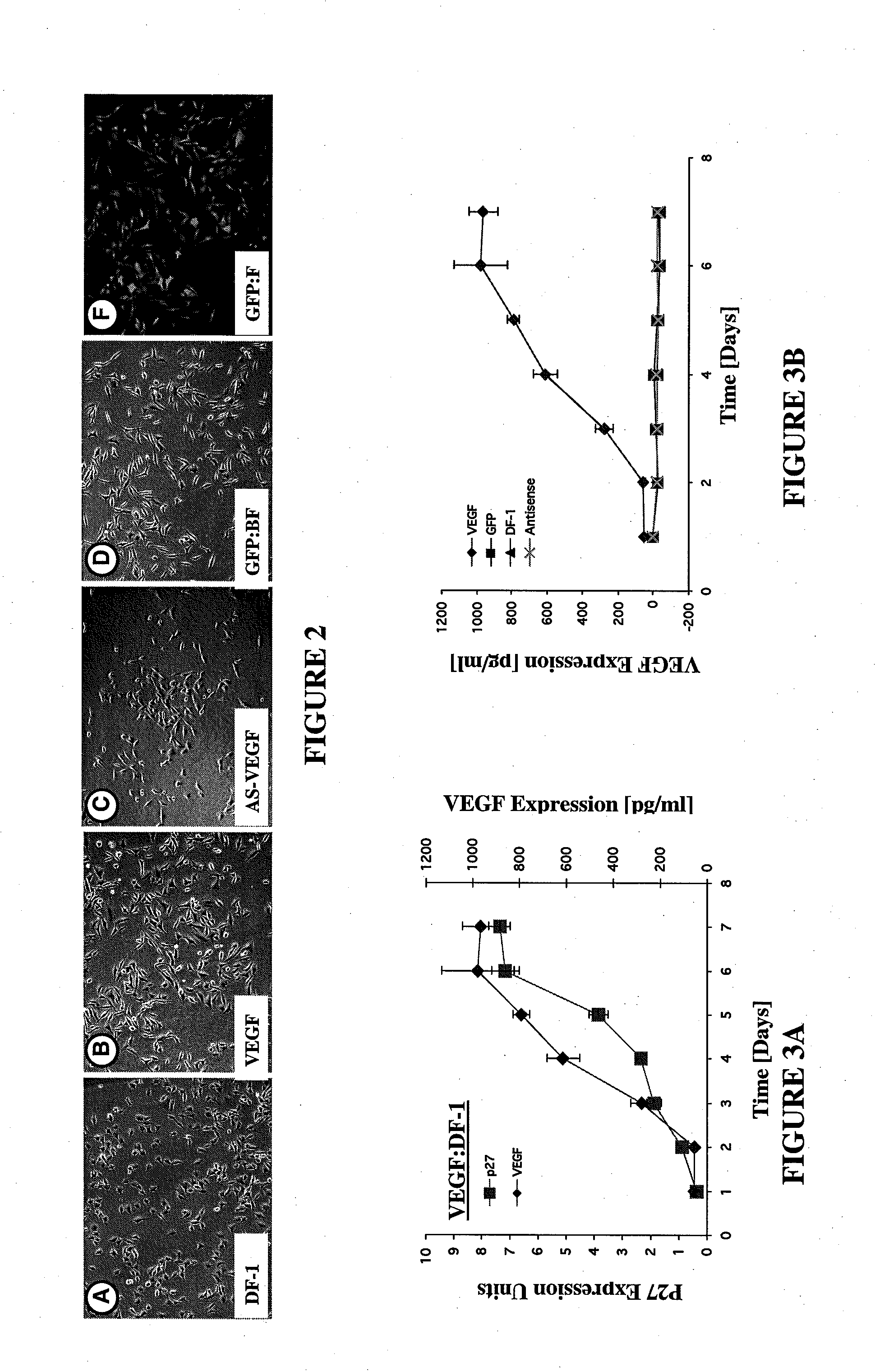
[0099]In one embodiment of the invention, a gene transfer system is included wherein a genetically engineered cell suitable for use in the ATS is produced. For example, as experimentally shown, a Rous Sarcoma Virus Vector Model for Gene Transfer was created wherein a helper-independent retroviral vector, RCAS, derived from Rous Sarcoma Virus (RSV) was used for gene transfer in the in vitro and ex ova CAM model studies. A mouse VEGF gene (mVEGF), genebank number M25200, said genebank disclosure incorporated fully herein by reference and associated with the sequence as shown in FIG. 22, was inserted into the RCAS proviral plasmid vector in both “sense” and “antisense” orientations using standard recombinant DNA manipulations. Specifically, a 908 by Taq I fragment containing the mVEGF open reading frame was mobilized from pBSK+mVEGF and ligated into the unique Cla I site of the RCAS-BP(A) proviral vector plasmid. The ligation products were screened by restriction mapping and both sense...


PUM

PropertyMeasurementUnit
timeaaaaaaaaaa
timeaaaaaaaaaa
timeaaaaaaaaaa

Abstract

An implant system and a method for controlling the natural and artificial microenvironments surrounding an implanted device using an artificial tissue system (ATS) and includes methods of diagnostic and testing related thereto. The ATS, among other things, induce better integration, function, and extended lifespan of the devices at the site of implantation. The ATS includes cells, such as naturally occurring, engineered, and / or artificial cells; matrices such as natural, engineered, artificial and / or hybrid matrices; tissue response modifiers (TRM); and / or cell response modifiers (CRM). The specific composition of the ATS is based on the nature of the tissue in which ATS-device combination is implanted and the nature of the implant device, as well as the required function and lifespan of the implanted device. Additionally, the ATS, as well as ATS-device combinations can be utilized in vitro to aid in the design of improved ATS, devices and ATS-device combinations for in vivo uses.

Description

CROSS REFERENCE TO RELATED APPLICATIONS [0001]This application is a divisional of co-pending application Ser. No. 10 / 578,171 filed May 4, 2006, which is the Section 371 US National Stage of PCT / US04 / 37302 filed Nov. 5, 2004, which claims the benefit of U.S. Provisional Application No. 60 / 518,412, filed Nov. 7, 2003, the content of which is incorporated by reference in their entirety.STATEMENT WITH REGARD TO FEDERAL SPONSORED RESEARCH & DEVELOPMENT[0002]This invention was made with Government support under Grants awarded by the National Institutes of Health (1RO1RR14171) the United States Department of Defense W81XWH-04-1-0002 and W81XWH-04-1-0313. The Government has certain rights in the invention.FIELD OF THE INVENTION[0003]The invention relates generally to the field of medical implants. In particular, this invention relates to apparatus and methods for better controlling the natural and artificial microenvironments surrounding implanted devices and sensors in biological systems.B...

Claims


Application Information

Patent Timeline
Patent Type & Authority Applications(United States)
IPC IPC(8): A61B5/00A61B5/145A61BA61K35/12A61L27/38C12N5/00
CPCA61B5/686A61B5/14503A61B5/14532A61B5/14546A61K35/12A61L27/38C12N5/0062
Inventor KLUEH, ULRIKE W.DORSKY, DAVID I.KREUTZER, DONALD L.
Owner UNIV OF CONNECTICUT
Why Eureka
Try Eureka