Method for culturing cardiac progenitor cells and use of cardiac progenitor cells

a technology of progenitor cells and culturing method, which is applied in the field of culturing cardiac progenitor cells and using cardiac progenitor cells, can solve the problems of difficult technical problems that must be solved, the heart is regenerated, and the heart function decreases, and achieves high yield, high industrial applicability, and great efficiency.

Inactive Publication Date: 2013-11-07
INJE UNIV IND ACADEMIC COOP FOUND +1
View PDF3 Cites 23 Cited by
  • Summary
  • Abstract
  • Description
  • Claims
  • Application Information

AI Technical Summary

Benefits of technology

[0015]It is therefore an object of the present invention to provide a method for isolating and culturing myocardium-resident cardiac progenitor cells stably and effectively, without tissue dissociation.
[0016]It is another object of the present invention to provide myocardium-resident cardiac progenitor cells, cultured using the method, which possess a potential for differentiation into multiple cardiogenic lineages and robust ex vivo expandability.
[0038]In addition to having the potential to differentiate into all cardiogenic lineages, the myocardium-resident cardiac progenitor cells of the present invention can be proliferated in vitro in a high yield. Further, the cardiac progenitor cells can spontaneously differentiate into cardiomyocytes even in the absence of a special differentiation inducing agent, and can survive in vivo with great efficiency after transplantation. Thanks to these advantages, the cardiac progenitor cells can be used to produce bio-active medicines such as cell therapeutics and tissue engineering therapeutics, with high industrial applicability. In addition, the cardiac progenitor cells of the present invention can find applications in various fields relevant to the mobilization, migration, growth, and differentiation of cardiac progenitor cells, including cell biological and molecular biological research and novel medicine development.

Problems solved by technology

When it is damaged by various factors, the heart is regenerated, although limitedly.
Ischemic heart diseases such as coronary artery disease and myocardial infarction cause death and irreversible loss of cardiomyocytes, resulting in a decrease in heart function, with the consequent onset of intractable myocardial diseases such as congestive heart failure.
Both ESCs and iPSCs have the potential to differentiate into all cardiogenic lineages, that is, cardiomyocytes (CMCs), vascular smooth muscle cells (vSMCs), and endothelial cells (ECs), but present difficult technical problems that must be solved before clinical application, such as immune rejection, tumorigenesis, and control of differentiation into cardiac muscle tissues.
However, hematopoietic stem cells are not easy to prepare sufficient therapeutic dose because of difficulty in in vitro proliferation.
In addition, hematopoietic stem cells lack the ability to directly differentiate into myocardial cells.
Mesenchymal stem cells derived from bone marrow, umbilical cord blood, skeletal muscle, and adipose tissue have an advantage over hematopoietic stem cells in terms of applicability as cell therapeutics thanks to how easily they can undergo in vitro amplification, but they are poor in biological effectiveness as a therapeutic for intractable myocardial diseases due to their lack of ability to directly differentiate into myocardial cells.
Since no markers specific solely for cardiac progenitor cells have been identified thus far, the immunological isolation of cardiac progenitor cells by using specific markers is always limited.
Moreover, the cells cannot be prevented from being damaged during the tissue dissociation process.
As a result, the tissue dissociation method has the disadvantage of being difficult to standardize, and of being inefficient due to low isolation yield.
Upon enzymatic treatment to produce myocardial fragments, extracellular matrixes and nucleic acids are also released, and interfere with the stable contact of the myocardial fragments with the culture vessel.
Thus, the substrata in which cells derived from the cardiac muscles can adhere and grow are not stably provided, resulting in the inhibition of the stable migration and growth of cardiac progenitor cells.
Moreover, a limited surface area of the substrata to which the seeded myocardial fragments are attached imparts a limitation to the migration and growth of cardiac progenitor cells in culture vessels.
However, this method also has the disadvantages raised by the two-dimensional seeding culture of myocardial fragments, and the complex multi-step procedure lowers the efficiency of culture.
So far, there have been very few in vitro experimental models that are useful for understanding the etiology of heart diseases and for developing therapeutics therefor.
However, the conventional method suffers from the disadvantage of requiring two or more weeks for the completion thereof and of inducing differentiation at limited efficiency.
Conventional methods cannot guarantee interactions between cells, or between cells and extracellular matrixes, which results in a failure to effectively and stably induce differentiation into cardiomyocytes.
Hence, the stable delivery of cell therapeutics into injured myocardium and the protection of delivered cell therapeutics from hemorrhage and excessive inflammation are significant challenges to be overcome.

Method used


Image

  • Method for culturing cardiac progenitor cells and use of cardiac progenitor cells
  • Method for culturing cardiac progenitor cells and use of cardiac progenitor cells
  • Method for culturing cardiac progenitor cells and use of cardiac progenitor cells

Examples


example 1

Construction of Hydrogel Microarchitecture with Various Concentrations of Fibrinogen

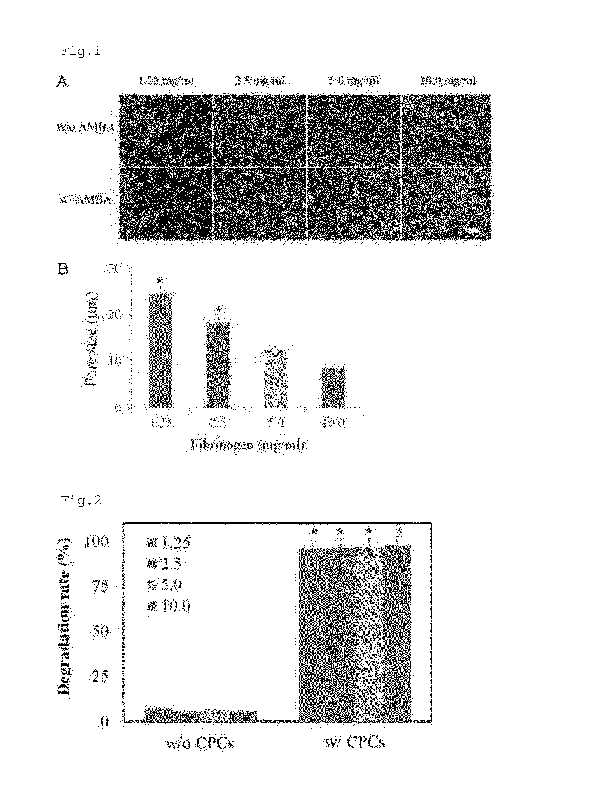
[0166]In order to examine the effect of fibrinogen on the microarchitecture thereof, fibrin hydrogels were constructed with various concentrations of fibrinogens. In this regard, fibrin was prepared from four concentrations of fibrinogen. Human plasma-derived fibrinogen (GreenCross, Seoul, Korea) was dissolved in DMEM containing 10 mM CaCl2 to give 2.5, 5.0, 10.0, and 20.0 mg / ml fibrinogen solutions. Alexa Fluor 488-conjugated fibrinogen (1:50 w / w) (Invitrogen, Carlsbad, Calif.) was added to the fibrinogen solutions. Separately, thrombin (Sigma, St. Louis, Mich.) was dissolved in DMEM to form a thrombin solution with a final concentration of 1 unit / ml. Each of the four fibrinogen solution was mixed at a ratio of 1:1 (v / v) with the thrombin solution, and 10 μl of each of the resulting mixtures was placed on a glass slide and incubated at 37° C. for 2 hrs for a cross-linking reaction. Finally, four fib...

example 2

Fibrinolysis of Cardiac Progenitor Cells

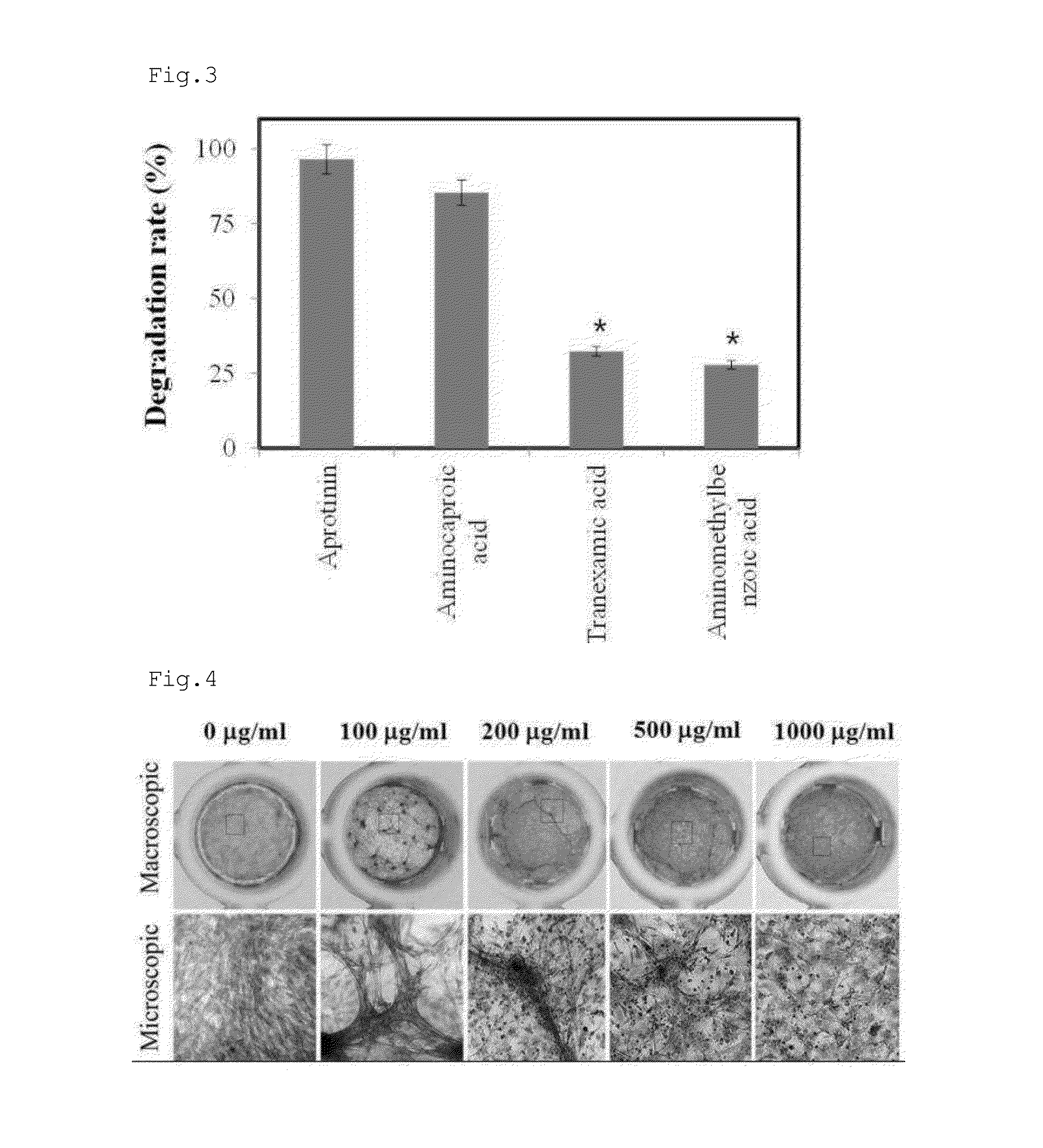
[0168]Four hydrogels with different fibrinogen concentrations were prepared in the same manner as in Example 1. Alexa Fluor 488-conjugated fibrinogen (1:50 w / w) (Invitrogen, Carlsbad, Calif.) was added to each of the four fibrinogen solutions. The cardiac progenitor cells were mixed at a density of 2×10⁵ cells per 100 μl of each of the four fibrinogen solutions with 100 μl of the thrombin solution, followed by a cross-linking reaction for 2 hrs. Following the formation of hydrogel, 300 μl of a cell culture was added to the hydrogel, and incubated for 1 day. A hydrogel void of cardiac progenitor cells was prepared for use as a control. Degradation rates of the fibrin contained in hydrogels were determined by measuring the Alexa Fluor 488-conjugate fibrinogen released to the culture medium by means of a fluorometer.

[0169]As can be seen in FIG. 2, the hydrogel embedded with cardiac progenitor cells (w / CPCs) started to degrade from 2 hrs after inc...

example 3

Inhibition of Antifibrinolytic Agents against Cardiac Progenitor Cell-Induced Fibrinolysis

[0170]Antifibrinolytic agents were purchased from Sigma. Cardiac progenitor cells were embedded in the same manner as in Example 2 into a hydrogel containing an antifibrinolytic agent, and incubated for 1 day before the analysis of fibrin degradation.

[0171]As can be seen in FIG. 3, fibrin hydrogels containing aprotinin or aminocaproic acid were degraded to the degrees of 95% and 80%, respectively, by the cardiac progenitor cells, indicating that aprotinin and aminocaproic acid are slightly inhibitory of cardiac progenitor cell-induced fibrinolysis. In contrast, the hydrogels containing tranexamic acid or aminomethylbenzoic acid were resistant to the fibrinolytic activity of cardiac progenitor cells, as demonstrated by the degradation of the hydrogel at a rate of less than 30%.


PUM


Abstract

Disclosed is a method for culturing myocardium-resident cardiac progenitor cells, comprising: embedding myocardial fragments into hydrogel; culturing the myocardial fragment into hydrogel; degrading only the hydrogel to recover cardiac progenitor cells grown out of the myocardial fragment to the hydrogel; and amplifying the cardiac progenitor cells in vitro. Also, the cardiac progenitor cells, a method for differentiating the same, and the use thereof as cell therapeutic agent for heart diseases are provided. In addition to possessing the potential to differentiate into cardiomyocytes, osteoblasts, adipocytes, chondrocytes, vascular endothelial cells, smooth muscle cells, neural cells, and skeletal muscle cells, the myocardium-resident cardiac progenitor cells can spontaneously differentiate into cardiomyocytes even in the absence of a special differentiation inducing agent. Thus, the cardiac progenitor cells can be used to produce bio-active medicines such as cell therapeutics and tissue engineering therapeutics with high industrial applicability.

Description

BACKGROUND OF THE INVENTION[0001]1. Field of the Invention[0002]The present invention relates to cardiac progenitor cells, a method for culturing the same, a method for differentiating the same, a cell therapeutic agent comprising the same, and a therapeutic agent for heart diseases comprising the same.[0003]2. Description of the Related Art[0004]When it is damaged by various factors, the heart is regenerated, although limitedly. Ischemic heart diseases such as coronary artery disease and myocardial infarction cause death and irreversible loss of cardiomyocytes, resulting in a decrease in heart function, with the consequent onset of intractable myocardial diseases such as congestive heart failure. Hence, there is a pressing need for a novel stem cell-based bio-active medicine that is clinically applicable to such intractable heart diseases.[0005]Stem cell therapeutics applicable to the therapy of intractable myocardial diseases may be sourced from embryonal stem cells (ESCs), induce...

Claims


Application Information

Patent Timeline
Patent Type & Authority Applications(United States)
IPC IPC(8): A61K35/34
CPCA61K35/34C12N5/0657C12N5/0692C12N2501/999C12N2533/52A61P9/00C12N5/00C12N11/04C12N2506/1315
Inventor YANG, YOUNG ILLEE, SEUNG JINKIM, HYEONG INKIM, JONG TAECHEONG, SOON HO
Owner INJE UNIV IND ACADEMIC COOP FOUND
Why Eureka
Try Eureka