Methods and Compositions for Diagnosis of Ovarian Cancer

a technology for ovarian cancer and compositions, applied in the field of methods and compositions for diagnosis of ovarian cancer, can solve the problems of difficult to identify cancer-specific changes in the context of this great complexity, and difficult to discover novel blood biomarkers for cancers

Inactive Publication Date: 2014-05-01
THE WISTAR INST OF ANATOMY & BIOLOGY
View PDF2 Cites 12 Cited by
  • Summary
  • Abstract
  • Description
  • Claims
  • Application Information

AI Technical Summary

Problems solved by technology

Epithelial ovarian cancer (EOC) is the fifth-leading cause of cancer-related deaths in women, with a higher fatality-to-case ratio than any other gynecologic malignancy in the United States.1-3 A major challenge in EOC is that greater than two-thirds of cases are diagnosed at advanced stages (Stages 3 or 4), when five-year survival is about 33%.
Although proteomic technologies have improved dramatically, discovering novel blood biomarkers for cancers remains formidable due to the vast complexity of the plasma proteome and the likelihood that any tumor-specific proteins will be present at very low abundance.
In addition, comparison of patient and control serum or plasma is complicated by the fact that EOC, as well as other cancers and many other conditions, induce an acute-phase reaction or inflammatory response that is not specific to a single disease.10 These changes affect levels of a substantial number of high- and medium-abundant plasma proteins.
Identifying cancer-specific changes in the context of this great complexity and inflammation-induced variability is very difficult when patient serum samples are directly analyzed to discover new biomarkers using proteomics.
Furthermore, changes in abundance levels of a protein in the tumor or shed by the tumor into the interstitial space do not necessarily correlate with their abundance levels in the blood.

Method used


Image

  • Methods and Compositions for Diagnosis of Ovarian Cancer
  • Methods and Compositions for Diagnosis of Ovarian Cancer
  • Methods and Compositions for Diagnosis of Ovarian Cancer

Examples


example 1

Materials and Methods

[0151]Reagents. Molecular-biology-grade ethanol (200 proof); LC-MS-grade formic acid; sodium phosphate monobasic; N,N-dimethylacrylamide (DMA), ammonium bicarbonate; and iodoacetamide were purchased from Sigma-Aldrich (St. Louis, Mo.). Sodium dodecyl sulfate (SDS), 2-mercaptoethanol, and Tris were purchased from Bio-Rad (Hercules, Calif.). ZOOM focusing buffers and thiourea were obtained from Invitrogen (Carlsbad, Calif.). PlusOne reagents dithiothreitol (DTT), 3-[(3-cholamidopropyl)dimethylammonio]-1-propanesulfonate (CHAPS), and urea were purchased from GE Healthcare (Piscataway, N.J.). HPLC-grade acetonitrile was purchased from Thomas Scientific (Swedesboro, N.J.). Tris(2-carboxyethyl)phosphine (TCEP) was obtained from Pierce (Rockford, Ill.), and sequencing-grade modified trypsin was purchased from Promega (Madison, Wis.).

[0152]Cell culture. The human EOC cell line OVCAR-3 was obtained from the American Type Culture Collection (ATCC, Manassas, Va.). The cell...

example 2

Xenograft Mouse Model

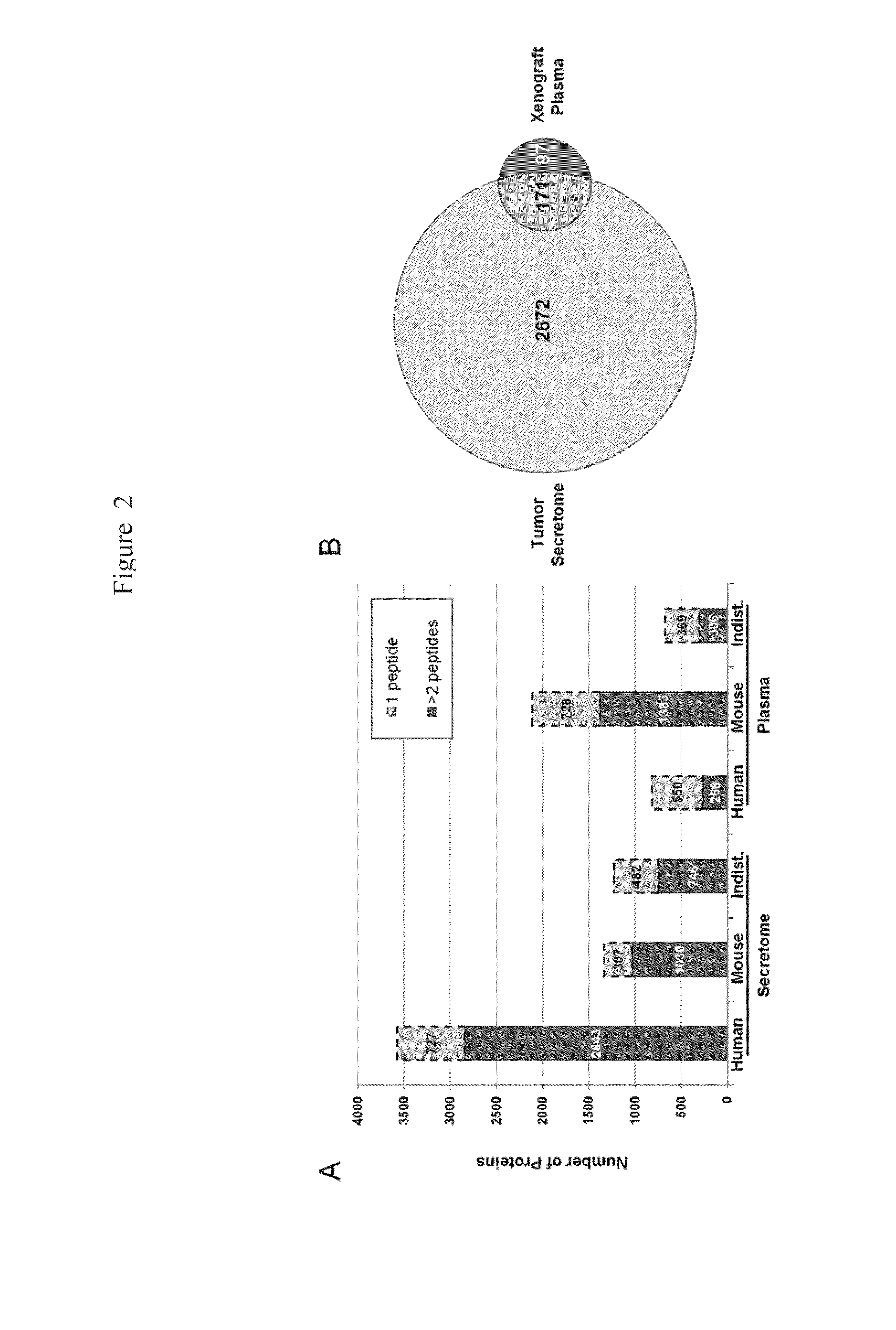
[0168]The primary experimental systems where migration into the blood is assured are mouse models. One of these approaches is to identity quantitative differences in plasma or serum of genetically engineered mice bearing murine ovarian tumors compared with appropriate controls.17 Importantly, an underutilized alternative is the xenograft mouse EOC model, where in-depth proteome analysis can unambiguously identify human proteins shed by the tumor into the murine blood based upon species differences in peptide sequences found in serum or plasma.

[0169]We recently demonstrated that an in-depth 4D analysis of serum from SCID mice bearing ovarian tumors formed by an endometrial ovarian cancer cell line could detect more than a hundred human proteins that were identified by two or more peptides per protein.18 Furthermore, pilot validation of selected candidate biomarkers demonstrated that many of these proteins could be detected in human serum using multiple reaction m...

example 3

Discovery, Verification, and Validation of EOC Biomarkers Using a Xenograft EOC Mouse Model

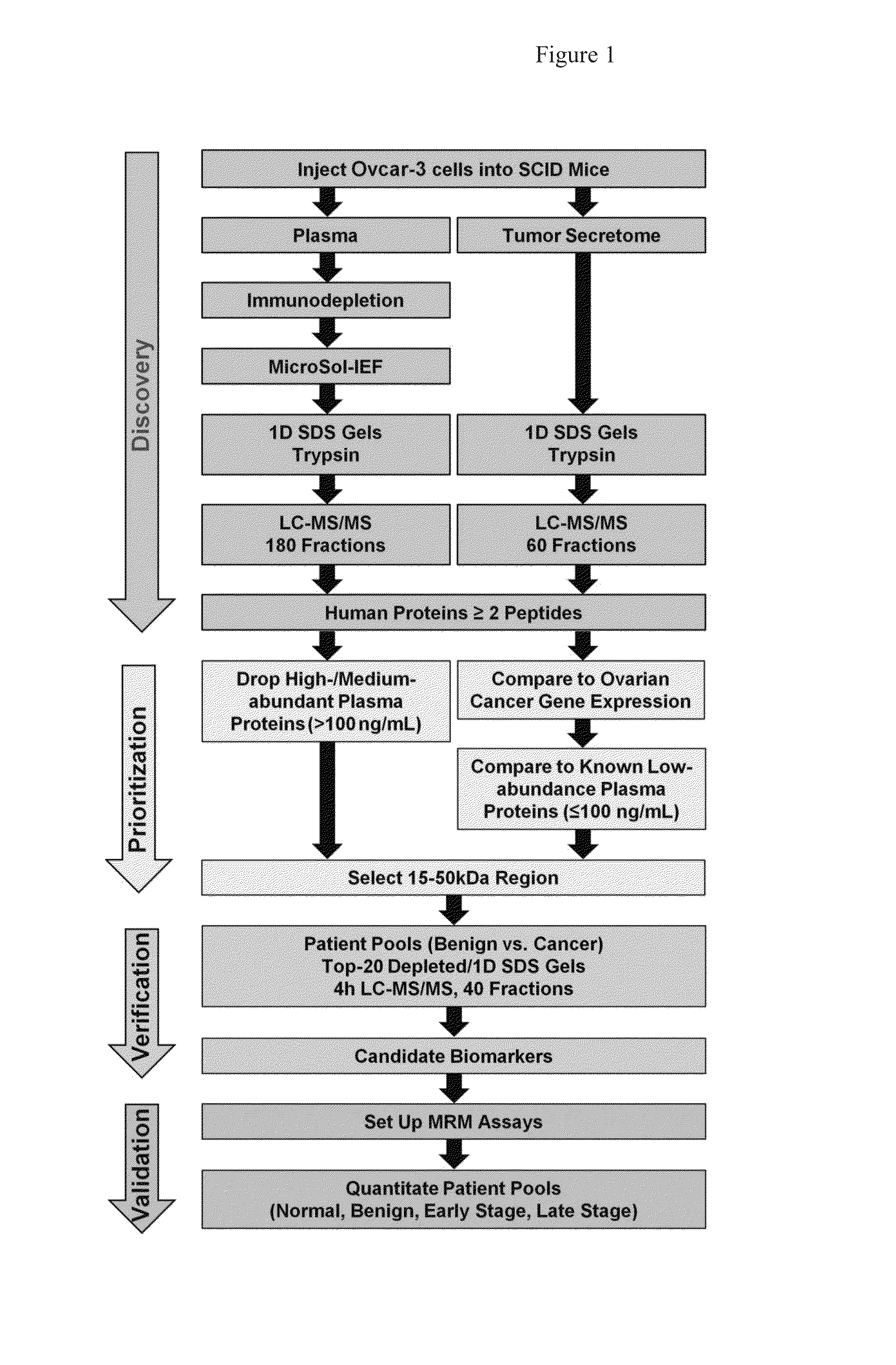
[0172]The strategies used to compare the merits of analyzing a xenograft mouse plasma proteome and / or the corresponding tumor secretome to discover novel ovarian cancer biomarkers, as well as downstream verification and initial validation, are outlined in FIG. 1. In the discovery phase, OVCAR-3, an established serous cell line, was grown in SCID mice. Tumors were excised, briefly cultured in serum-free media, and supernatants were divided into 60 fractions and analyzed using GeLC-MS / MS. Due to its greater complexity, the xenograft mouse plasma was subjected to more extensive fractionation (180 fractions) using a 4D plasma proteome separation method developed in our laboratory.22 Human proteins from the xenograft plasma and tumor secretome were prioritized, as described below, and the 15-50 kDa region of the gel was selected for proof-of-principle verification and initial validation studies bec...


PUM

PropertyMeasurementUnit
pHaaaaaaaaaa
particle sizeaaaaaaaaaa

Abstract

Methods and compositions are provided for diagnosing ovarian cancer in a mammalian subject, preferably in a serum or plasma sample of a human subject. The methods and compositions enable the detection or measurement in the sample or from a protein level profile generated from the sample, the protein level of one or more specified biomarkers. Comparing the protein level(s) of the biomarker(s) in the subject's sample or from protein abundance profile of multiple biomarkers, with the level of the same biomarker(s) or profile in a reference standard, permits the determination of a diagnosis of ovarian cancer, or the identification of a risk of developing ovarian cancer, or enables the monitoring of the status of progression or remission of ovarian cancer in the subject followed during a therapeutic protocol.

Description

CROSS-REFERENCE TO RELATED APPLICATIONS[0001]This application claims the benefit of the priority of U.S. Provisional Patent Application No. 61 / 720,616, filed Oct. 31, 2012, which application is incorporated herein by reference in its entirety.STATEMENT REGARDING FEDERALLY SPONSORED RESEARCH OR DEVELOPMENT[0002]This invention was made with government support under Grant Nos. CA131582 and CA10815 awarded by the National Institutes of Health. The government has certain rights in this invention.BACKGROUND OF THE INVENTION[0003]Epithelial ovarian cancer (EOC) is the fifth-leading cause of cancer-related deaths in women, with a higher fatality-to-case ratio than any other gynecologic malignancy in the United States.1-3 A major challenge in EOC is that greater than two-thirds of cases are diagnosed at advanced stages (Stages 3 or 4), when five-year survival is about 33%. In contrast, when the disease is diagnosed at Stage 1, five-year survival is approximately 90%.2 CA125 is the biomarker ...

Claims


Application Information

Patent Timeline
Patent Type & Authority Applications(United States)
IPC IPC(8): G01N33/68
CPCG01N33/6893G01N33/57449G01N2800/52G01N2800/60
Inventor SPEICHER, DAVID W.TANG, HSIN YAOBEER, LYNN A.
Owner THE WISTAR INST OF ANATOMY & BIOLOGY
Why Eureka
Try Eureka