Method of preparing a growth factor concentrate derived from human platelets

a technology of growth factor and concentrate, which is applied in the direction of growth factor/regulator, animal/human protein, pharmaceutical non-active ingredients, etc., can solve the problems of non-specific tissue degradation, inability to heal wounds, and ‘off-target’ destruction of growth factors

Inactive Publication Date: 2015-08-13
KASIAK RES PVT
View PDF3 Cites 11 Cited by
  • Summary
  • Abstract
  • Description
  • Claims
  • Application Information

AI Technical Summary

Problems solved by technology

However, in vivo, matrix metalloproteinases (MMPs), which usually play an important role in protein turnover during tissue formation, can also cause non-specific tissue degradation if a high concentration of MMPs is present in the wound site for a prolonged duration.
This can result in ‘off target’ destruction of growth factors that are essential for healing.
Proteases present in the wound exudate and bacterial contamination in the wound further degrade the growth factors and can make wound healing impossible leading to a chronic, un-healing wound.
However, usually bovine thrombin is used for this purpose which may cause immune reactions and also carries a threat of transmission of bovine viruses or diseases to humans.
The use of human thrombin makes the product uneconomical due to its high cost.
Further, clinical use of PRP for nerve injury and sports medicine has produced inconsistent results in early trials.
In fact there are very few controlled clinical trials that have adequately evaluated the safety and efficacy of PRP treatments and these trials have generally concluded that PRP is a ‘promising’ but not ‘proven’ treatment option for joint, tendon, ligament, and muscle injuries.
The reason for such inconsistent results and negative outcome for PRP clinical studies are associated with poor quality of PRP produced by non-standardized methods.
Another problem is that most PRP preparing devices do not count the platelets and produce PRP with only a fixed percentage of platelets which is directly related to the percentage present in the original blood sample.
This variable may affect the results since, on one hand, higher concentration of growth factors is shown to have inhibitory effects, and on the other hand PRP with lower platelet numbers may not show optimum results.
One of the major drawbacks of methods that are currently being used is that PRP can only be used as an autologous therapy and cannot be used as an allogeneic therapy because of immunological factors such as ABO incompatibility wherein donor plasma or donor platelet cell membranes may be immunogenic and cause serious adverse events in patients.
Further, when PRP is administered to a patient, it is the entire intact platelets that are injected and hence the existing platelet cell membranes may cause allo-immunization and adverse reactions.
Another drawback of the existing method for preparing PRP is that it is a single blood draw procedure for a single dose and hence multiple blood draws are needed in case multiple doses are required.
Yet another drawback is that PRP has to be used within four hours of processing and cannot be stored because storage causes flocs and fibrin clots to be formed in the plasma.
Another theory behind why PRP does not work as well as expected is that the concentration of platelets in the PRP obtained from automated machines is too low to achieve the desired clinical effect, probably because 30 to 35% of platelets are lost during processing in automated machines.
Although recently, a new substance called human platelet lysate (HPL) has been developed which has similar characteristics of PRP, it has not been used for clinical applications because it has several drawbacks including variable platelet numbers and growth factor levels.
Preparation of HPL is time consuming as it involves multiple freeze-thaws of PRP for activation of the platelets to release the growth factors as a low amount of growth factors are released in a single cycle of freezing and thawing.
Multiple freeze-thaws and long-term storage cause denaturation of the growth factors and also causes a fibrin clot to be formed on storage, making HPL unsuitable for use.
Majority of the growth factors get trapped in the clot formed, thus, leaving insufficient growth factors in the plasma lysate prepared for downstream applications.
Recently, a technology for removal of fibrin present in the plasma has been identified, however the method for fibrin removal is complicated and time consuming and hence makes the product unaffordable.
Animal blood has also been used to make HPL and this is unsuitable for human use as it can cause xenogeneic immune reactions.
Also, HPL is conventionally used in cell culture media and is not used for any therapeutic purposes / clinical applications.
Further, solvent or detergents are often used for making the platelet concentrates before lysis, and the use of such solvents or detergents also makes these methods unacceptable for use of HPL in injectables for treatment of humans.

Method used


Image

  • Method of preparing a growth factor concentrate derived from human platelets
  • Method of preparing a growth factor concentrate derived from human platelets
  • Method of preparing a growth factor concentrate derived from human platelets

Examples


example 1

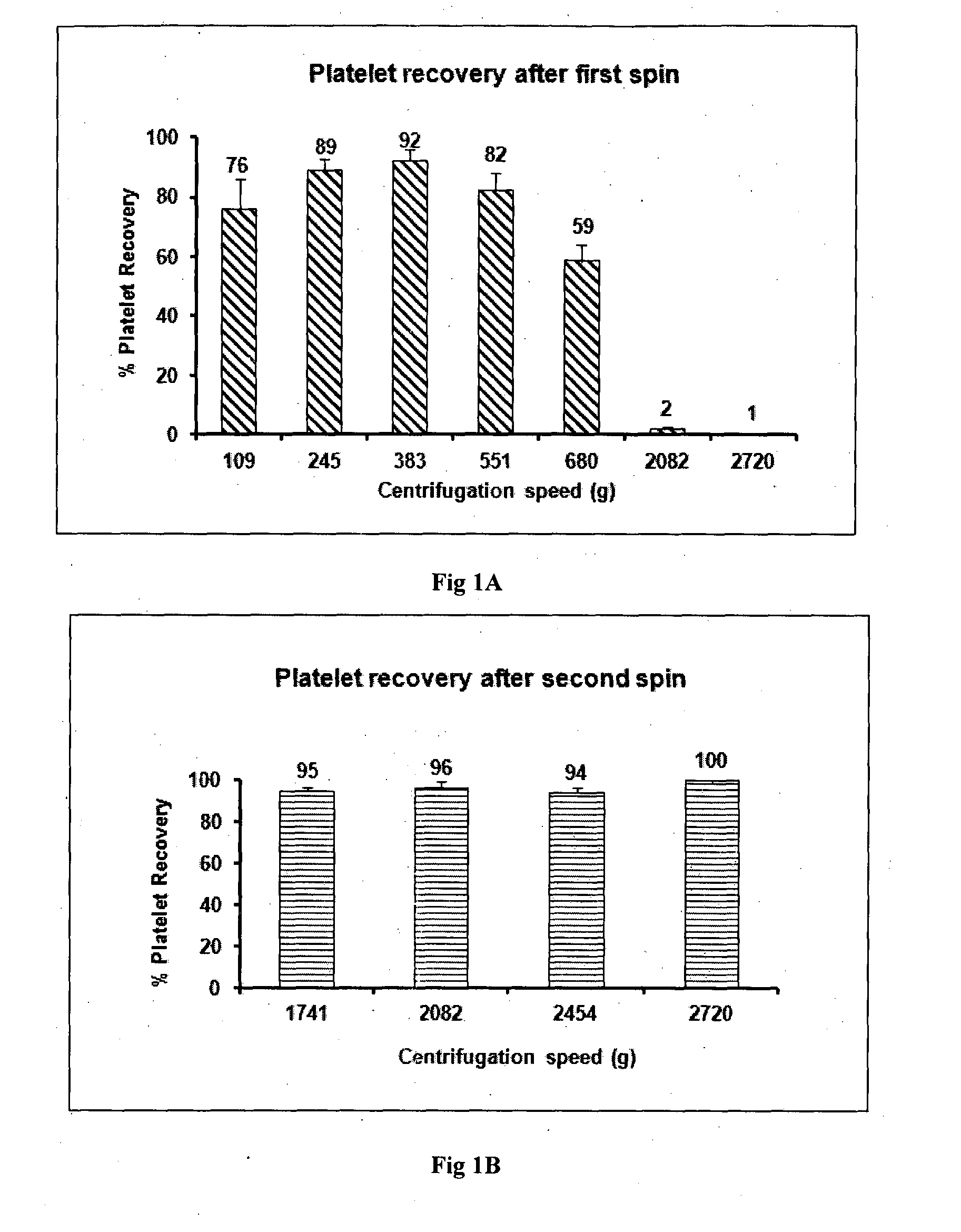
[0080]Human blood was withdrawn into vacutainers after getting informed consent from the patient. About 50-60 ml blood is collected in two types of vacutainers: 50 ml in vacutainers containing ACD-A for preparing GFC and 5-10 ml blood in EDTA tubes for infectious disease marker testing and complete blood parameter testing. Minimum 50 ml of blood was taken for 10 ml of GFC preparation. Blood was transported at 15 to 30° C. preferably at 22° C. within 4 hours of withdrawal. The first centrifugation was done at 382 g for 15 minutes since platelet recovery at 382 g for 15 minutes is optimum with a loss of only 8-10% platelets as shown in FIG. 1A. Platelet loss is significantly greater at lower or higher centrifugation speeds. Upon completion of first centrifugation three layers were formed. At the bottom were packed red blood cells, in the middle were leukocytes and the upper layer had plasma containing platelets. Plasma containing platelets was aspirated and transferred in another ster...

example 2

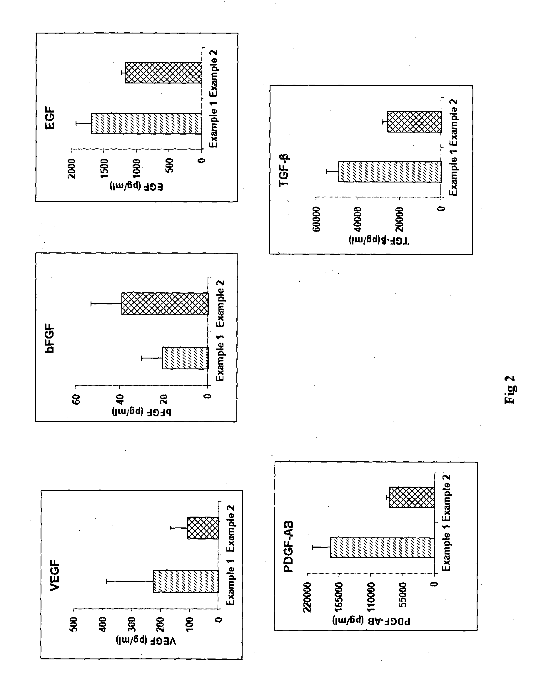
[0082]The process of Example 1 was followed except that the platelet pellet was suspended in 1 ml of multiple electrolyte isotonic solution and after the freeze-thaw, the thawed solution was then mixed with 9 ml more of multiple electrolyte isotonic solution. FIG. 2 shows a comparison of the concentrations of various growth factors obtained by the process of Example 1 versus Example 2 for blood obtained from the same donor. The graphs show that levels of various growth factors like Vascular Endothelial Growth Factor (VEGF), basic Fibroblast Growth Factor (bFGF), Platelet Derived Growth Factor-AB (PDGF-AB), Epidermal Growth Factor (EGF), Transforming Growth Factor-beta (TGF-β) as determined by Enzyme linked immunosorbent assay (ELISA) were almost comparable in GFC prepared with plasma (Example 1) and GFC which is plasma free (Example 2).

example 3

[0083]The processes of Example 1 and Example 2 were separately followed using blood obtained from the same donor. Further, the suspension containing GFC was mixed with 10% mannitol. The solution was then lyophilized. The lyophilized product was then packed in vials with flip off caps so as to make an “off the shelf” product. FIG. 3 is a comparison of the levels of growth factors obtained by the process of Example 1 and 2 vis-à-vis the process of Example 3 as determined by ELISA. It is evident that the levels of growth factors are almost the same or marginally reduced in the lyophilized GFC product as compared to the GFC prepared by Example 1 and Example 2.


PUM

PropertyMeasurementUnit
temperatureaaaaaaaaaa
volumeaaaaaaaaaa
temperatureaaaaaaaaaa

Abstract

The invention relates to a method of preparing an intra-dermally, intra-articularly, sub-dermally or topically administrable growth factor concentrate derived from human platelets. The method comprises the steps of suspending human platelets in multiple electrolyte isotonic solution; snap-freezing the suspension; thawing the frozen suspension; and sterile-filtering the suspension. In particular, in this method, a fixed number of platelets is suspended in a fixed volume of multiple electrolyte isotonic solution to obtain the required concentration of growth factors in the growth factor concentrate, snap-freezing of the suspension is carried out at a temperature of −120° C. to −200° C., thawing of the frozen suspension is carried out at 25° C. to 37° C., and cellular debris are separated from the thawed suspension and the resultant suspension of growth factors is diluted with an isotonic medium before sterile-filtering.

Description

FIELD OF THE INVENTION[0001]This invention relates to a method of preparing a growth factor concentrate derived from human platelets. The invention also relates to the growth factor concentrate, a lyophilized preparation thereof, a composition comprising the growth factor concentrate, a method of treating dermatological, orthopedic, neurological and endocrinological conditions, and use of the growth factor concentrate.BACKGROUND OF THE INVENTION[0002]Platelets, on activation, are known to release growth factors, cytokines, chemokines and adhesive proteins. These growth factors, cytokines, chemokines and adhesive proteins play an important role in reducing inflammation and increasing proliferative events that help in tissue remodeling and regeneration of tissue in case of injuries or wounds. However, in vivo, matrix metalloproteinases (MMPs), which usually play an important role in protein turnover during tissue formation, can also cause non-specific tissue degradation if a high conc...

Claims


Application Information

Patent Timeline
Patent Type & Authority Applications(United States)
IPC IPC(8): A61K38/18A61K45/06A61Q7/00A61K8/64A61Q19/08A61K35/19A61K8/98
CPCA61K38/1858A61K38/1866A61K38/1808A61K38/1825A61K38/1841A61K2800/84A61K45/06A61K8/983A61K8/64A61Q19/08A61Q7/00A61K35/19C07K14/475A61K9/19A61Q19/02A61K2300/00
Inventor TOTEY, SATISH MAHADEORAOMANIYAR, RACHANA RAJIVFONSECA, LYLE CARLLAGHATE, SNEHA DEEPAKKOSHY, NICOLE
Owner KASIAK RES PVT
Why Eureka
Try Eureka