Detection of membrane proteins

a membrane protein and detection technology, applied in the field of detection of membrane proteins, can solve the problems of reducing the charge state of membrane proteins, reducing the activation energy needed to liberate membrane proteins from detergent micelles during detection, and reducing the activation energy needed to liberate membrane proteins from detergent micelles

Inactive Publication Date: 2015-12-03
OXFORD UNIV INNOVATION LTD
View PDF1 Cites 7 Cited by
  • Summary
  • Abstract
  • Description
  • Claims
  • Application Information

AI Technical Summary

Benefits of technology

[0007]The present invention is based, at least in part, on a surprising discovery that by using polyoxyalkylene glycol detergents to form micellar solutions of membrane proteins, the charge state of the membrane proteins may be significantly reduced. This is turn means that the activation energy n...

Problems solved by technology

As a significant biological target in disease and cancer, their study by traditional structural biology approaches, such as X-ray crystallography and nuclear magnetic resonance, has been frustrated by limitations relating to their expression and solubility.
Furthermore, X-ray analysis...

Method used


Image

  • Detection of membrane proteins
  • Detection of membrane proteins
  • Detection of membrane proteins

Examples


example 1

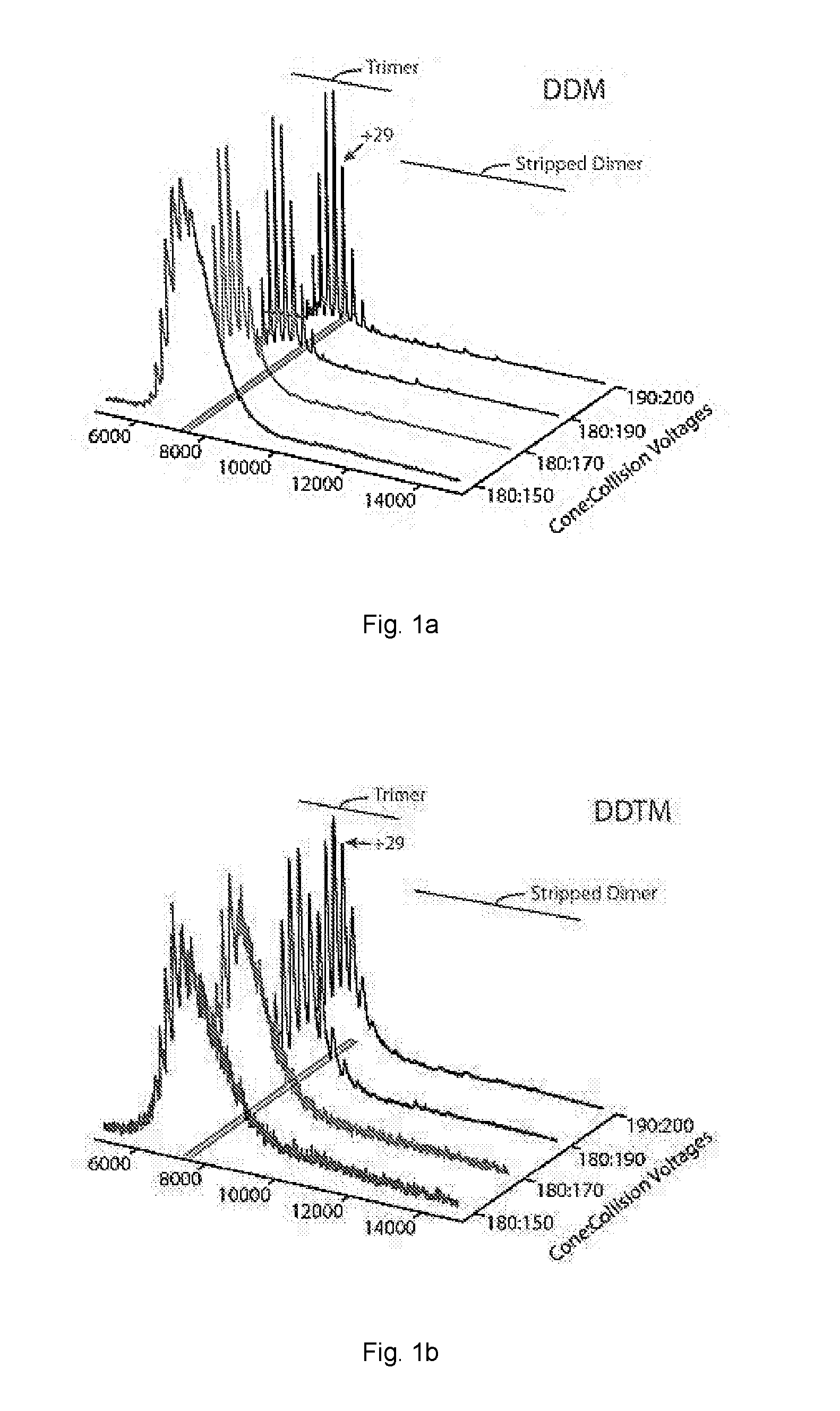
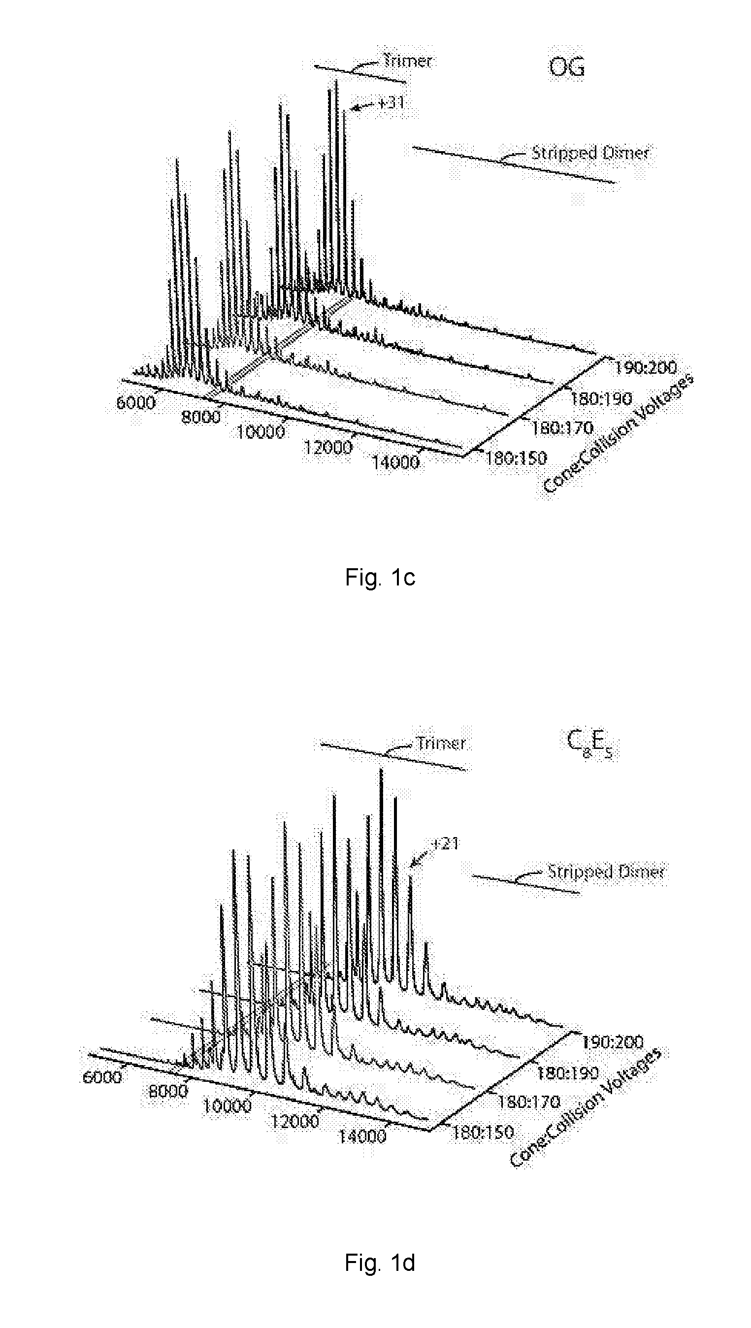
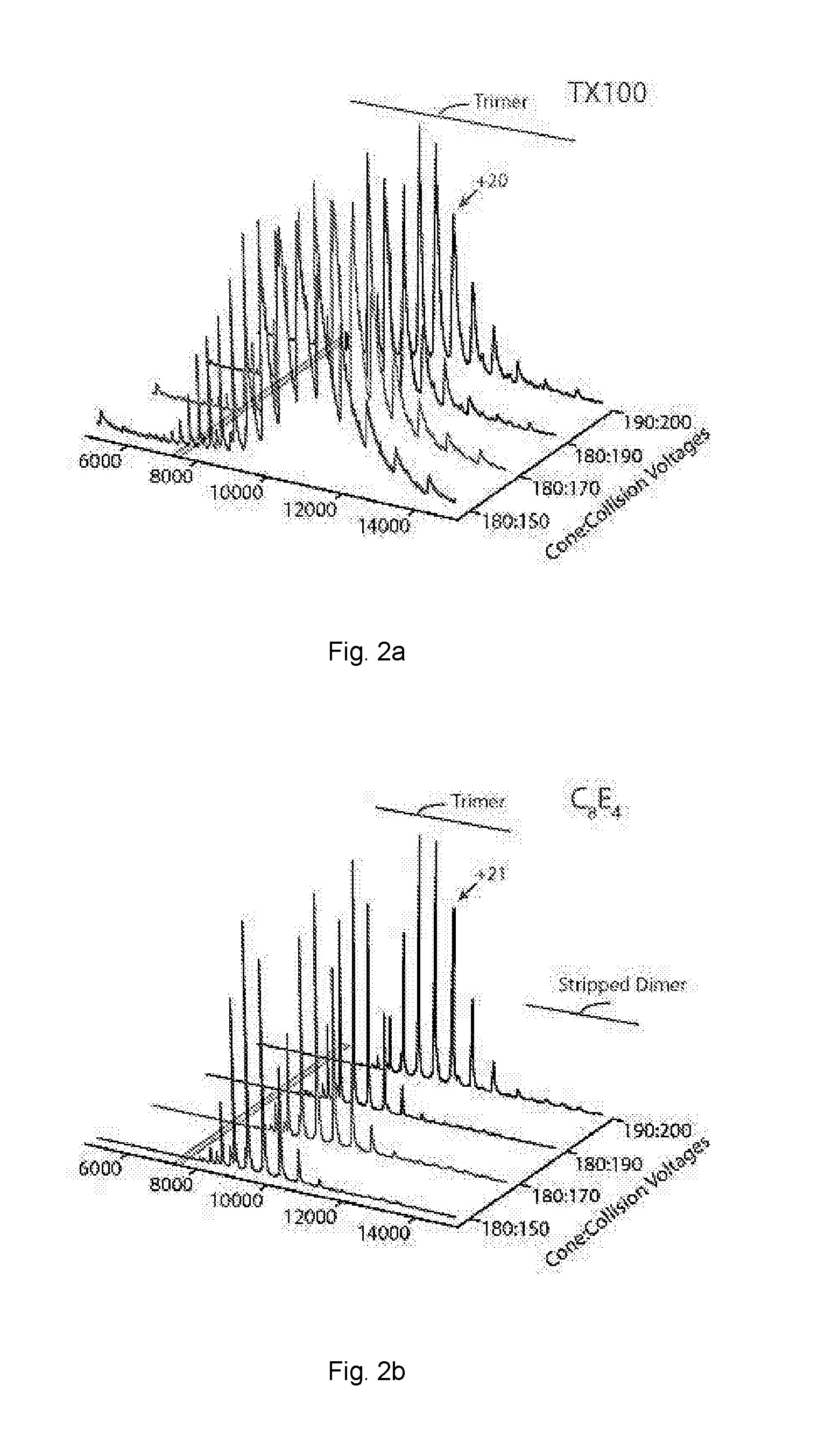
[0063]The activation energy required to achieve resolved peaks of E. coli ammonium channel C-terminally fused to green fluorescent protein (AmtB-GFP) was assessed for a variety of non-ionic detergents.

[0064]A green fluorescent protein (GFP) expression plasmid was constructed by subcloning the multiple cloning site region from XbaI (New England Biolabs) and BlpI (New England Biolabs) of pET23b (Novagen) into the backbone pET15b (Novagen). The resulting engineered vector was linearized by Ndel (New England Biolabs) and Nhel (New England Biolabs) such that an Infusion cloning reaction (Clonetech) resulted in a TEV cleavable C-terminal fusion to superfolder GFP (subcloned from Gandhi et al., Protein Science 2011, 20, 313-326) followed by a 6× His-tag.

[0065]The AmtB and AQPZ genes were amplified by polymerase chain reaction (PCR) with Phusion high-fidelity DNA polymerase (New England Biolabs) from E coli genomic DNA with primers designed for an Infusion (Clonetech) reaction using the man...

example 2

[0072]Mass spectra activation series for AmtB-GFP and Aquaporin Z (AQPZ) were obtained using a variety of non-ionic detergents.

[0073]AmtB-GFP and AQPZ-GFP were extracted from purified membranes in 2% OG in Buffer B and incubated overnight at 4° C. Extracted membrane proteins were clarified by centrifugation at 20,000 g for 25 minutes at 4° C. The clarified supernatant was filtered before loading onto a 5 mL HisTrap-HP column (GE Healthcare, Piscataway, NJ) equilibrated in Buffer E (200 mM sodium chloride, 10% glycerol, 20 mM Imidazole, 0.025% DDM, and 50 mM TRIS, pH 7.4 at room temperature). After the clarified supernatant was loaded, the column was initially washed with 40-50 mL of Buffer E containing 1% OG instead of DDM followed by several column volumes of Buffer E until a steady baseline was reached. Protein was eluted with a linear gradient to 100% in two column volumes of Buffer F (100 mM sodium chloride, 10% glycerol, 500 mM Imidazole, 0.025% DDM, and 50 mM TRIS, pH 7.4 at r...

example 3

[0080]A mass spectrometry approach was used to monitor the effects of individual lipid binding events on various membrane protein complexes anticipated to respond differently to the phospholipid environment. Three membrane protein complexes were selected to give a range of topologies, oligomeric states and anticipated selectivity towards phospholipids: (i) the pentameric mechanosensitive channel of large conductance (MscL) from Mycobacterium tuberculosis with two transmembrane helices (TMH) and an intimate relationship with phospholipids; (ii) the tetrameric water efflux channel Aquaporin Z (AQPZ) from E. coli with six TMH for which associated phospholipid or detergent molecules have been revealed in crystal structures and in related homologues; and (iii) the trimeric ammonia channel (AmtB) from E. coil, with eleven TMH involved in the transport of ammonia / ammonium, for which no phospholipid binding has previously been observed in crystal structures.

[0081]To study the membrane prote...


PUM


Abstract

According to the present invention, there is provided a method of detecting a membrane protein by mass spectrometry, the method comprising the steps of: (a) providing a solution comprising a detergent micelle in which said membrane protein is contained, wherein said solution contains a polyoxyalkylene glycol detergent; (b) providing a mass spectrometer comprising a nanoelectrospray ionisation source, a mass analyser and a detector; (c) vaporising the solution using the nanoelectrospray ionisation source under conditions such that the membrane protein is released from the micelle; (d) ionising the membrane protein; (e) resolving the ionised membrane protein using the mass analyser; and (f) detecting the resolved membrane protein using the detector. Also provided are reagents for use in said method.

Description

FIELD OF THE INVENTION[0001]The present invention relates to the detection of membrane proteins. More particularly, the present invention relates to methods for the detection of membrane protein complexes by mass spectrometry. The methods disclosed herein may be used to detect membrane proteins, including intact membrane protein complexes and complexes of membrane proteins with ligands such as therapeutic agents.BACKGROUND TO THE INVENTION[0002]Membrane proteins are responsible for a wide range of biological functions. Some of the most prevalent human diseases, including some cancers, result from their dysfunction. Despite representing around a third of the human genome, membrane proteins represent targets for more than half of all current therapeutic agents. As a significant biological target in disease and cancer, their study by traditional structural biology approaches, such as X-ray crystallography and nuclear magnetic resonance, has been frustrated by limitations relating to th...

Claims


Application Information

Patent Timeline
IPC IPC(8): G01N33/68
CPCG01N2333/705G01N33/6848
Inventor LAGANOWSKY, ARTHURREADING, EAMONN
Owner OXFORD UNIV INNOVATION LTD
Why Eureka
Try Eureka