Diagnosis of viral infections by detection of genomic and infectious viral DNA by molecular combing

a technology diagnostics, which is applied in the field of diagnostics of viral infections by detection of genomic and infectious viral dna by molecular combing, can solve the problems of virus useless or uncompetitive, technology can be used, and errors in the genome, etc., and achieves the effects of reliable, simple, fast and inexpensiv

Inactive Publication Date: 2016-02-18
GENOMIC VISION
View PDF2 Cites 6 Cited by
  • Summary
  • Abstract
  • Description
  • Claims
  • Application Information

AI Technical Summary

Benefits of technology

[0062]The invention provides a reliable, simple, fast, and inexpensive way to detect infectious or pathogenic polynucleotide sequences, including viral genomic sequences, in a sample from a subject or patient using a Molecular Combing and / or DNA stretching techniques in combination with specific probes which are able to bind with at least 5%, 10%, 25%, 50%, 75%, especially sets of probes that bind to 80-100% of the infectious or pathogenic polynucleotide sequence.
[0064]Unlike prior art techniques for detecting virus in infected cells or tissues, the Molecular Combing techniques of the invention can be performed simultaneously and do not require multiple time consuming and expensive procedures. The methods of the invention are easily applicable as diagnostic tools for detecting viral contamination or infection in cells or tissues, useful for determining the efficacy of antiviral treatments by quantifying or otherwise evaluating the effects or efficacies of such treatments on the quantity of infectious virus or infectious viral polynucleotides or viral replication in infected mammalian cells or tissues. The inventors have found that Molecular Combing overcomes many of the problems associated with past methods of detecting or diagnosing a viral infection, including PCR-based methods, and offers an easy and rapid way to do so. Specific embodiments of the invention include the following.

Problems solved by technology

However, up to now, it was not shown that this technology can be used to detect the infectious forms of viruses in an infected eukaryotic cell or viral sequences or genome of a derived mutated viral sequence infecting a cell or tissue or present in a fluid of mammals.
RNA viruses generally have smaller genome sizes than DNA viruses because of a higher error-rate when replicating, and have a maximum upper size limit.
Beyond this limit, errors in the genome when replicating render the virus useless or uncompetitive.
When this happens with influenza viruses (Hampson and Mackenzie 2006), pandemics might result.
Like other enveloped viruses, herpes viruses are prone to deactivation by organic solvents or detergents and are unstable outside the host's body.
Many types of viral infections, such as a common cold, are self limiting in generally healthy people.
This means that the viral infection causes illness for period of time, then it resolves and symptoms disappear.
However, some people are at risk for developing serious complications of viral infection.
As an example of serious damage, some forms of measles virus infection lead to brain's inflammation, and consequently permanent brain injury.
Although viruses cause disruption of healthy homeostasis, resulting in disease, they can also exist relatively harmlessly within an organism.
This results in inflammation, edema, cell lysis, and formation of a characteristic thin-walled vesicle.
In 25% to 50% of cases, keratitis is recurrent and chronic and can interfere with vision.
As a consequence, HSV-1 is a leading cause of blindness throughout the world.
2009), a condition in humans in which the immune system begins to fail, leading to life-threatening opportunistic infections.
When CD4+ T cell numbers decline below a critical level, cell-mediated immunity is lost, and the body becomes progressively more susceptible to opportunistic infections.
This method, however, will not distinguish among HSV-1, HSV-2, or other herpesviruses, which require more specific subtyping.
Direct tests on specimens or cell cultures using fluorescent antibodies or detection of DNA using specific probes or amplification by Polymerase Chain Reaction (PCR) can differentiate among HSV-1, HSV-2, and closely related HSV, but are not useful tools to confirm that the genomes of said viruses found in a sample are intact or infectious.
While serological analysis is useful for primary infection, it is inconclusive for recurrent illness because the antibody titer to HSV upon recurrence of the infection usually does not increase.
However, current treatments do not reduce the load of the DNA matrix, and thus are unable to reduce the risk of further viral reactivation.
However, none of these methods allowed for both the detection and the nature of an infectious HSV genome in infected cells in a single analysis.
The most reliable method of diagnosis of the HSV infectious disease is to isolate the virus for determination, but this method required culture cells and requires several days for the determination.
There are also immunological methods but they are mostly unreliable and difficult to perform, especially during the latency phase.
A large number of PCR-based methods have been develop for enhanced sensitivity and faster time to result than is possible by conventional means but these methods do not allow analysis of the whole HSV genome and cannot predict the infectivity of the sample containing nucleotide sequences of the virus because the complete genome of the said virus is not tested.
1992) have been reported but these methods are not able to determine the type of HSV.
However, this technique does not discriminate between the isomer of HSV genome in infected cells or tissues.
This family of tests is highly time-consuming and in all cases requires southern blotting, implying manipulation of radioactivity, long migration and / or exposure times.
However, in spite of its clinical success, HAART cannot eradicate the virus, mainly due to the persistence of various viral reservoirs including latently infected resting CD4+ cells (Hermankova, Siliciano et al.
HAART failure as a result of development of drug-resistant HIV-1 strains is a common problem (del Rio 2006).
However, systematic studies of the relationships between the cellular HIV-1 RNA / DNA levels and therapy outcome are hindered by the extremely low copy numbers of HIV-1 RNA / DNA in PBMC under HAART.
However, despite their accuracy and specificity, single-step real-time RT-PCR methods using the TaqMan detection chemistry are unable to reliably quantify <100 copies of HIV-1 RNA / DNA target per reaction in the context of total cellular RNA / DNA (Espy, Uhl et al.
This evokes the possibility of yielding false-negative results when PBMC material from patients under HAART is studied, especially when limited amounts of clinical material are available for analysis.
2006) but are prone to false-positive results because DNA binding dyes do not bind in a sequence-specific manner.
However, only semi-quantitative data can be produced with this method.
In addition, it requires labor-intensive and time-consuming experimental procedures.
A significant limitation of PCR is that it does not allow one to confirm the integrity of the complete genome detected and consequently cannot be a standard of characterization of complete infectious viral genome found in a tested sample.
PCR thus also lacks the specificity required for testing the efficiency of an antiviral treatment against infectious virus particles because its results do not indicate whether the viral polynucleotides detected are infectious.
Serological methods do not directly detect infectious viral polynucleotides, such as chromosomally-integrated viral genomic DNA, and require that a subject mount an immune response to a virus prior to detection or that a sufficient amount of viral antigen be present in a sample.
However, only a minority of persons or animals will go on to develop cancers after infection.
The viral promoter or other transcription regulation elements, in turn, cause over-expression of that proto-oncogene, which, in turn, induces uncontrolled cellular proliferation.
One of the problems of gene therapy that can result from the use of lentiviruses or retroviruses is that the integrase enzyme can insert the genetic material of the virus into any arbitrary position in the genome of the host; it randomly shoves the genetic material into a chromosome.
If the gene happens to be one regulating cell division, uncontrolled cell division (i.e., cancer) can occur.
But the recombinant AAV, which does not contain any viral genes and only the therapeutic gene, does not integrate into the genome.

Method used


Image

  • Diagnosis of viral infections by detection of genomic and infectious viral DNA by molecular combing
  • Diagnosis of viral infections by detection of genomic and infectious viral DNA by molecular combing
  • Diagnosis of viral infections by detection of genomic and infectious viral DNA by molecular combing

Examples


example 1

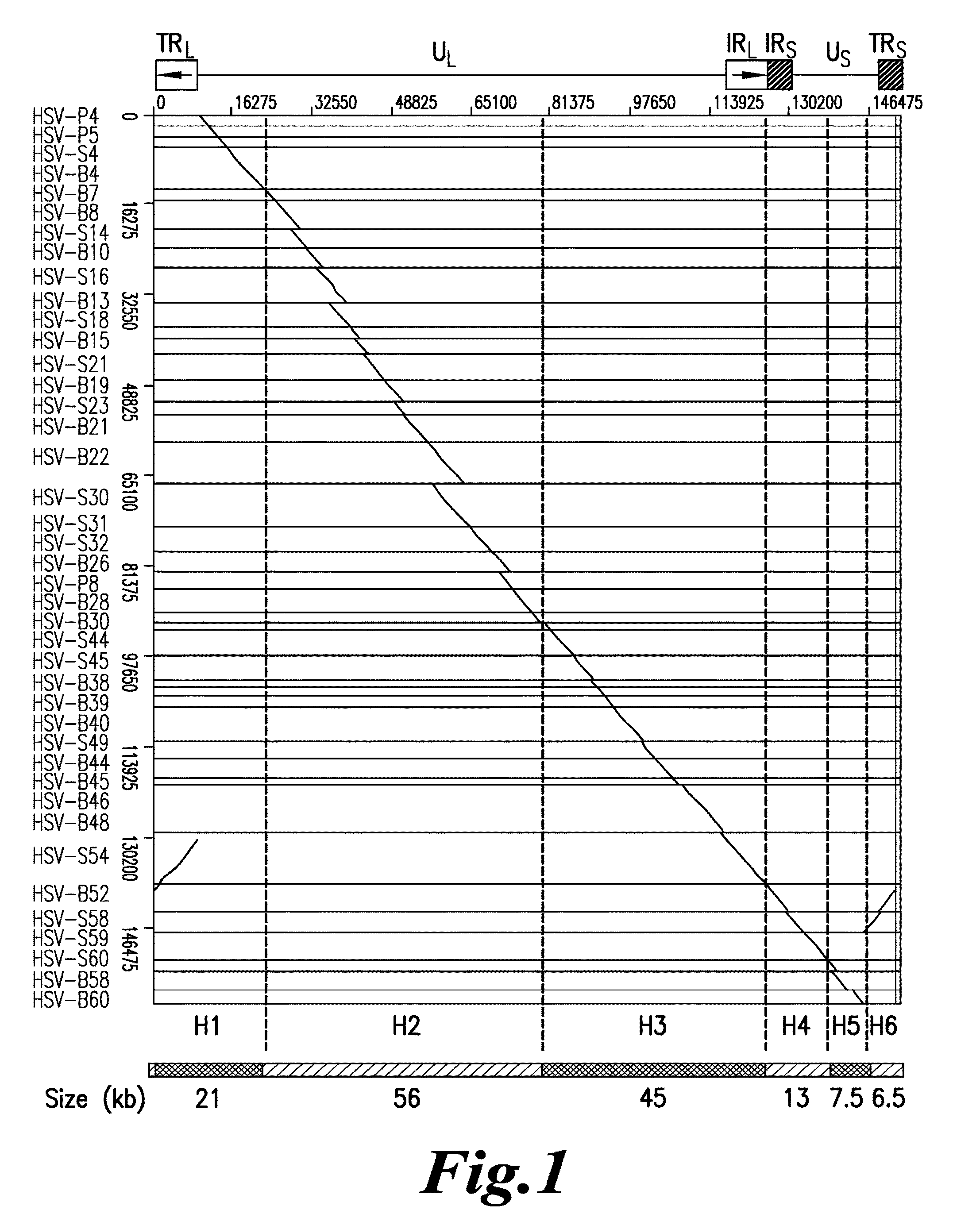
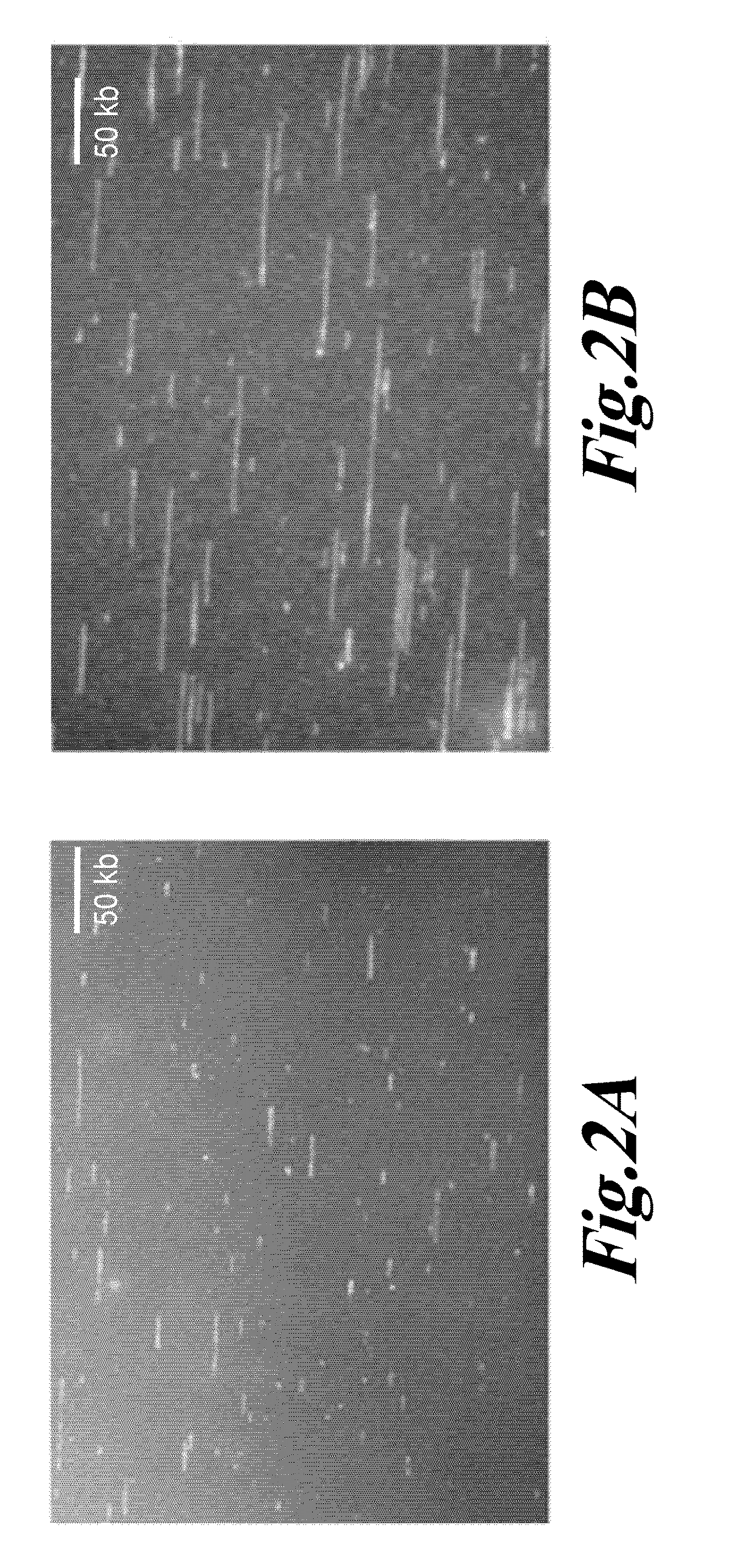
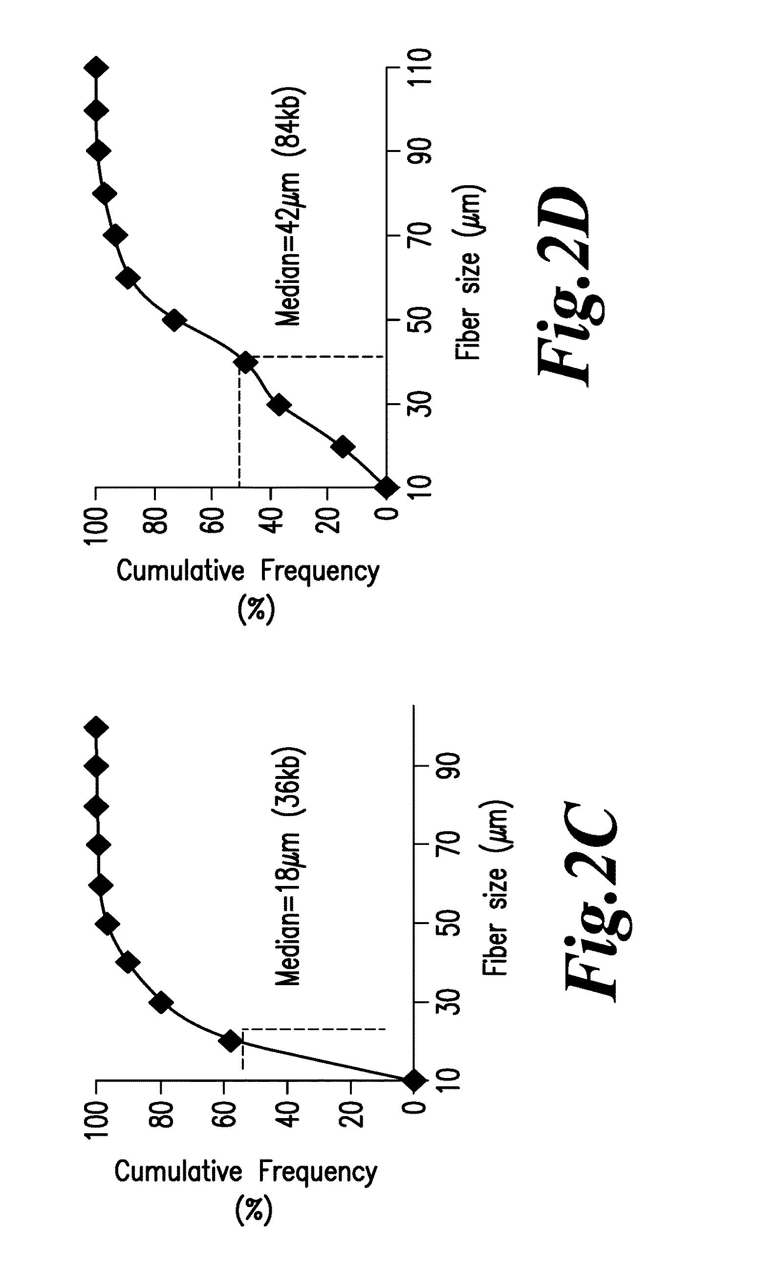
Herpes Simplex Virus Detection

[0115]Preparation of Embedded DNA Plugs from Viral Particles

[0116]HSV-1 DNA was extracted from viral particles by standard phenol:chloroform extraction (Ben-Zeev, Weinberg et al. 1974) or by a modified procedure described in Lebofsky et al. (Lebofsky, Heilig et al. 2006) which are both incorporated by reference. Briefly, HSV-1 particles were resuspended in 1×PBS at a concentration of 5′106 viral particles / mL, and mixed thoroughly at a 1:1 ratio with a 1.2% w / v solution of low-melting point agarose (Nusieve GTG, ref. 50081, Cambrex) prepared in PBS, at 50° C. 90 μL of the viral particles / agarose mix was poured in a plug-forming well (BioRad, ref. 170-3713) and left to cool at least 30 min at 4° C. Embedded viral particles were lysed in 0.1% SDS-0.5M EDTA (pH 8.0) solution at 50° C. for 30 minutes. After three washing steps in 0.5M EDTA (pH 8.0) buffer of 10 minutes at room temperature, plugs were digested by overnight incubation at 50° C. with 2 mg / mL Pr...

example 2

Human Immunodeficiency Virus Detection

[0170]Preparation of Embedded DNA Plugs from ACH-2 Cells Culture

[0171]ACH-2 cell lines (Clouse, Powell et al. 1989) were cultivated according to the authors' instructions. DNA was extracted as described in (Schurra and Bensimon 2009). Briefly, cells were resuspended in 1×PBS at a concentration of 107 cells / mL mixed thoroughly at a 1:1 ratio with a 1.2% w / v solution of low-melting point agarose (Nusieve GTG, ref. 50081, Cambrex) prepared in 1×PBS at 50° C. 90 μL of the cell / agarose mix was poured in a plug-forming well (BioRad, ref. 170-3713) and left to cool down at least 30 min at 4° C. Agarose plugs were incubated overnight at 50° C. in 250 μL of a 0.5M EDTA (pH 8), 1% Sarkosyl, 250 μg / mL proteinase K (Eurobio, code: GEXPRK01, France) solution, then washed twice in a Tris 10 mM, EDTA 1 mM solution for 30 in at room temperature.

[0172]Final Extraction of DNA and Molecular Combing

[0173]Plugs of embedded DNA from ACH-2 cells were treated for combi...

example 3

Detecting an Oncogene by Molecular Combing

[0196]In a manner to analogous to the detection of HSV genomic DNA in Example 1, probes are designed to detect the presence of a viral oncogene. Probes are designed to complement 80-100% of the active viral oncogene of interest and Molecular Combing is performed. The results indicate the presence of an active oncogene in a subject, leading to diagnosis and therapeutic intervention.


PUM

PropertyMeasurementUnit
diameteraaaaaaaaaa
survival timeaaaaaaaaaa
widthaaaaaaaaaa

Abstract

A method for detecting in vitro the presence of a genome of a DNA virus or a viral derived DNA in an infected eukaryotic cell, tissue or biological fluid using Molecular Combing or other nucleic acid stretching methods together with probes, especially nucleic acid probes, having a special design. A method for monitoring in vitro the effects of anti-viral treatment by following the presence of genomic viral or viral derived DNA polynucleotides in a virus-infected cell, tissue or biological fluid. Detection of an infectious form of a virus using Molecular Combing and DNA hybridization. A kit comprising probes used to carry out these methods and a composition comprising the probes.

Description

CROSS-REFERENCE TO RELATED APPLICATIONS[0001]This application is a continuation of U.S. Ser. No. 13 / 092,210, filed Apr. 22, 2011, and claims priority under 35 U.S.C. 119(e) to U.S. Provisional Application 61 / 327,397 filed Apr. 23, 2010 which is incorporated by reference in its entirety.STATEMENT REGARDING FEDERALLY SPONSORED RESEARCH OR DEVELOPMENT[0002](None)REFERENCE TO MATERIAL ON COMPACT DISK[0003](None)BACKGROUND OF THE INVENTION[0004]1. Field of the Invention[0005]A method for easily, rapidly and accurately detecting the presence of infectious viral DNA or other infectious or pathogenic DNA sequences in a viral host cell, tissue or biological fluid obtained from a subject or patient using Molecular Combing and / or DNA stretching in combination with specially designed probes for the infectious viral or pathogenic DNA sequence. A method for monitoring in vitro the effects of anti-viral treatment by following the presence of genomic viral polynucleotides in a virus-infected cell, ...

Claims


Application Information

Patent Timeline
Patent Type & Authority Applications(United States)
IPC IPC(8): C12Q1/70
CPCC12Q2600/156C12Q1/708C12Q1/6816C12Q2533/101C12Q1/703C12Q1/705A61P31/20C12Q1/6883
Inventor MAHIET, CHARLOTTESALVAIRE, FABRICECONSEILLER, EMMANUELBARRADEAU, SEBASTIEN
Owner GENOMIC VISION
Why Eureka
Try Eureka