Detection of alpha, beta-dicarbonyl compounds with fluorogenic probes

Inactive Publication Date: 2016-06-23
UNIVERSITY OF WARWICK
View PDF0 Cites 0 Cited by
  • Summary
  • Abstract
  • Description
  • Claims
  • Application Information

AI Technical Summary

Benefits of technology

[0011]The emerging interest in identifying compounds which decrease or increase in the concentrations of α-oxoaldehydes for therapeutic applications requires technology that is adaptable for screening libraries of chemical compounds in high throughput analysis. The fluorescent characteristics of the fluorogenic probes described herein, in particular, 4,5-diaminofluorescein

Problems solved by technology

Modifications by MG can lead to loss of function of the protein and result in DNA strand breaks and mutations at chromosomal hotspots.
α-Oxoaldehyde compounds such as MG and glyoxal are not chromophoric nor fluorescent and therefore are not readily detectable.
Current analytic methods of MG and related α-oxoald

Method used


Image

  • Detection of alpha, beta-dicarbonyl compounds with fluorogenic probes
  • Detection of alpha, beta-dicarbonyl compounds with fluorogenic probes
  • Detection of alpha, beta-dicarbonyl compounds with fluorogenic probes

Examples


example 1

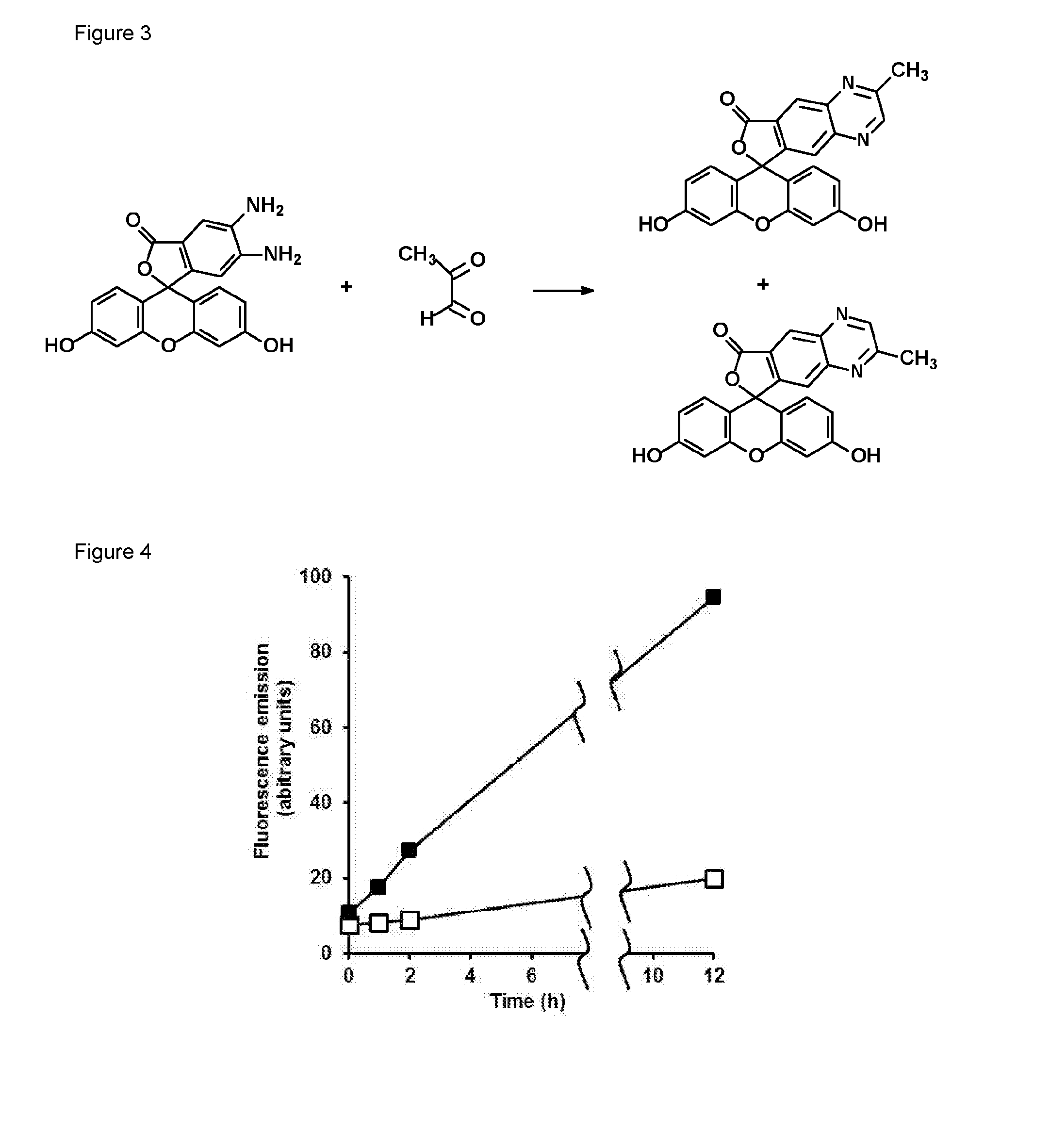
Reaction of methylglyoxal with 4,5-diaminofluorescein (DAF-2)

[0109]4,5-Diaminofluorescein (DAF-2) was a fluorogenic probe developed for the detection of nitric oxide [6]. 4,5-Diaminorhodamine (DAR) [7], 8-(3,4-diaminophenyl)-2,6-bis(2-carboxyethyl)-4,4-difluoro-1,3,5,7-tetramethyl-4-bora-3a,4a-diaza-s-indacene (DAM BO-P″) [8] and o-diaminocyanine [5] are analogous o-diamino derivatizing agents based on different intense fluorophores. DAF-2 reacts with nitric oxide to form the fluorophore triazolofluorescein [6]. For example, incubation of DAF-2 (50 μM) with the NO donor (Z)-1-[2-(2-aminoethyl)-N-(2-ammonioethyl)amino]diazen-1-ium-1,2-diolate (DETA-nonoate), 10 μM, in 10 mM sodium phosphate buffer, pH 7.4 at 37° C., for one hour gave characteristic fluorescence of triazolofluorescein—excitation and emission maximum wavelengths of 491 and 515 nm, respectively. A further metabolite that reacts with DAF-2 is ascorbic acid which forms adducts DAF-2-DHA-5008 and DAF-2-DHA-518 which are fl...

example 2

Dose Response Curve for the Reaction of DAF-2 with Methylglyoxal

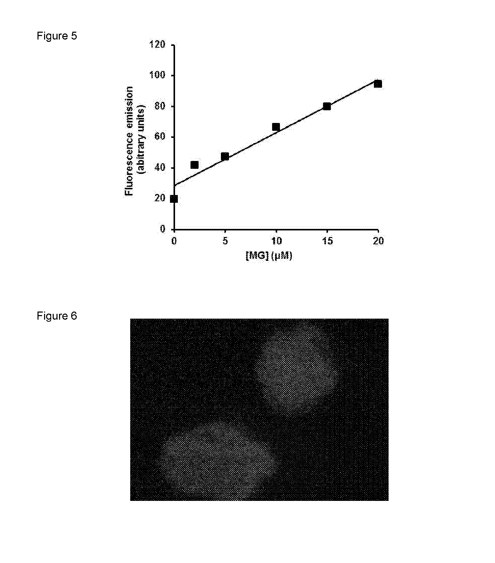
[0112]DAF-2 (50 μM) was incubated with and without 20 μM MG in 10 mM phosphate buffer at pH 7.4 at 37° C. for 24 h. There was a marked increase in fluorescence (excitation wavelength 441 nm and emission wavelength 533 nm) over the incubation period—FIG. 4. When DAF-2 (50 μM) was incubated with 2, 5, 10, 15, and 20 μM MG in 10 mM phosphate buffer at pH 7.4 at 37° C. for 24 h and fluorescence spectra were recorded for excitation and emission wavelength maxima for MG-DAF2, 441 nm and 533 nm respectively, after 24 h the results indicate that the formation of MG-DAF2 fluorescence is proportional to initial MG concentration—FIG. 5.

example 3

Fluorescence Imaging of MG in Isolated Human Leukaemia

[0113]When human leukaemia 60 cells (1×10⁵ cells per ml) in RPMI 1640 with 10% fetal calf serum were incubated with DAF-2 (10 μM) for 40 min at 37° C., washed twice with phosphate buffered saline and re-suspended in fresh medium and incubated for 30-120 min, subsequent detection of fluorescence with excitation laser 457 nm, emission filter 470-500 nm gave fluorescence indicate of the DAF-2 MG adduct and hence an image of endogenous MG in HL60 cells—FIG. 6.


PUM


Abstract

The disclosure relates to a fluorescence based assay for the detection of alpha, beta-dicarbonyl containing compounds, for example alpha-oxoaldehyde compounds such as glyoxal, methylglyoxal, hydroxypyruvaldehyde, erythrosone, 3-deoxy-erythrosone, ribosone, 3-deoxyribosone, glucosone, 3-deoxyglucosone and stereoisomers thereof and butan-2,3-dione that uses a fluorogenic probe which is stable under ambient conditions. The fluorogenic probes comprise a 1,2-diaminophenyl moiety. The diaminophenyl probes yield a 20-fold increase in quantum yield when they are a derivative of fluorescein (DAF), rhodamine (DAR), BODIPY or cyanine. The assay is preferably carried out at pH 4-8. Corresponding screening methods for modulators of alpha, beta-dicarbonyl compound concentrations are also disclosed.

Description

[0001]The disclosure relates to a fluorescence based assay for the detection of α,β-dicarbonyl containing compounds, for example α-oxoaldehyde compounds such as methylglyoxal, glyoxal and 3-deoxyglucosone, in a sample using fluorogenic probes comprising a 4,5-diaminofluorescein, 4,5-diaminorhodamine, 3,4-diaminophenyl boron dipyrromethene or o-diaminocyanine moiety.BACKGROUND TO THE INVENTION[0002]The dicarbonyl, methylglyoxal (MG), is a highly cytotoxic metabolite formed mainly from the degradation of triosephosphate intermediates of glycolysis. It is a minor product formed by spontaneous degradation of triosephosphates in mammalian metabolism—accounting for approximately 0.1% triosephosphate flux, increasing in ageing and disease, and may account for much higher triosephosphate flux in microbial metabolism where MG is from enzymatically from dihydroxyacetonephosphate by methylglyoxal synthase. MG is a potent glycating agent modifying mainly arginine residues in proteins and deoxyg...

Claims


Application Information

Patent Timeline
IPC IPC(8): G01N33/64G01N33/50G01N33/58
CPCG01N33/64G01N33/582G01N33/502G01N2800/7042G01N2800/042G01N2800/044G01N2800/323G01N33/5008G01N33/52G01N2800/102G01N2800/16G01N2800/166G01N2800/2821G01N2800/2835G01N2800/2871G01N2800/301G01N2800/302G01N2800/32G01N2800/321G01N2800/347G01N2800/368
Inventor THORNALLEY, PAULRABBANI, NAILA
Owner UNIVERSITY OF WARWICK
Why Eureka
Try Eureka