Cell-Based Device For Local Treatment With Therapeutic Protein

a technology of local treatment and cell-based devices, which is applied in the direction of pharmaceutical delivery mechanisms, peptide/protein ingredients, surgery, etc., can solve the problems of inability to improve patient medical condition, high preparation costs, and inability to successfully conduct clinical trials of factors, so as to reduce the possibility of adverse side effects, and facilitate the modification of the combination

Inactive Publication Date: 2017-09-21
KEMIJSKI INST
View PDF3 Cites 0 Cited by
  • Summary
  • Abstract
  • Description
  • Claims
  • Application Information

AI Technical Summary

Benefits of technology

[0069]The main advantage of the present invention is that it is possible to easily modify the combination of therapeutic proteins to suit for the treatment of each separate condition or even patient while the costs of production are not significantly increased.
[0070]The implantation of the device of the invention enables local (in situ), continuous and simultaneous delivery of physiological amounts of therapeutic proteins at site where needed most, while avoiding frequent and systemic applications of therapeutic proteins which often led to serious adverse effects. Additionally, several therapeutic proteins in low amounts are used to achieve the maximal healing effect, which also lowers the possibility of adverse side-effects caused by application of non-physiological amounts of single therapeutic protein.
[0071]Another advantage is that the production of therapeutic proteins by the used cells is high, thus lower numbers of cells secreting therapeutic proteins are needed to achieve physiological concentrations of therapeutic proteins, consequently leading to lower amounts of other (non-monitored) proteins secreted.
[0072]The cell lines to be used in the present invention (“carrier cell lines”) are preferably immortal or immortalized cell lines, which can be easily genetically manipulated and banked, which is much cheaper in contrast to the isolation and manipulation of stem cells. Additional advantage of our system over stem cells is that the composition of therapeutic protein mixture is defined and controlled, while for stem cells it is not possible to define how they react in vivo.
[0073]Therapeutic proteins in the sense of the present invention are either proteins, which exist in nature, such as unmodified growth factors, or are designed therapeutic proteins, such as single-chain variable fragments of naturally occurring proteins or a variants thereof. Therapeutic proteins are preferably introduced into a carrier cell line via genetic manipulation techniques well known to the experts in the field.
[0074]Therapeutic proteins exert their biological activity via different healing mechanisms. For example, the epidermal growth factor is known to stimulate keratinocyte and fibroblast proliferation, transforming growth factor alpha (TGF-α) is chemotactic for keratinocytes and fibroblasts, TGF-β1 and TGF-β2 promote angiogenesis and up-regulate the production and inhibit degradation of collagen, while their antagonist TGF-β3 promotes scarless wound healing. TGF-β1 suppresses immune system. The therapeutic proteins of the vascular endothelial growth factor (VEGF) family promote angiogenesis, fibroblast growth factors (FGFs) promote angiogenesis, granulation, and epithelialization. The platelet-derived growth factor (PDGF) is chemotactic for granulocytes, macrophages and fibroblasts. The keratinocyte growth factor (KGF) stimulates keratinocyte migration, proliferation and differentiation. The hepatocyte growth factor (HGF) promotes progenitor cell mobilization, induces angiogenesis and cell proliferation. The insulin-like growth factor (IGF) family induces cell proliferation and inhibits apoptosis, monocyte chemotactic protein-1 induces angiogenesis, and inhibits apoptosis. The brain-derived neurotrophic factor (BDNF), ciliary neurotrophic factor (CNTF), glial cell-derived neurotrophic factor (GDNF) and the nerve growth factor (NGF) promotes neuronal cell survival. The biological activity of the therapeutic proteins as well as functional variants thereof can be measured by assays well-known to the person skilled in the art, such as various migration and proliferation assays in vitro (as described by Schreier et al., 1993), measuring of binding protein activity, their effect on DNA synthesis, protein accumulation and mouse models of particular condition.

Problems solved by technology

However, some individual soluble factors failed to improve medical condition of patients such as the combination therapy of IGF-1 and PDGF for treatment of diabetic foot ulcers or even had severe adverse effects, for example CNTF in treatment of ALS.
WO 2011 / 123779, US 2011 / 0020291 etc. disclose the use of stem cells for wound healing, however, there are some mayor issues connected to stem cells regarding the safety, cellular retention and high preparation costs.
U.S. Pat. No. 5,487,889 describes a bandage containing a container with cells, which are engineered to secrete human PDGF, human EGF, human TGF, bovine GH and combinations thereof to improve wound healing, yet some of those factors have already been shown not to be successful in clinical trials and additional trophic factors seem to be required for the effective therapy.
Additionally, the use of bandage of U.S. Pat. No. 5,487,889 is limited to topical applications.
However, in many human diseases, conditions and as a consequence of treatments the normal wound healing process is disrupted resulting in a chronic wound.
Although such treatment was effective in animal models of neurodegeneration, it did not work when it was tested in clinical trials.
In mentioned studies, where they showed synergistic effect, they used high dosage of recombinant protein, the dosage, which is unphysiological and therefore could present a possible risk for tissue hyperproliferation and also tumor development.
Diabetes is associated with micro- and macrovascular complications, which result in heart conditions and ischemic infarction (heart, brain, and kidney).
Problem of normal blood flow occurs in all sorts of diseases, like in Berger's disease, coronary heart disease, ischemic infarction, that all can result in death.

Method used


Image

  • Cell-Based Device For Local Treatment With Therapeutic Protein
  • Cell-Based Device For Local Treatment With Therapeutic Protein
  • Cell-Based Device For Local Treatment With Therapeutic Protein

Examples


example 1

Preparation of DNA Constructs

[0101]DNA sequences for therapeutic proteins described above were either ordered from supplier, e.g. “Sino biological Inc.”, or designed from amino-acid sequences of the selected protein domains using tool Designer from DNA2.0 Inc. that enables the user to design DNA fragments and optimize expression for the desired hosts (e.g. human cells) using codon optimization. DNA encoding the genes were then ordered from GeneArt or GeneScript, cut out with restriction endonucleases (restriction enzymes) and cloned into the appropriate vector containing necessary regulatory sequences known to the experts in the field. Vectors used include commercial vectors of pcDNA, pMXs etc. carrying all necessary features such as antibiotic resistance, origin of replication and multiple cloning site.

[0102]Molecular biology methods (DNA fragmentation with restriction endonucleases, DNA amplification using polymerase chain reaction-PCR, PCR ligation, DNA concentration detection, a...

example 2

Preparation of Transient and Stable Cell Lines

[0103]Selected carrier cell lines HEK293 (ATCC CRL-1573), NIH 3T3 (ATCC CRL-1573) and ARPE-19 were transfected with prepared constructs for the constitutive production of therapeutic proteins via lipofectamine 2000 and polyethylene imine reagents. 24 h to 36 h post transfection the production of therapeutic proteins was assessed in cell culture supernatant by commercially available ELISA tests.

[0104]Therapeutic cell lines were generated by the addition of selective marker (antibiotic such as neomycin, puromycin, zeocin or blasticidine). Several clones exhibiting high therapeutic protein secretion level, growth characteristics and stability were selected for each therapeutic protein. Stocks of therapeutic cell lines were frozen and stored in liquid nitrogen vapor phase.

[0105]Mouse fibroblasts NIH 3T3 were plated at low density in 10 cm petri dish and transfected with plasmids encoding soluble factors (EGFL7, FGF-2, IGF-I, PDGF-B, TGF-β1 a...

example 3

Gap Migration Assay of Therapeutic Protein Combinations

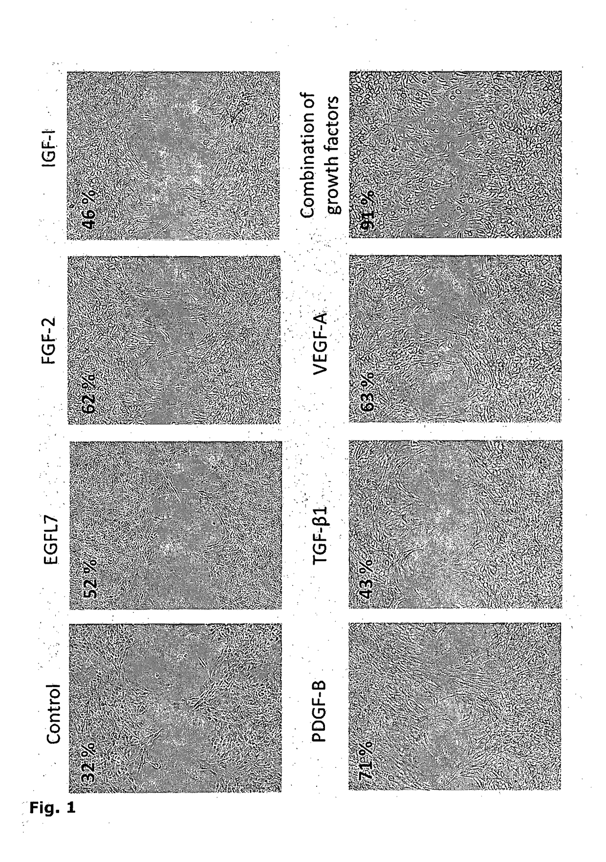
[0107]The effect of each therapeutic protein and their combinations was first tested in gap migration assay on established cell line NIH 3T3.

[0108]One day before the wound scratch assay, NIH 3T3 cells (5*104 cells / well) were seeded onto 8 well μ-slide with inserted culture insert (Ibidi). Day after seeding a confluent monolayer of cells was formed. Insert was removed leaving a 500 μm gap. Supernatants containing a single therapeutic protein or a combination in concentrations ranging from 1 pg / ml to several ng / ml were added. Gap closure was measured after 6, 12 and 24 h. As seen in FIG. 1, the addition of therapeutic proteins had a positive effect on wound closure compared to control without growth factors. Addition of a combination of 6 growth factors (EGFL7, FGF-2, IGF-I, PDGF-B, TGF-β1 and VEGF-A) had the best effect on gap closure.


PUM

PropertyMeasurementUnit
concentrationaaaaaaaaaa

Abstract

The present invention provides a therapeutic device that comprises of mixture of cells secreting combination of therapeutic proteins, where cells producing therapeutic proteins are sealed in container which enables the exchange of nutrient and therapeutic proteins. The cells inside the therapeutic device produce and secrete certain amounts of therapeutic proteins. Cells are prepared by introducing genes encoding therapeutic proteins under the control of a constitutive or inducible promoter. The combination and concentration of therapeutic proteins is defined by the ratio of cells secreting different therapeutic proteins and/or by the gene expression ratio of the therapeutic proteins in the cells incorporated into the semi-permeable container. The therapeutic device can be used for treatments of various diseases and injuries for instance enhancement of wound healing and angiogenesis.

Description

FIELD OF THE INVENTION[0001]The field of invention is directed at a therapeutic device comprising cells secreting a combination of therapeutic proteins, wherein the cells preferably are located in a container which enables the exchange of nutrients and therapeutic proteins.[0002]The cells producing therapeutic proteins are present in the device in ratios and / or show expression and secretion of the therapeutic proteins which are appropriate for therapeutic application.[0003]The present invention provides a device comprising cells secreting a combination of therapeutic proteins, and the use of said device for the treatment of diseases and injuries in which said therapeutic proteins are effective.BACKGROUND OF THE INVENTION[0004]Individual growth factors have been tested for treatment of various diseases and injuries of humans and animals. PDGF-BB is useful in the treatment of diabetic foot ulcers, GM-CSF in the treatment of venous and diabetic foot ulcers and HGH in the treatment of p...

Claims


Application Information

Patent Timeline
Patent Type & Authority Applications(United States)
IPC IPC(8): A61L31/16A61K38/18A61L31/00
CPCA61L31/16A61L31/005A61K38/1808A61K38/1825A61L2430/02A61K38/1858A61K38/1841A61K38/1866A61L2300/414A61K38/18A61K9/0024A61K9/0092
Inventor KADUNC, LUCIJALAINSCEK, DUSKOHORVAT, SIMONJERALA, ROMANHAFNER BRATKOVIC, IVA
Owner KEMIJSKI INST
Why Eureka
Try Eureka