
Collagen Targeted Imaging Probes

Active Publication Date: 2017-12-21
THE GENERAL HOSPITAL CORP
View PDF1 Cites 2 Cited by
  • Summary
  • Abstract
  • Description
  • Claims
  • Application Information

AI Technical Summary

Benefits of technology

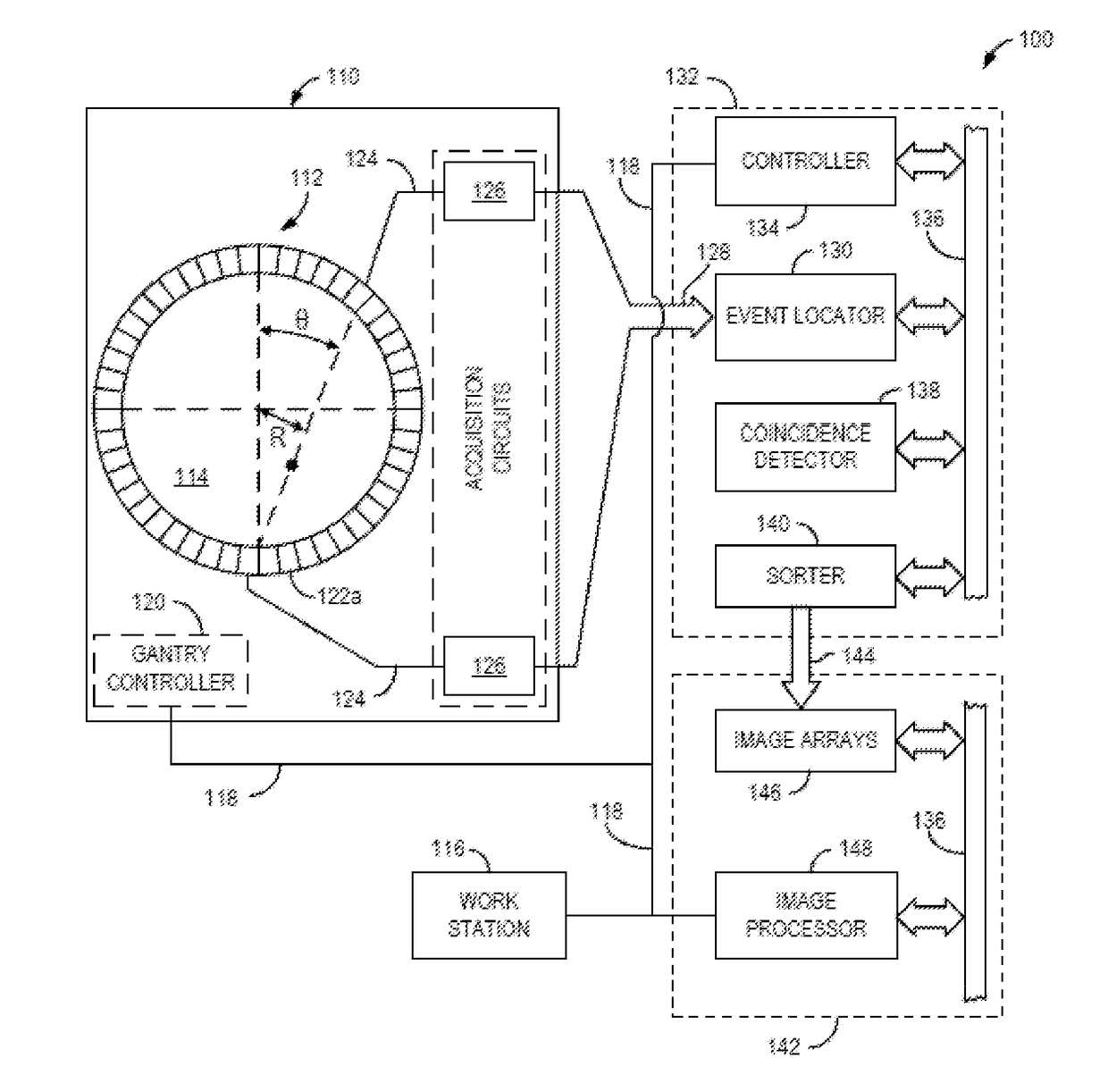
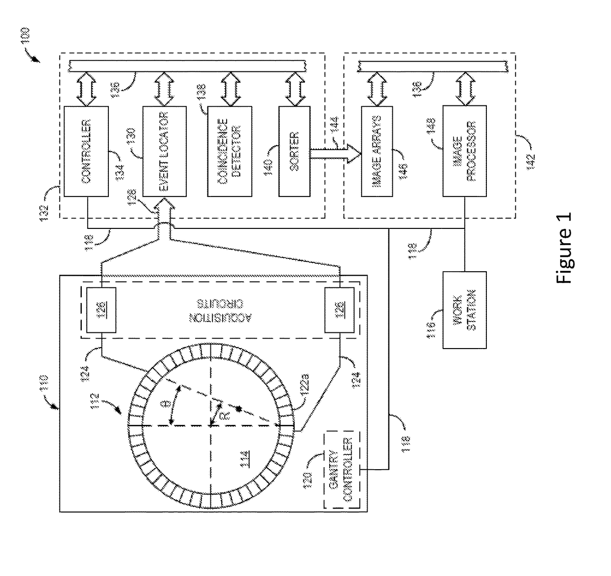
The present invention provides a method for detecting fibrotic pathologies in mammals using a collagen-binding PET probe. The method involves administering an imaging agent with a collagen-targeting peptide and a radioactive element to the mammal, and then using PET imaging to capture images of the newly formed and still disorganized collagen. These images are then combined with anatomical images obtained through computed tomography (CT) to accurately locate the collagen image within the mammal's anatomical image. Fibrotic pathologies exhibit higher uptake of the imaging agent compared to non-fibrotic pathologies, making the method effective for identifying these pathologies.

Problems solved by technology

However in chronic tissue injury, ongoing cycles of injury and repair lead to accumulation of scar tissue and disruption of normal tissue architecture and function, which ultimately can result in organ failure.
Many therapeutic interventions can reverse fibrosis if detected early; however, current radiological techniques only detect later stage disease where tissue damage may be irreversible.
However, depending on the organ of interest this can be challenging.
Similar rates arise with renal biopsy, and the risk of complications is much higher in lung biopsies.
In addition, biopsy can suffer from sampling error, inter-observer variability, and does not sample differences in fibrosis across the organ.
Repeated biopsies to evaluate disease progression or response to treatment are unattractive due to patient compliance and the increased risk of complications.
Abnormal or excessive accumulation of collagen in organs such as the liver, lungs, kidneys, or breasts, and vasculature can lead to fibrosis of such organs (e.g., myocardial fibrosis, heart failure, nonalcoholic steatohepatitis of the liver (also known as NASH), cirrhosis of the liver, primary biliary cirrhosis), lesions in the vasculature or breasts, collagen-induced arthritis, Muscular dystrophy, scleroderma, Dupuytren's disease, rheumatoid arthritis, and other collagen vascular diseases.

Method used


Image

  • Collagen Targeted Imaging Probes
  • Collagen Targeted Imaging Probes
  • Collagen Targeted Imaging Probes

Examples


example 1

Synthesis of 64Cu-CBP1

[0264]Pep(1).

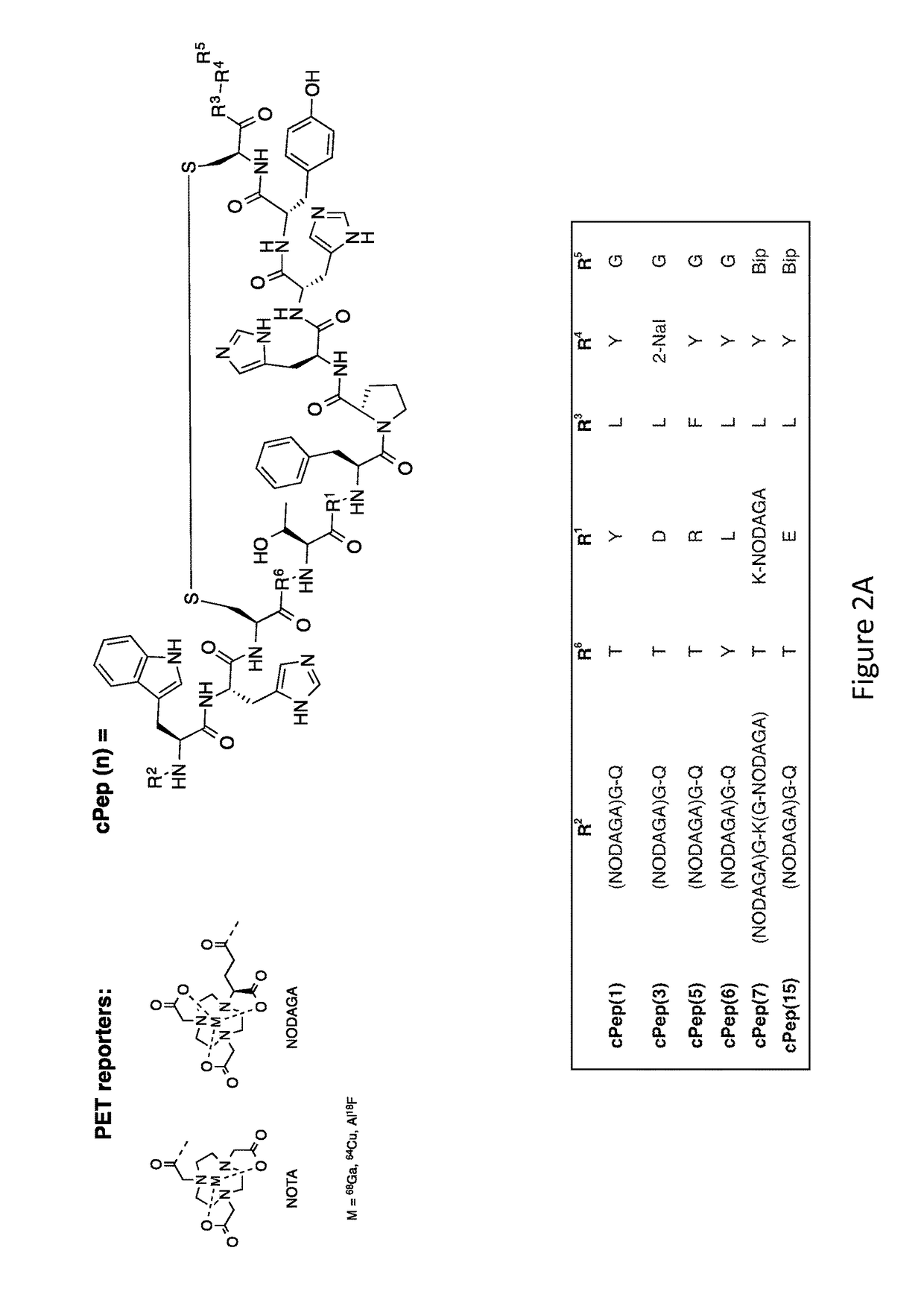
[0265]The CPB1 peptide (see FIG. 2A) was prepared on resin and synthesized by standard Fmoc chemistry using solid-phase peptide synthesis on a OEM microwave peptide synthesizer (Matthews, N.C.) on a 0.1 mmol scale using microwave assisted solid phase synthesis using Rink amide MBHA resin (EMD Millipore), FMOC protected amino acids (Novabiochem) and 2-(1H-benzotriazole-1-yl)-1,1,3,3-tetramethyl-uronium hexafluorophosphate (HBTU) coupling chemistry. Side chain protections of the amino acids used were the following: glutamine, cysteine, histidine: trityl; arginine: 2,2,4,6,7-pentamethyldihydro-benzofuran-5-sulfonyl; aspartic acid: tert-butyl; lysine, tryptophane: tert-butoxycarbonyl; and threonine: tert-butyl. The protected amino acids were dissolved in dimethylformamide (DMF), (0.2 M) the activator HBTU solution was prepared in DMF (0.45 M) and the activator base, diisopropylethylamine (DIPEA), was prepared in N-Methylpyridinone NMP (2M). The deprote...

example 2

Synthesis of 64Cu-CBP3

[0274]Pep(3).

[0275]The CPB3 peptide (see FIG. 2A) was prepared using the same conditions as in Example 1. Pep(3): Molecular weight for C98H124N26O23S2, MS(ESI) calc: 1050.2 [(M+2H) / 2]2+; found: 1050.5.

[0276]cPep(3).

[0277]The linear Pep(3) peptide, about 30 mg, was cyclized and purified using the same conditions as in Example 1, (yield>90%, purity 99%). cPep(3): Molecular weight for C98H122N26O23S2, MS(ESI) calc: 1049.2 [(M+2H) / 2]2+; found: 1049.2.

[0278](tBu)NODAGA-Pep(3).

[0279](tBu)NODAGA-NHS was conjugated to the N-terminus of cPep(3) using the same conditions as in Example 1. The product eluted from the column at approximately 33-36% B. (tBu)NODAGA-cPep(3): Molecular weight for C125H169N29O30S2. MS(ESI): calc: 1312.0 [(M+2H] / 2]2+; found: 1311.3.

[0280]NODAGA-Pep(3).

[0281](tBu)NODAGA-Pep(3) was deprotected using the same conditions as in Example 1. NODAGA-cPep(3): Molecular weight for C113H145N29O30S2. MS(ESI): calc: 1227.9 [(M+2H] / 2]2+; found: 1227.1. 64Cu-CBP...

example 3

Synthesis of 64Cu-CBP5

[0282]Pep(5).

[0283]The CPB5 peptide (see FIG. 2A) was prepared using the same conditions as in Example 1. Pep(5): Molecular weight for C99H127N29O22S2, MS(ESI) calc: 1070.7 [(M+2H) / 2]2+; found: 1070.2.

[0284]cPep(5).

[0285]The linear Pep(5) peptide, about 30 mg, was cyclized and purified using the same conditions as in Example 1, (yield>90%, purity 99%). cPep(5): Molecular weight for C99H125N29O22S2, MS(ESI) calc: 1069.7 [(M+2H) / 2]2+; found: 1069.6.

[0286](tBu)NODAGA-Pep(5).

[0287](tBu)NODAGA-NHS was conjugated to the N-terminus of cPep(5) using the same conditions as in Example 1. The product eluted from the column at approximately 33-36% B. (tBu)NODAGA-cPep(5): Molecular weight for C126H172N32O29S2. MS(ESI): calc: 1332.5 [(M+2H] / 2]2+; found: 1331.7.

[0288](tBu)NODAGA-Pep(5).

[0289](tBu)NODAGA-Pep(5) was deprotected using the same conditions as in Example 1. NODAGA-cPep(5): Molecular weight for C114H148N32O29S2. MS(ESI): calc: 1248.4 [(M+2H] / 2]2+; found: 1247.5.

[029...


PUM

PropertyMeasurementUnit
Structureaaaaaaaaaa

Abstract

Disclosed herein are collagen-targeted imaging agents for positron emission tomography and related imaging methods using the collagen-targeted imaging agents. The collagen-targeted imaging agent is a cyclic polypeptide comprising a cyclic main body, wherein the cyclic main body comprises at least one S—S bond; at least two branches, wherein each of the at least two branches comprises at least three amino acids; and a linker, wherein the linker is capable of linking an imaging reporter.

Description

CROSS-REFERENCES TO RELATED APPLICATIONS[0001]This application claims priority from U.S. Patent Application No. 62 / 014,744 filed Jun. 20, 2014.STATEMENT REGARDING FEDERALLY SPONSORED RESEARCH[0002]This invention was made with government support under grant number NIH HL116315 awarded by National Institutes of Health. The government has certain rights in the invention.BACKGROUND OF THE INVENTION1. Field of the Invention[0003]The invention relates to collagen targeted imaging agents for positron emission tomography.2. Description of the Related Art[0004]There is a major unmet medical need to noninvasively quantify fibrosis. Fibrosis quantification would enable early diagnosis of many chronic diseases and provide a means to monitor response to therapy, thus speeding evaluation of novel treatments. Noninvasive identification and staging of fibrosis would impact tens of millions of Americans with some form of chronic liver (e.g., hepatitis C), lung (e.g., idiopathic pulmonary fibrosis), ...

Claims


Application Information

Patent Timeline
IPC IPC(8): A61K51/08A61K38/00C07K14/00
CPCA61K51/08C07K14/00A61K38/00
Inventor DESOGERE, PAULENECARAVAN, PETER
Owner THE GENERAL HOSPITAL CORP
Learn More