Microchip high density hanging drop three-dimension culture platform

a three-dimensional culture and microchip technology, applied in the field of microchip high density hanging drop three-dimensional culture platform, can solve the problems of complex and potential toxic effects, limited broad practical application, and inability to replicate the important features of three-dimensional (3d) conditions in conventional two-dimensional monolayer cultures, etc., to facilitate tissue physiological function, rapid test of many drugs in real time, and wide range of effects

Pending Publication Date: 2021-11-04
THE BOARD OF TRUSTEES OF THE UNIV OF ILLINOIS +1
View PDF4 Cites 1 Cited by
  • Summary
  • Abstract
  • Description
  • Claims
  • Application Information

AI Technical Summary

Benefits of technology

[0015]Any of a number of immiscible fluids may be used, so long as the fluid does not substantially mix with the fluid in the microwells, including a culture media. Preferably, the immiscible fluid is relatively high viscosity and surface tension so that upon inversion, the immiscible fluid does not significantly flow or otherwise move off the microwells top surface. In this manner, the fluid in the microwells is effectively isolated from the surrounding environment and undesirable evaporation or leakage prevented or minimized. For example, the immiscible fluid may comprise mineral oil and the cells in the microwells are provided in a culture media. As desired, a removable oxygen permeable cover slide may be provided over the immiscible fluid, particularly for those immiscible fluids that may be prone to evaporation and / or flow off the surface of the microwells, including via the force due to gravity.
[0018]The partitioning step may comprise providing a hydrophobic removable reservoir and contacting a top surface of the hydrophobic removable reservoir with mineral oil. This can further assist in ensuring the culture media liquid in the microwells remains in the microwells.
[0023]The methods provided herein have a number of advantages, including any one or more of: a time to form a multicellular spheroid in a plurality of microwells is one-day or less; the partitioning and immiscible layer avoids or minimizes selective pressure on the cell and tissue culture; the array of microwells number between 100 and 1,000,000 (providing reliable and readily controlled high-throughput); and / or each microwell has a volume that is less than 10 μL with an independently-controllable shape parameter testing of the cell and tissue culture geometry on a drug interaction. The combination of small volume, high-throughput, and reliable shape control is a powerful platform that provides a number of benefits not feasible with conventional culture systems.
[0024]For example, the methods are particularly well-suited for screening a drug for biological efficacy. “Biological efficacy” refers to the impact of a drug candidate on a biological cell, including a cancer cell or other biological disease condition. The impact may be in cell growth, cell death, cell proliferation, toxicity, morphology, phenotype and the like. The high-throughput, need for small cell and tissue volumes, and ability to reliably to control shape, is a particularly advantageous combination for rapid, efficient and increasingly reliable drug screen.
[0033]As described herein, microcancer size and volume can be controlled and readily varied through both chip design and cell seeding densities. Formed three-dimensional cultured cells and tissues, including spheroids, may be characterized in real-time without removing the culture from the microwells through upright fluorescence microscopy for tumor spheroid formation, cell death, and other processes. Exemplified herein are LN229 Human Glioblastoma cells and patient derived xenograft (PDX) Glioblastoma cells, also demonstrating compatibility with automated live cell imaging showing formation of the microcancers. Of course, the methods and devices provided herein are compatible with any of a range of cell types and tissues. Also illustrated is the capability to perform high-resolution confocal analysis to study the expression of protein markers directly on a chip without the need for extraction of the cultured cells or tissue, including tumor spheroids. To demonstrate that the platform does not bias tumor evolution by putting selective pressure, it is shown through the use of mate-pair sequencing, a complete overlap in chromosomal rearrangement events between the patient derived xenograft and the corresponding microcancers grown in the present platform. In one embodiment of the present technology, results are shown for direct drug testing on LN229 and PDX glioblastoma tumor spheroids formed in the hang drop culture platform as characterized by real-time optical microscopy. The nano-droplet culture model allows simultaneous testing of hundreds to hundreds of thousands of 3D microcancer cultures in real time where each microcancer captures the salient characteristics of the tumor. Because of the small size of the platform, smaller biopsies can be tested with many drugs. Drug testing can be done in days and the system also allows for sequential (staggered time point) drug testing. Therefore, this system makes it possible to rapidly test many drugs in real time even from clinical scenarios where only small biopsies are available.
[0034]Furthermore, geometric control in a 3D culture platform can be advantageous since tissue structures including size and shape facilitate the physiological function of the tissue by allowing cells and their microenvironment to exchange chemical, electrical and mechanical cues. The instant methods and systems are validated using human cells, including through the use of hanging drop culture, for geometric control of 3D cultured structures, including from Glioblastoma cells. Provided are microchips with etched channels, thereby facilitating chip fabrication with different well shapes, including circular, square, and triangular annular wells. This allows the media droplet shape to conform to the shape of channel cavity due to capillary forces. Using this approach, dense and continuous annular circle, square, and triangle 3D shapes are generated ranging in sizes from greater than one hundred micrometers to several millimeters, including reliably on the order of a few hundred micrometers. The present platform opens up applications of hanging drop culture where different cell types can be co-cultured in 3D on-chip to generate self-assembled and self-organized tissues with the salient in vivo-like features naturally formed via cell-cell interactions.

Problems solved by technology

However, conventional two dimensional monolayer cultures do not replicate the important features of three dimensional (3D) conditions and much work has been done to elucidate the significant differences in cellular morphology, behavior and molecular signaling between the classic cell monolayer approaches and analogous 3D cultures.
Although several 3D culture techniques exist that deploy protein-based or synthetic polymer-based gel environments, rotation-based bioreactors, magnetic levitation and hanging drop techniques, their broad practical application has been limited due to several reasons.
Matrix and hydrogel-based techniques are often used to generate organotypic cultures from isolated stem cells or tumor-initiating cells, but those techniques require cell proliferation and matrix engineering, as the scaffolds can introduce unpredictable cell-polymer interactions that influence and bias downstream applications of the organoids.
Aggregation techniques such as magnetic levitation using nanoparticles to cause spheroid formation have demonstrated a limited number of spheroids and require cells to be pre-treated with magnetic beads at high concentrations adding complexity and potential toxicity.
However, the conventional technology for performing hanging drop cultures is very laborious and low throughput, and requires the disruption of culture conditions and transfer of formed spheroids into a secondary plate for endpoint analysis.
Those techniques are not compatible with direct live microscopy-based characterizations and also suffer from elevated osmolarity caused by evaporation of media from the droplets, which also limits the time for growth and analysis.
Engineering of tissues with tunable morphologies has been studied in 3D culture techniques such as bioprinting and micropatterning, but those techniques are known to cause high shear stress on cells during seeding.
However, the formed structures in that platform are unable to retain shape during cellular compaction, and, pre-treatment with magnetic beads at high concentrations adds complexity and potential toxicity.
Although three-dimensional culture techniques are available, no single platform allows high replicates and high throughput for drug screening, ability to engineer interconnections between different 3D cultured microtissues, compatibility with direct on-chip real-time or high-resolution confocal microscopy, and geometric control of formed cell mass in 3D.

Method used


Image

  • Microchip high density hanging drop three-dimension culture platform
  • Microchip high density hanging drop three-dimension culture platform
  • Microchip high density hanging drop three-dimension culture platform

Examples


example 1

Hang Drop Culture Platform

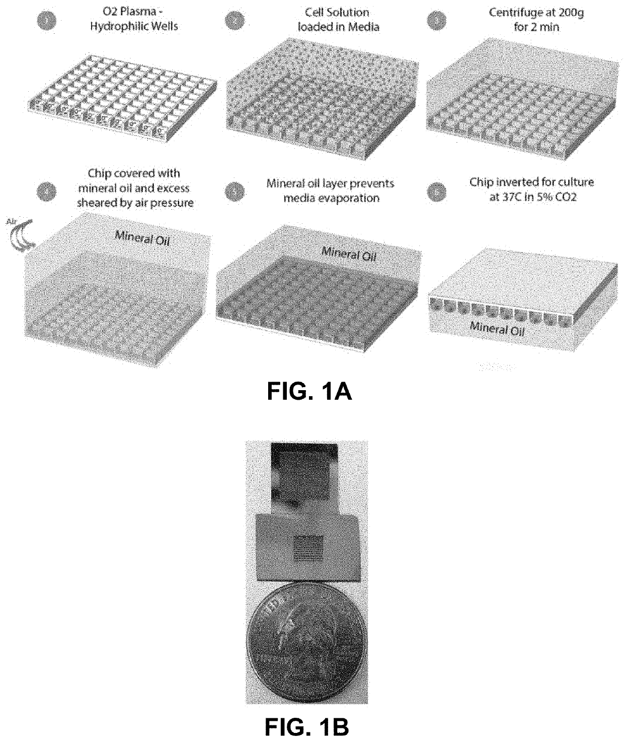
[0078]Several microchip designs are fabricated based on a silicon substrate. The microchips contain an array of silicon microwells of varying dimensions etched to a depth of 120 μm. The silicon surface was oxidized after the formation of the wells. FIG. 1A shows the platform design and schematic of on-chip cell seeding protocol. Panel 2, for clarity, does not illustrate the removable reservoir used to reliably position the cell solution loaded in media over the microwells. The entire chip is less than the size of a quarter and FIG. 1B shows two chip designs, one with an array of 900 microwells with a constant well size of 300 μm (side length of well) and the other with a well size gradient in which the well side lengths were varied from 100 to 500 μm. This system is readily scaled to a larger size and a higher number of individual wells. The spacing between each well was maintained at 20 μm. Each individual well in this array will downstream act as an isola...

example 2

Cell and Tissue Culture Size Control

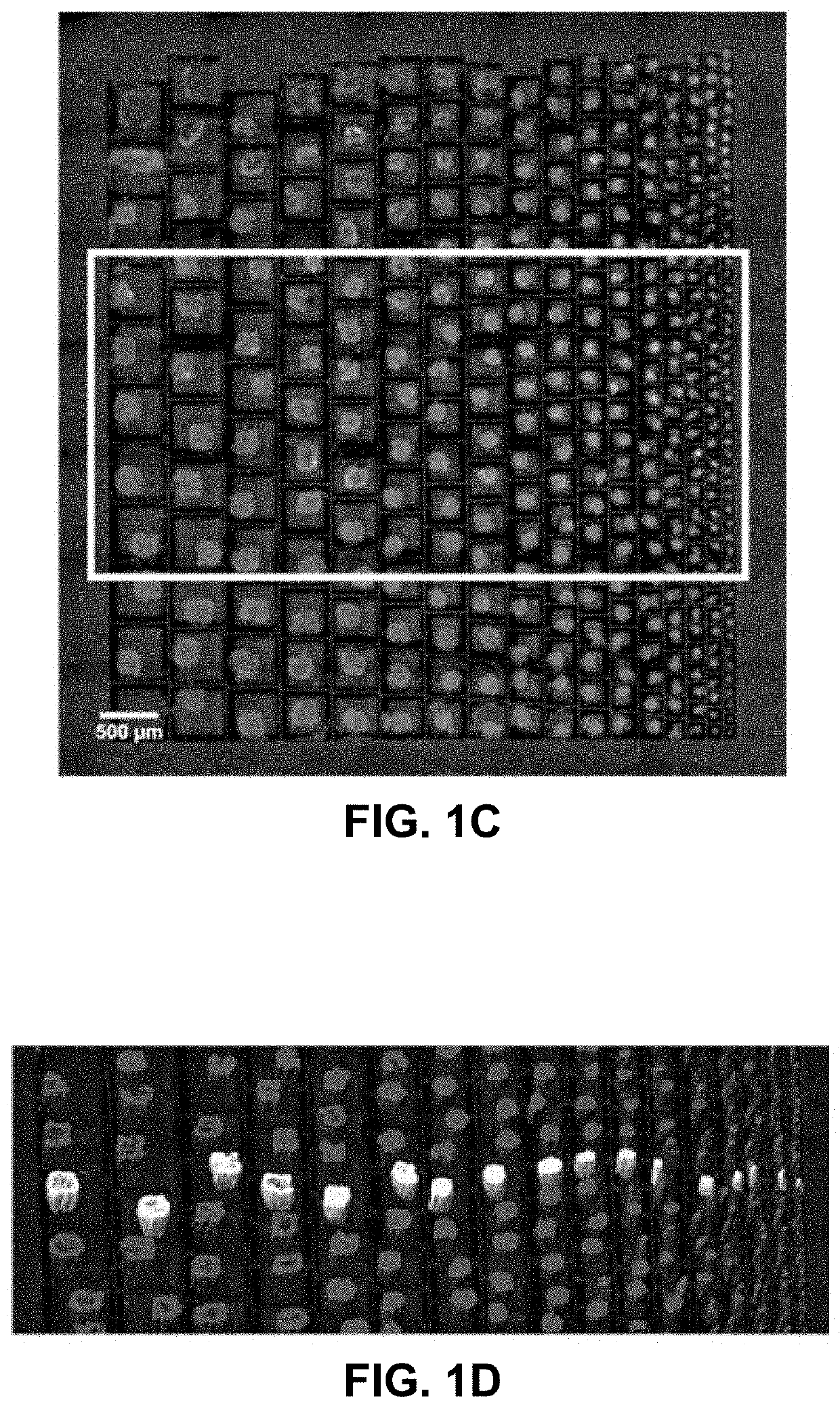
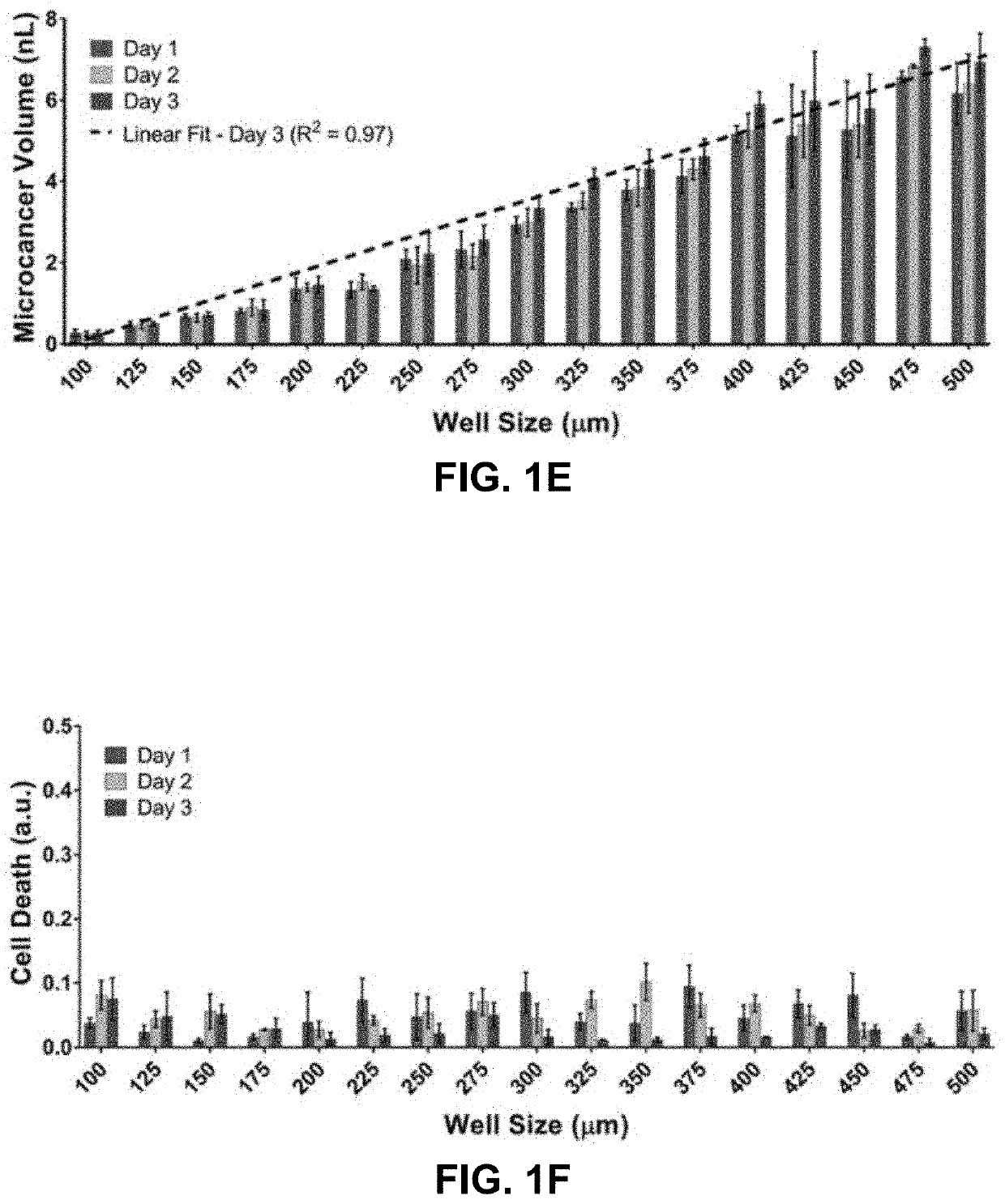
[0079]To evaluate the platform's ability to form and culture microcancer spheroids on chip of different sizes, LN229 cells were first seeded on the well size gradient chip and examined the survival profiles of the formed microcancers over time. A real time imaging compatible cell death indicator green dye (Celltox, Promega) and a cell membrane staining red dye (PKH, Sigma Aldrich) were added to the media to quantify cell death in the microcancer tumor volume with time. Tile and z stack imaging was performed for each day of culture to volumetrically map the green and red volumes within a micro-well. Volumetric mapping of the green and red dyes was performed on Imaris software, and final individual well tracking was performed using MATLAB. The cell death for each microcancer was normalized to its own culture volume to account for well-to-well variations in cell numbers. FIG. 1C shows a fluorescence microscope image of a high throughput culture of LN...

example 3

Harvesting 3D Cultures from Chip for Genomic Analysis

[0083]The present system is in an open format with an immiscible layer of fluid over the array. The system, therefore, is compatible with harvesting the formed 3D cultures from the chip. To do this, the chip is first submerged in media (e.g., on the order of 1 mL) to remove the residual oil layer from the top of the array. Then, the 3D cultures are removed such as by pipetting and aspirating the volume from the chip. In this manner, as desired, any cell and tissue culture can be removed without disturbing any of the other cell and tissue culture contained in other microwells. Of course, the system provided herein is also advantageously configured to obtain a number of real-time parameters to characterize cell and tissue parameters (e.g., growth, response to drug, etc.) without having to remove the cell and tissue from the microwell.


PUM

PropertyMeasurementUnit
depthaaaaaaaaaa
volumeaaaaaaaaaa
depthaaaaaaaaaa

Abstract

Provided are methods and related devices for preparing a cell and tissue culture, including a hanging drop culture. Microwells are specially loaded with cell mixtures using a removable reservoir and forcing cells into the underlying microwells. The removable reservoir is removed and the cells partitioned into the individual microwells and covered by an immiscible layer of fluid. The microwells and immiscible layer is inverted and the cells in the microwells cultured. The microwells may have shape-controlling elements to control the three-dimensional shape of the culture.

Description

CROSS-REFERENCE TO RELATED APPLICATIONS[0001]This application claims the benefit of priority to U.S. Provisional Patent Application No. 63 / 019,823, filed May 4, 2020, which is hereby incorporated by reference in its entirety to the extent not inconsistent herewith.STATEMENT REGARDING FEDERALLY SPONSORED RESEARCH OR DEVELOPMENT[0002]This invention was made with Government support under R01 GM129709 awarded by the National Institutes of Health. The government has certain rights in the invention.BACKGROUND OF INVENTION[0003]Cell culture has been extensively applied in drug discovery, tissue engineering, and stem cell research. However, conventional two dimensional monolayer cultures do not replicate the important features of three dimensional (3D) conditions and much work has been done to elucidate the significant differences in cellular morphology, behavior and molecular signaling between the classic cell monolayer approaches and analogous 3D cultures. 3D culture systems offer the uni...

Claims


Application Information

Patent Timeline
Patent Type & Authority Applications(United States)
IPC IPC(8): B01L3/00B01L3/02B01L9/00C12M3/00
CPCB01L3/5088B01L3/0262B01L2400/024C12M21/08B01L9/523C12M23/12C12M25/01B01L2300/161B01L2300/165
Inventor GANGULI, ANURUPBASHIR, RASHIDANASTASIADIS, PANAGIOTIS Z.VASMATZIS, GEORGE
Owner THE BOARD OF TRUSTEES OF THE UNIV OF ILLINOIS
Why Eureka
Try Eureka