Prediction of prostate cancer progression by analysis of selected predictive parameters

a predictive parameter and prostate cancer technology, applied in the field of prostate cancer progression prediction by selecting predictive parameters, can solve the problems of inability to accurately and specifically measure the sensitivity of markovian texture measurement, inability to explain the complex etiology, and inability to accurately and specifically measure the sensitivity of the markovian texture measurement,

Inactive Publication Date: 2000-02-15
CYTODIAGNOSTICS +3
View PDF0 Cites 240 Cited by
  • Summary
  • Abstract
  • Description
  • Claims
  • Application Information

AI Technical Summary

Benefits of technology

The present invention provides new and improved methods for determining the biological potential for progression of treatable, localized prostate cancers using core biopsies, fine needle aspirates, or radical prostatectomy specimens in order to: (1) better evaluate which patients have tumors that need any treatment, (2) determine the prognosis of patients with prostate cancer pathologi

Problems solved by technology

These causal correlations, though impressive, can not yet explain the complex etiology, biologic heterogeneity, and rapidly increasing incidence of this disease, and await further investigations of genetic, epigenetic and environmental factors.
Unfortunately, no accurate and specific methods presently exist to distinguish

Method used


Image

  • Prediction of prostate cancer progression by analysis of selected predictive parameters
  • Prediction of prostate cancer progression by analysis of selected predictive parameters
  • Prediction of prostate cancer progression by analysis of selected predictive parameters

Examples


example i

DNA Staining Procedure Using CAS Quantitative DNA Staining Kit (Elmhurst, Ill.; Catalog #102300-01)

Preparation of Feulgen Stain Solution:

Place 90 ml of Type I H.sub.2 O in a volumetric flask and add 10 ml of 1N HCL. Place a stir bar in a 125 ml Erlenmeyer flask and add the above solution. Add 1 vial of DNA stain reagent to the flask while stirring the solution. Place a rubber stopper in the flask, and stir the contents for at least 1 hour. This Feulgen stain solution should be filtered through a Whatman No. 1 filter immediately before staining of the specimen.

Preparation of Feulgen Rinse Solution:

Place 285 ml of Type I H.sub.2 O in a 500 ml graduated cylinder and add 15 ml of 1N HCL. Pour this solution into a 500 ml bottle. Immediately before rinsing, place 1 vial of DNA rinse reagent into the bottle and mix the contents by swirling. This solution is stable for 2-3 hours.

Preparation of Calibration Slides:

To prepare the control cells, place two (2) CAS calibration slides (Elmhurst, I...

example ii

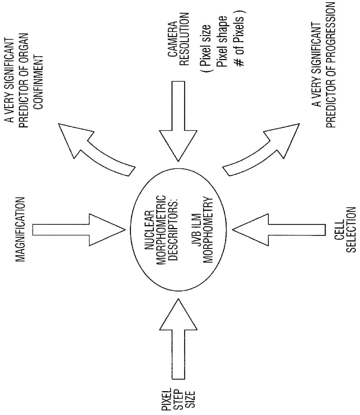
Collection and Processing of CAS-200 CMP v3.0 Nuclear Morphometric Descriptors (40.times. Objective)

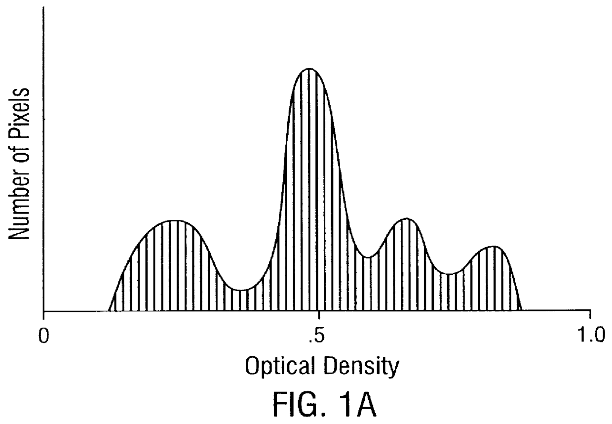
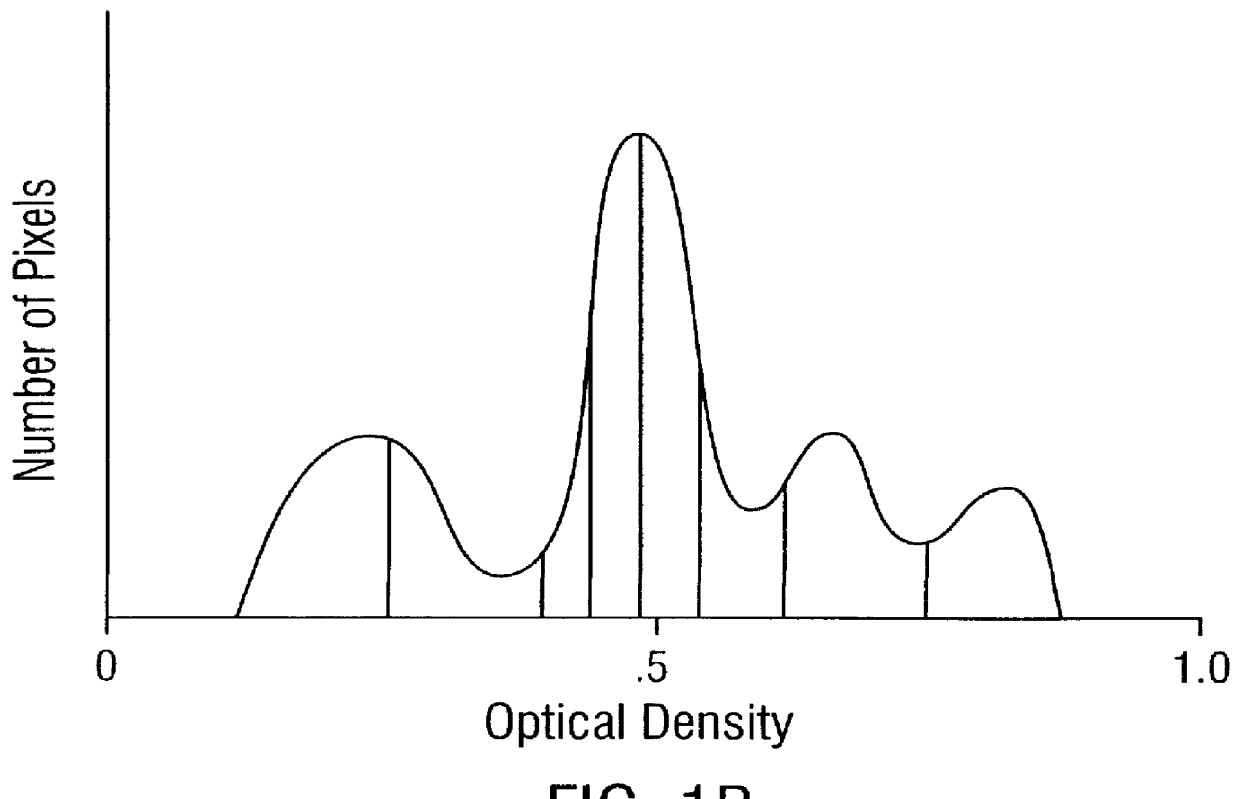
The morphometry data from the radical prostatectomy specimens is captured using the Cell Measurement Program v3.0 (CMP v3.0) software from a CAS-200 Image Analysis System. First, a study is set up in CMP v3.0 using the QDA Morphology Mode. The QDA Morphology Mode of CMP v3.0 allows the measurement of the Sum O.D., size, shape, cell class, and the 22 Markovian texture features (a step size of 1 was used in this invention) for each cell (see Table I), as well as the generation of a DNA histogram through the use of the QDA v3.0 software program on the CAS-200 Image Analysis System. Once the study is set up, the CMP v3.0 program (under the QDA Morphology Mode) activates the QDA v3.0 program, and the optical system is calibrated using the CAS calibration slides that were stained with the specimen slides. At least 20 calibration cells are measured, with a calibration peak percent coefficien...

example iii

Collection and Processing of JVB ILM Morphometry v1.0 Nuclear Morphometric Descriptors

The morphometry data from radical prostatectomy specimens is captured from the saved listmode files (*.ILM) using the JVB ILM Morphometry v1.0 software program, which allows the measurement and calculation of up to 36 different features. The listmode files (*.ILM) are created using the QDA v3.0 software from a CAS-200 Image Analysis System. The optical system is calibrated using the CAS calibration slides that were stained with the specimen slides by measuring at least 20 calibration cells, with a calibration peak percent coefficient of variation (% C.V.) of less than 2.0%. (NOTE: If the % C.V. is greater than 2.0%, a problem has occurred in the staining process.) Next, at least 125 cancer cells are analyzed using the method described in Example IV, and the cell nuclear images captured from each 5 .mu.m Feulgen stained tissue section. The DNA content information and cell nuclear images are saved to...


PUM

PropertyMeasurementUnit
Volumeaaaaaaaaaa
Volumeaaaaaaaaaa
Volumeaaaaaaaaaa

Abstract

A method for screening individuals at risk for prostate cancer progression is disclosed. The method is useful for evaluating cells from patients at risk for recurrence of prostate cancer following surgery for prostate cancer. Specifically, the method uses specific Markovian nuclear texture factors, alone or in combination with other biomarkers, to determine whether the cancer will progress or lose organ confinement. In addition, methods of predicting the development of fatal metastatic disease by statistical analysis of selected biomarkers is also disclosed. The invention also contemplates a method that uses a neural network to analyze and interpret cell morphology data. Utilizing Markovian factors and other biomarkers as parameters, the network is first trained with a sets of cell data from known progressors and known non-progressors. The trained network is then used to predict prostate cancer progression in patient samples.

Description

BACKGROUND OF THE INVENTION1. Field of the InventionThe present invention relates to fields of computer-assisted quantitative image analysis and methods to classify cells related to cancer progression. More specifically, it concerns methods to detect patients at risk for progression following radical surgery that have been diagnosed with localized prostate cancer by current state of the art clinical pathology and clinical laboratory methodology. Additionally, it concerns using the same approach to predict organ confined disease status using pre-treatment information extracted from the core biopsy materials.2. Description of the Related ArtProstate cancer is diagnosed in 100 / 100,000 white males and in 70.1 / 100,000 black males in the United States. It is the second leading cause of male cancer deaths and the most commonly diagnosed cancer in men in the United States representing 21% of all newly diagnosed cancers. In 1993 an estimated 165,000 men in the United States were diagnosed wi...

Claims


Application Information

Patent Timeline
IPC IPC(8): C12Q1/68G01N33/574
CPCC12Q1/68G01N33/574Y10S436/813G16B40/20G16B40/00G16H50/20G16B20/00G16H50/50G01N33/57434G16B25/10
Inventor VELTRI, ROBERT WBACUS, MICHAEL P.MILLER, M. CRAIGASHENAYI, KAVEHCOFFEY, DONALD P.PARTIN, ALAN W.EPSTEIN, JONATHAN I.
Owner CYTODIAGNOSTICS
Why Eureka
Try Eureka