Treatment of skin disorders
a skin disorder and skin technology, applied in the field of skin disorders, can solve the problems of significant cosmetic problems, source of discomfort, keloids and hypertrophic scars can become so large that they are crippling, and achieve the effect of reducing the number of patients
- Summary
- Abstract
- Description
- Claims
- Application Information
AI Technical Summary
Problems solved by technology
Method used
Image
Examples
example 2
Inhibitory Effect of Halofuginone on Collagen Synthesis in Keloid-Derived Tissue
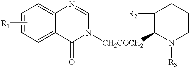
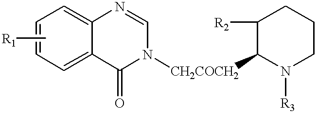
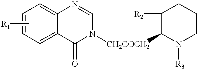
Halofuginone was shown to specifically inhibit collagen synthesis in keloid-derived tissue. The results are shown in FIG. 2. The experiment was conducted as follows.
The keloid was removed as described in Example 1 above. The keloid-derived cells were incubated with and without Halofuginone for 24 hours in 0.5 ml glutamine-free DMEM containing 5% FCS (Fetal Calf Serum), ascorbic acid (50 .mu.g / ml), B-aminopropionitrile (50 .mu.g / ml) and 2 .mu.Ci of [.sup.3 H]proline. At the end of incubation, the medium was decanted and the cells were incubated with or without collagenase for 18 hours, followed by precipitation of proteins by the addition of TCA (trichloroacetic acid). The amount of radiolabelled collagen was estimated as the difference between total labelled-proline containing proteins and those left after collagenase digestion [Granot, I. et al., Mol. Cell Endocrinol., Vol. 80, p. 1-9, 1991].
The ratio o...
example 3
Halofuginone Inhibition of Sulfate Incorporation into ECM of Cultured Endothelial Cells
As noted above, skin cell hyperproliferation is enabled by the deposition of ECM components. The following examples illustrate the ability of Halofuginone to inhibit such deposition of ECM components, further supporting the use of Halofuginone as a treatment for psoriasis. These examples illustrate the unexpected finding that Halofuginone completely abolishes deposition of all ECM components, and not just collagen, thereby preventing cell proliferation which is enabled by the formation of ECM.
Cultures of bovine corneal endothelial cells were established from steer eyes and maintained as previously described [D. Gospodarowicz, et al., Exp. Eye Res., No. 25, pp. 75-99 (1977)]. Cells were cultured at 37.degree. C. in 10% CO.sub.2 humidified incubators and the experiments were performed with early (3-8) cell passages.
For preparation of sulfate-labelled ECM (extra-cellular matrix), corneal endothelial ...
example 4
Inhibition of Incorporation of Sulfate, Proline, Lysine and Glycine into ECM of Bovine Corneal Endothelial Cells
Corneal endothelial cells were seeded at a confluent density and grown as described in Example 3 above. The cells were cultured with or without Halofuginone in the presence of either Na.sub.2.sup.35 SO.sub.4 (FIG. 4A), .sup.3 H-proline (FIG. 4B), .sup.14 C-lysine (FIG. 4C) or .sup.14 C-glycine (FIG. 4D). Eight days after seeding, the cell layer was dissolved substantially as described in Example 3 above. The underlying ECM was then either trypsinized to determine the effect of Halofuginone on incorporation of labeled material into total protein, substantially as described in Example 3 above, or subjected to sequential digestions with collagenase and trypsin to evaluate the effect of Halofuginone on both collagenase-digestible proteins (CDP) and non-collagenase digestible proteins (NCDP).
As FIGS. 4A-4D show, Halofuginone inhibited the incorporation of sulfate, proline, lysi...
PUM
 Abstract
Description
Claims
Application Information
 - R&D
- Intellectual Property
- Life Sciences
- Materials
- Tech Scout
- Unparalleled Data Quality
- Higher Quality Content
- 60% Fewer Hallucinations
Browse by: Latest US Patents, China's latest patents, Technical Efficacy Thesaurus, Application Domain, Technology Topic, Popular Technical Reports.
© 2025 PatSnap. All rights reserved.Legal|Privacy policy|Modern Slavery Act Transparency Statement|Sitemap|About US| Contact US: help@patsnap.com


