Electrosurgical system

a surgical system and electro-optical technology, applied in the field of electro-optical systems, can solve the problems of high power with a monopolar arrangement, severe skin burns, and the risk of capacitive coupling between the instrument and the patient's tissues at the entry point into the body cavity,

Inactive Publication Date: 2002-01-08
GYRUS MEDICAL LTD
View PDF23 Cites 1333 Cited by
  • Summary
  • Abstract
  • Description
  • Claims
  • Application Information

AI Technical Summary

Problems solved by technology

Using high power with a monopolar arrangement is also hazardous, due to the tissue heating that occurs at the return plate, which can cause severe skin burns.
There is also the risk of capacitive coupling between the instrument and patient tissues at the entry point into the body cavity.
However, the depth of effect is directly related to the distance between the two electrodes; and, in applications requiring very small electrodes, the inter-electrode spacing becomes very small, thereby limiting tissue effect and output power.
Spacing the electrodes further apart would often obscure vision of the application site, and would require a modification in surgical technique to ensure correct contact of both electrodes with tissue.
When either bipolar or monopolar electrosurgery is employed on the skin surface, there is a high risk of excessive thermal damage and tissue carbonisation.
Such thermal damage and carbonisation can lead to delayed healing, wound infection and excessive scar formation.
In addition to these problems, when using bipolar arrangements, the impedance of the electrical contact between the skin and the return electrode can significantly reduce effectiveness.
One of the obvious limitations with such a design is that the active electrode (such as a needle) must be completely buried in the tissue to enable the return electrode to complete the circuit.
Another problem is one of orientation: eve

Method used


Image

  • Electrosurgical system
  • Electrosurgical system
  • Electrosurgical system

Examples


first embodiment

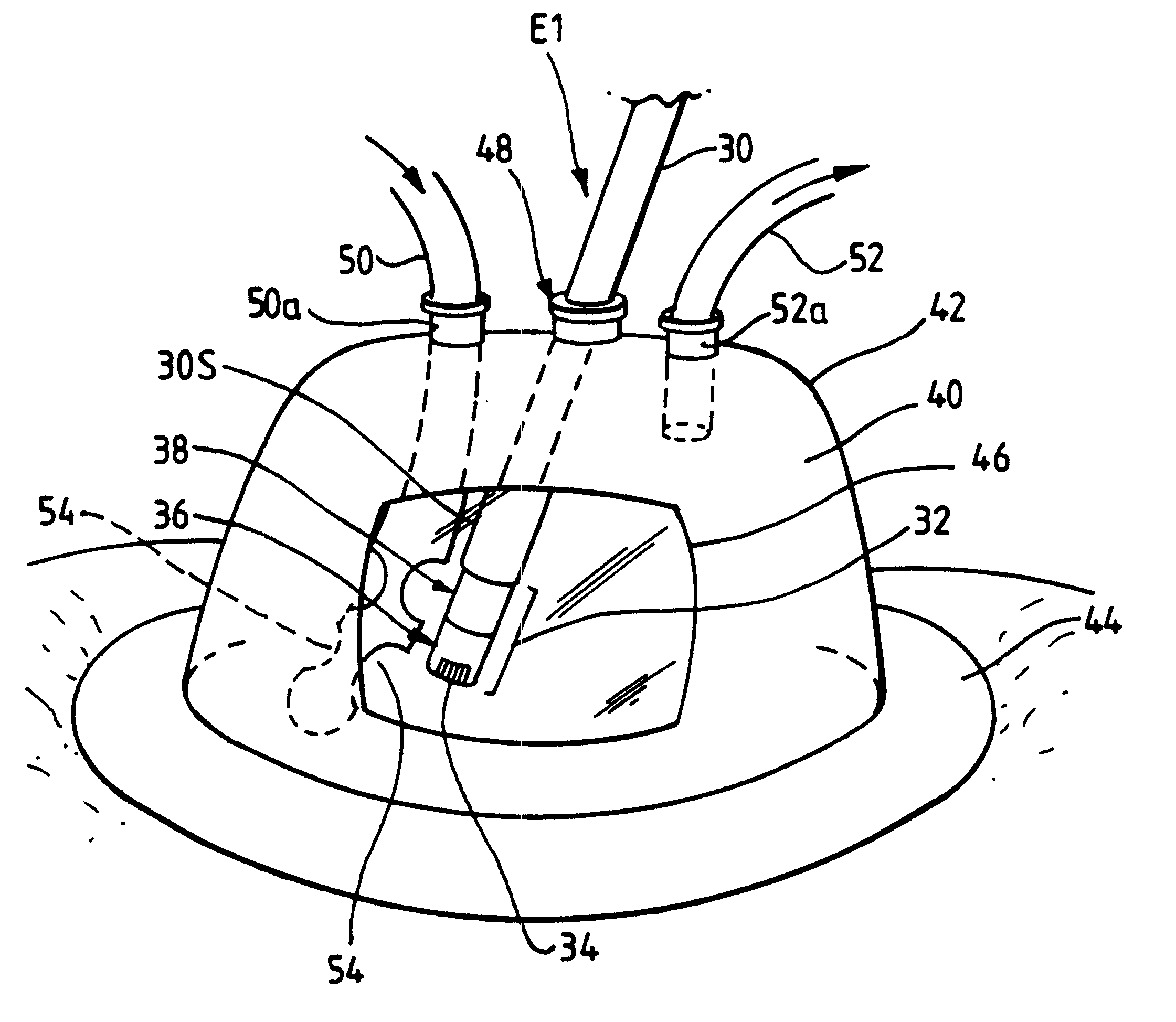
In a first embodiment, shown in FIG. 2, an electrode unit E1 is detachably fastened to the handpiece 3 (not shown). The electrode unit E1 comprises a shaft 30 which may be a conductive (e.g. metallic) tube covered with an insulating sheath 30S, with an electrode assembly 32 at the distal end of the shaft. At the other end of the shaft 30 (not shown), means are provided for connecting the unit E1 to the handpiece 3 both mechanically and electrically.

The electrode assembly 32 is bipolar, having an active (tissue treatment electrode) 34 which is axially spaced from a return electrode 38 by means of an insulator 36. The return electrode 38 is constituted by the distal end portion of the tube 30, the portion not being covered by insulating sheet material. In use, the active electrode 32 is positioned in contact with, or in close proximity to, the tissue to be treated. This means that, in normal use when the electrode assembly 32 is immersed in a conductive fluid medium 40, the return ele...

second embodiment

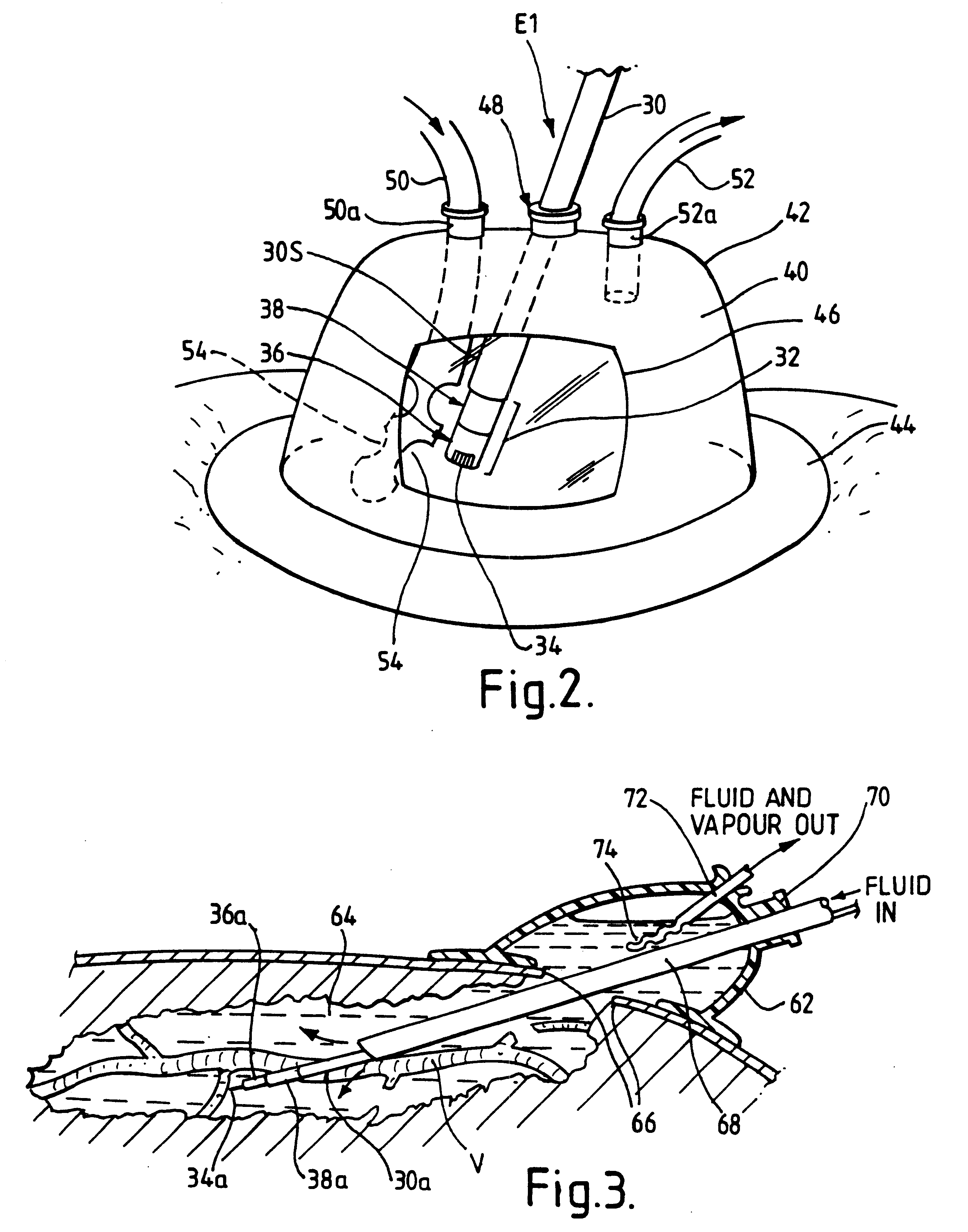
In a second embodiment, shown in FIG. 3, a fluid enclosure 62 is positioned over a site on the patient's body wherein a space 64 has been surgically created in the tissues through an incision 66. This space 64 may be created using a dissecting instrument under endoscopic visualisation prior to application of the fluid enclosure 62, or may be created under a fluid-filled environment using an electrosurgical instrument or instruments based on the electrode assembly 32, or may be based on a combination of the two. Advantageously, the fluid-filled environment, combined with the generator 1 and instrument system for use with the invention, allows the use of tissue treatment electrodes commonly used for dissection, for example, the hook electrode 34a shown in FIG. 3 or a needle electrode. Such electrodes allow the sealing of larger blood vessels than would normally be treated in this way in a gaseous environment. This is particularly beneficial when sealing, for example a large vein such ...


PUM


Abstract

An electrosurgical system comprises a radio frequency generator (1), an electrosurgical instrument (E1), and a fluid enclosure (42). The generator (1) has a radio frequency output for delivery of power to the electrosurgical instrument (E1) when immersed in an electrically-conductive fluid. The electrosurgical instrument (E1) has an electrode assembly (32) at the distal end thereof, the electrode assembly comprising a tissue treatment electrode (34), and a return electrode (38) axially spaced therefrom in such a manner as to define, in use, a conductive fluid path that completes an electrical circuit between the tissue treatment electrode and the return electrode. The fluid enclosure (42) is adapted to surround an operation site on the skin of a patient or an incision leading to a cavity surgically created within the patient's body. The fluid enclosure (42) includes sealing means (44) for sealing against the patient's tissue, and the fluid enclosure includes at least one port (50a, 52a) through which the electrosurgical (E1) is insertable, and through which the electrically-conductive fluid can enter and/or leave the enclosure.

Description

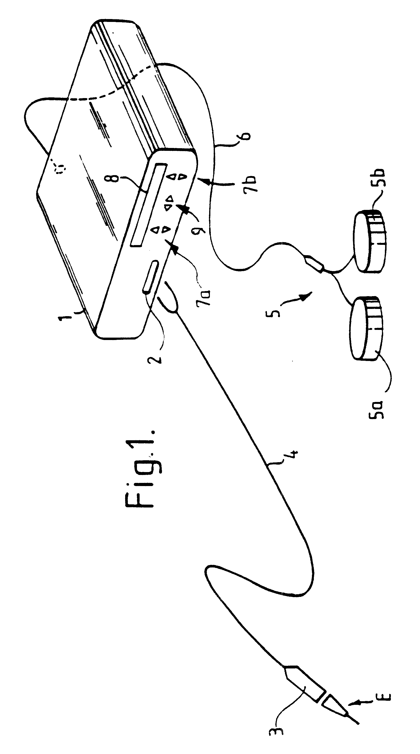
BACKGROUND TO THE INVENTIONThis invention relates to an electrosurgical system for the treatment of tissue in the presence of an electrically-conductive fluid medium, and in particular to such a system including a fluid isolation enclosure for facilitating the immersion of tissues on, or within, a patient's body, such that the system can be operated to vaporise, coagulate, desiccate or otherwise thermally modify such tissues.Endoscopic electrosurgery is useful for treating tissue in cavities of the body, and is normally performed in the presence of a distension medium. When the distension medium is a liquid, this is commonly referred to as underwater electrosurgery, this term denoting electrosurgery in which living tissue is treated using an electrosurgical instrument with a treatment electrode or electrodes immersed in liquid at the operation site.Underwater surgery is commonly performed using endoscopic techniques, in which the endoscope itself may provide a conduit (commonly refe...

Claims


Application Information

Patent Timeline
IPC IPC(8): A61B18/14A61B17/34A61B19/00A61B18/00
CPCA61B17/3423A61B18/1482A61B19/38A61B2218/008A61B2018/162A61B2218/002A61B2018/1472A61B90/40
Inventor GOBLE, NIGEL M
Owner GYRUS MEDICAL LTD
Why Eureka
Try Eureka