Capillary array-based sample screening

a sample and capillary array technology, applied in the field of capillary array-based sample screening, can solve the problems of inability to efficiently apply the current available methods for screening and producing lead compounds to these under-explored resources, difficult or impossible culture of organisms for screening or re-supply, synthesis and development of lead compounds, etc., and achieves rapid and efficient methods.

Inactive Publication Date: 2005-03-15
BASF ENZYMES
View PDF37 Cites 25 Cited by
  • Summary
  • Abstract
  • Description
  • Claims
  • Application Information

AI Technical Summary

Benefits of technology

FIG. 3 depicts a vertical cross-section of a capillary of the invention. Depicted is capillary (20) having a first wall (30) and a second wall of black EMA glass (50) defining a lumen (40). A cell, clone or molecule (70) is depicted within the lumen (40). During use an excitation light is directed into the lumen (40) contacting the cell, clone or molecule (70) and exciting a reporter fluorescent material causing emission of light. The emitted light travels the length of the capillary until it reaches a detector. One advantage of the present invention is that the excitation light and emitted light cannot cross contaminate adjacent capillary tubes in a capillary array due to the black EMA glass (50). In addition, the black EMA glass refracts and directs the emitted light towards either end of the capillary tube thus increasing the signal detected by an optical detector (e.g., a CCD camera and the like).
is that the excitation light and emitted light cannot cross contaminate adjacent capillary tubes in a capillary array due to the black EMA glass (50). In addition, the black EMA glass refracts and directs the emitted light towards either end of the capillary tube thus increasing the signal detected by an optical detector (e.g., a CCD camera and the like).
FIG. 4 depicts a method of using a capillary array of the invention. In this depiction, capillary array (10) is immersed or contacted with a container (100) containing cells, clones, molecules or compounds suspended in media. The media and cells are then wicked up the capillary tubes by capillary action. The natural wicking that occurs as a result of capillary forces obviates the need for pumping equipment and liquid dispensers. Substrates, as described above, for measuring biological activity (e.g., enzyme activity) can be contacted with the cells either before or after introduction of the cells into the capillaries in the capillary array. The substrate and at least a subset of the clones can be introduced simultaneously into capillaries in the capillary array by placing open ends of the capillaries in a reservoir containing a mixture of the substrate and clones. Alternatively, a solution of cells may be wicked into the capillaries before the capillary array is placed in a reservoir containing substrate, where the substrate is then wicked into the capillaries that already contain the cells to be screened for the desired activity. As depicted in FIGS. 7A and 7B, and discussed more fully below, various methods can be used to mix or stir the substrate with the cells present in the capillaries including, for example, pareamagnetic beads.
The substrate solution can then be incubated with the cells for a period of time and at an appropriate temperature necessary for cell growth and, for example, to allow the substrate to permeabilize the cell membrane to produce an optically detectable signal or for a period of time and temperature for optimum enzymatic activity. The incubation can be performed, for example, by placing the capillary array in a humidified incubator or at ambient temperature in an apparatus containing a water source to ensure reduced evaporation within the capillary tubes. The evaporative flow rate may be reduced by increasing the humidity (e.g., by placing the capillary array in a humidified chamber). The evaporation rate can also be reduced by capping the capillaries with an oil, wax, membrane or the like. Alternatively, a high molecular weight fluid such as various alcohols, or molecules capable of forming a molecular monolayer, bilayers or other thin films (e.g., fatty acids), or various oils (e.g., mineral oil) can be used to reduce evaporation.
The concentration ranges for substrate solutions will vary according to the substrate utilized. Commercially available substrates will generally contain instructions on concentration ranges to be utilized in, for example, cell staining. These ranges may be employed in the determination of an optimal concentration or concentration range to be utilized in the present invention. Such determination is within the ability of one of skill in the art.
In one embodiment, a first fluid containing cells or clones is wicked into the capillary until only about 50% of the capillary is filled. A small air-bubble is then introduced into the capillary. The air-bubble can be introduced by any number of means including, for example, contacting the capillary with a compositions that creates a seal at the capillary opening on the opposite end of the capillary and then removing the composition thereby causing a vacuum to move the fluid further into the capillary. The capillary or capillary array is then contacted with a second fluid, typically a substrate. The second fluid may contain paramagnetic beads or particles in addition to any substrate. The second fluid is then wicked into the capillary or capillary array and is separated from the fluid containing the cell or clone by the air-bubble. The capillary or capillary array can then be incubated for a period of time to allow the first and second media to reach optimal temperature or for sufficient time to allow cell growth or for sufficient time to allow the first and second media to reach the same temperature. The air-bubble separating the two media types can be disrupted to allow mixing of the media and enzymatic activity. In one embodiment, paramagnetic beads are used to disrupt and / or mix the contents of the capillary tube or capillary array. For example, FIGS. 7A and 7B depicts an embodiment of the invention in which paramagnetic beads are magnetically attracted from one area of the capillary or capillary array to another area of the capillary or capillary array. The paramagnetic beads are attracted by electric or magnetic fields produced at various locations around the capillary or capillary array. Accordingly, alternating an electrical field or magnetic field having a polarity opposite that of the paramagnetic beads from one end of a capillary to another would cause the paramagnetic beads to move within the capillary causing movement of the fluid within the capillary.

Problems solved by technology

However, the currently available methods for screening and producing lead compounds cannot be applied efficiently to these under-explored resources.
For instance, it is estimated that at least 99% of marine bacteria species do not survive on laboratory media, and commercially available fermentation equipment is not optimal for use in the conditions under which these species will grow, hence these organisms are difficult or impossible to culture for screening or re-supply.
Recollection, growth, strain improvement, media improvement and scale-up production of the drug-producing organisms often pose problems for synthesis and development of lead compounds.
Furthermore, the need for the interaction of specific organisms to synthesize some compounds makes their use in discovery extremely difficult.

Method used


Image

  • Capillary array-based sample screening
  • Capillary array-based sample screening
  • Capillary array-based sample screening

Examples


example 1

DNA isolation. DNA is isolated using the IsoQuick Procedure as per manufacture's instructions (Orca Research Inc., Bothell, Wash.). The isolated DNA can optionally be normalized according to Example 2 (below). Upon isolation, the DNA is sheared by pushing and pulling the DNA through a 25-gauge double-hub needle and a 1-cc syringe about 500 times. A small amount is run on a 0.8% agarose gel to make sure the majority of the DNA is in the desired size range (about 3-6 kb).

Blunt-ending DNA. The DNA is blunt-ended by mixing 45 μl of 10× Mung Bean Buffer, 2.0 μl Mung Bean Nuclease (1050 u / μl) and water to a final volume of 405 μl. The mixture is incubated at 37° C. for 15 minutes. The mixture is phenol:chloroform extracted, followed by an additional chloroform extraction. One ml of ice cold ethanol is added to the final extract to precipitate the DNA. The DNA is precipitated for 10 minutes on ice. The DNA is removed by centrifugation in a microcentrifuge for 30 minutes. The pellet is wash...

example 2

Normalization

Prior to library generation, purified DNA can be normalized. DNA is first fractionated according to the following protocol. A sample composed of genomic DNA is purified on a cesium-chloride gradient. The cesium chloride (Rf=1.3980) solution is filtered through a 0.2 um filter and 15 ml is loaded into a 35 ml OptiSeal tube (Beckman). The DNA is added and thoroughly mixed. Ten micrograms of bis-benzimide (Sigma; Hoechst 33258) is added and mixed thoroughly. The tube is then filled with the filtered cesium chloride solution and spun in a Bti50 rotor in a Beckman L8-70 Ultracentrifuge at 33 k rpm for 72 hours. Following centrifugation, a syringe pump and fractionator (Brandel Model 186) are used to drive the gradient through an ISCO UA-5UV absorbance detector set to 280 nm. Peaks representing the DNA from the organisms present in an environmental sample are obtained. Eubacterial sequences can be detected by PCR amplification of DNA encoding rRNA from a 10 fold dilution of t...

example 3

Evaporation

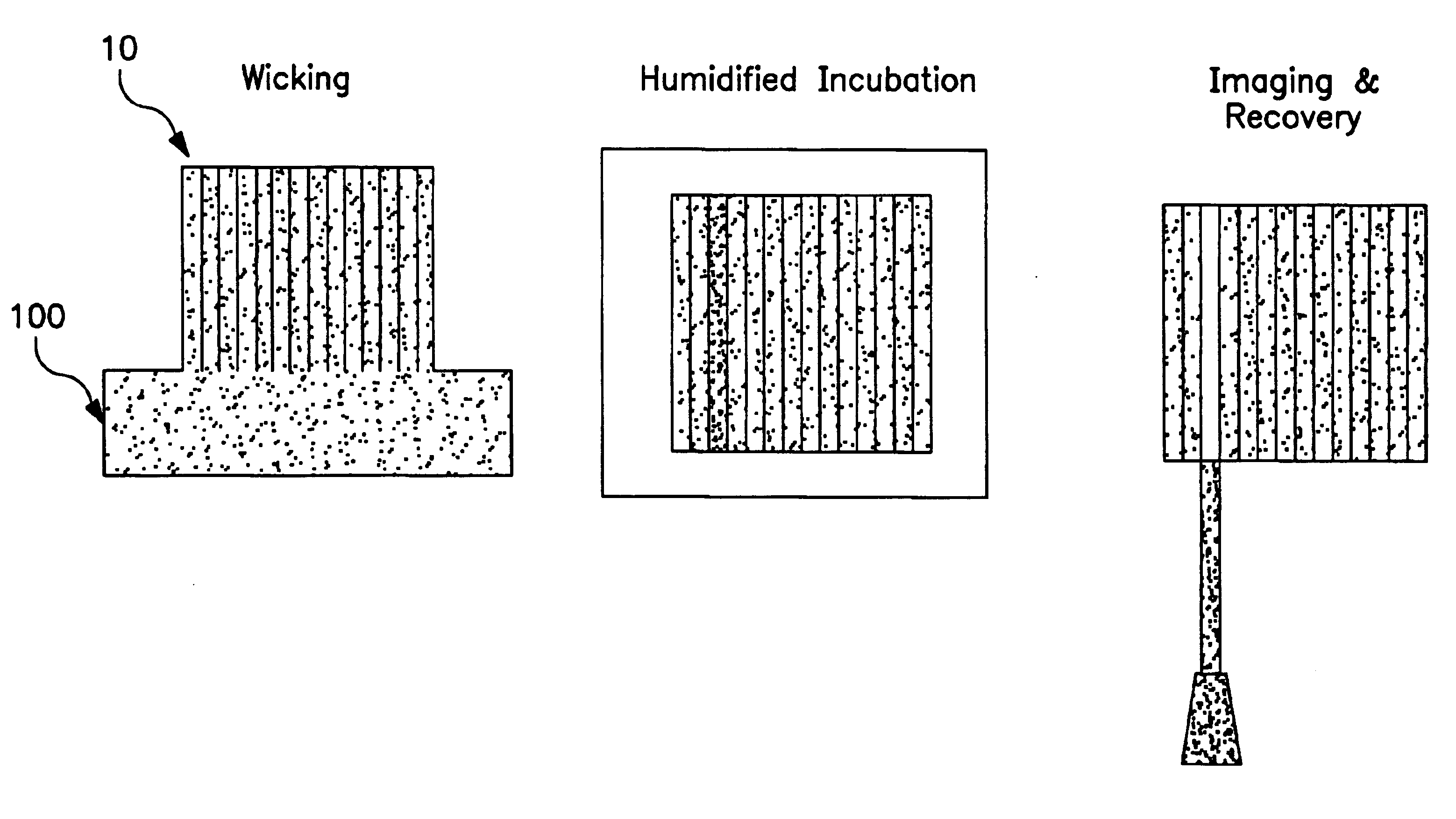
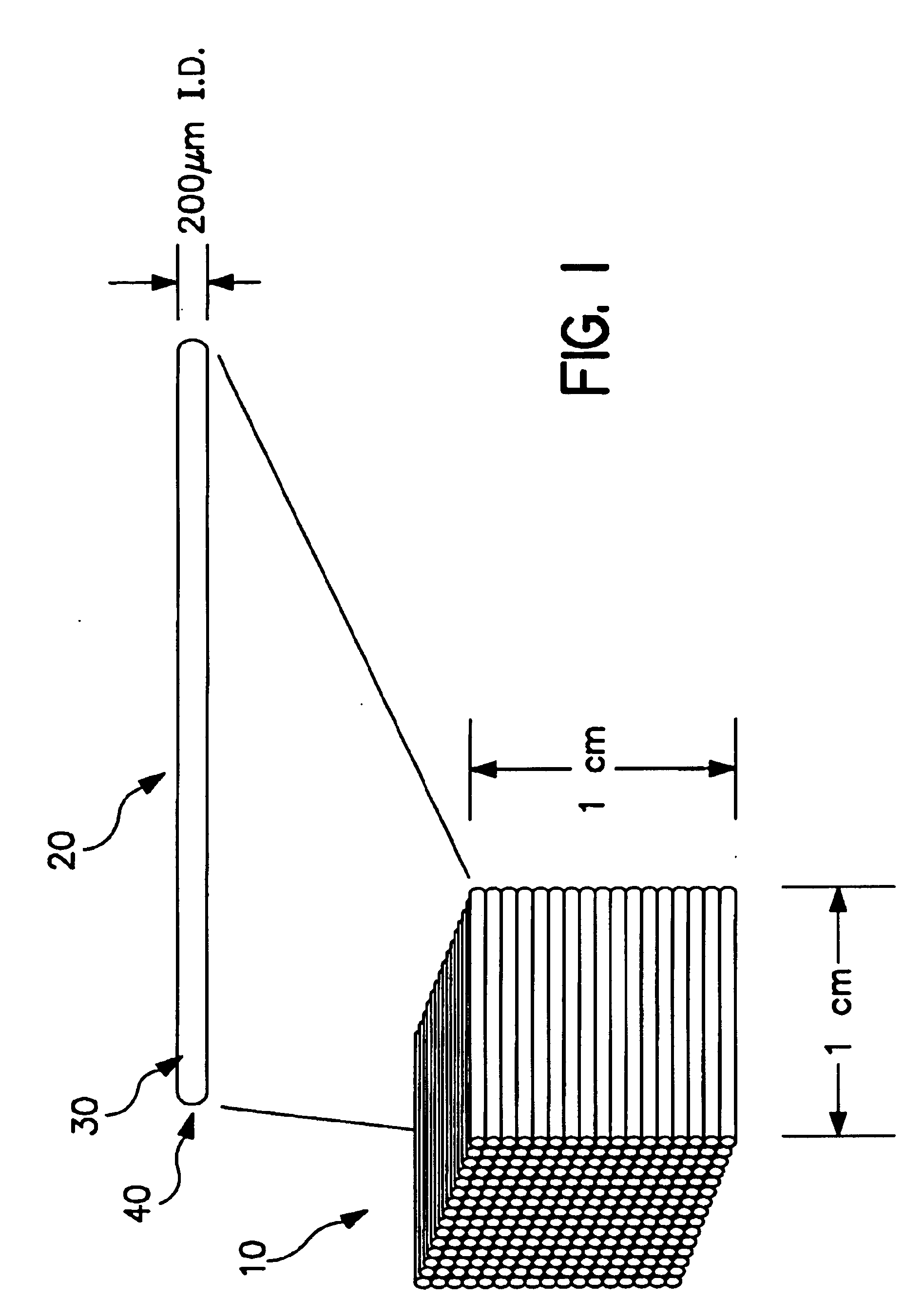
Water was wicked into arrays comprised of capillaries having 100 μm and 200 μm diameter and that were 25 and 20 mm long respectively. The arrays were weighed initially (t=0) and subsequent time periods. The arrays were incubated at 37° C. and 85% humidity in a humidified incubator. Volumes were calculated from the weights. The experiment demonstrated that 25% of the volume was loss in 5 hours and 37% in 17 hours (FIG. 8).

In a subsequent experiment a humid chamber was made from a pipette tip box with the arrays on the tip rack and water in the bottom of the box (FIG. 9A). The arrays were placed on the tip rack on their sides (capillaries oriented horizontally) during incubation. This incubation method provided a 7-10% loss over 5 hours and a 30% loss over 48 hours (FIG. 9B).


PUM

PropertyMeasurementUnit
temperaturesaaaaaaaaaa
temperatureaaaaaaaaaa

Abstract

Provided are methods of screening and identification of bioactivities and bioactive molecules of interest using a capillary array system. More specifically, disclosed are methods of using optical detection and capillary array-based techniques for screening libraries and recovering bioactive molecules having a desired activity or a nucleic acid sequence encoding such bioactive molecules.

Description

FIELD OF THE INVENTIONThe present invention relates generally to screening and identification of new bioactive molecules. More specifically, the present invention relates to methods of using optical detection and capillary array-based techniques for screening samples or libraries and recovering bioactive molecules having a desired activity or nucleic acid sequences encoding bioactive molecules.BACKGROUNDThere has been a dramatic increase in the need for bioactive compounds with novel activities. This demand has arisen largely from changes in worldwide demographics coupled with the clear and increasing trend in the number of pathogenic organisms that are resistant to currently available antibiotics as well as the need for new industrial processes for synthesis of compounds. For example, while there has been a surge in demand for antibacterial drugs in emerging nations with young populations, countries with aging populations, such as the U.S., require a growing repertoire of drugs aga...

Claims


Application Information

Patent Timeline
Patent Type & Authority Patents(United States)
IPC IPC(8): B01L3/00B01J19/00C12N15/10C12Q1/68C40B40/02C40B40/06C40B60/14
CPCB01J19/0046B01L3/5085B01L3/50857C12N15/1037C12N15/1055C12Q1/6811C40B40/02C40B60/14B01J2219/00367B01J2219/00416B01J2219/00479B01J2219/00481B01J2219/00522B01J2219/00585B01J2219/00596B01J2219/00605B01J2219/0061B01J2219/00612B01J2219/00619B01J2219/00621B01J2219/00626B01J2219/0063B01J2219/00637B01J2219/00659B01J2219/00677B01J2219/00686B01J2219/00689B01J2219/00702B01J2219/00722B01J2219/0074B01J2219/00743B82Y30/00C12Q1/6837C40B40/06B01J2219/00351
Inventor LAFFERTY, WILLIAM MICHAELSHORT, JAY M.KELLER, MARTIN
Owner BASF ENZYMES
Why Eureka
Try Eureka