Microbial protein expression system

a protein expression and microorganism technology, applied in the field of biotechnology, can solve the problems of no major system for secretion of extracellular products from i>e. coli, incorrect folding, degradation or formation of misfolded proteins as inclusion bodies,

Inactive Publication Date: 2005-07-19
BIOTECNOL SA
View PDF4 Cites 5 Cited by
  • Summary
  • Abstract
  • Description
  • Claims
  • Application Information

AI Technical Summary

Problems solved by technology

Commercial production of various medically and industrially valuable recombinant proteins by microbes is one the key challenges of modem biotechnology.
Even though such systems are known, there are severe technical problems which are encountered within large-scale exploitation of microbial cell machinery.
A major problem on recombinant expression in E. coli is incorrect folding with accompanying protein degradation or accumulation in an insoluble and inactive form as inclusion bodies.
Incorrect folding of proteins in the cytosol may lead to degradation or formation of misfolded protein as inclusion bodies.
Although there are quite a few systems available for periplasmic localisation of proteins, there is no major system for secretion of extracellular products from E. coli.
Although there has been some success in expressing of proteins, there are a number of limitations within the existing systems as outlined below.
The carrier domain may interfere with folding of the recombinant protein resulting in protein degradation, insolubility of the protein due to membrane association or formation of insoluble inclusion bodies at higher concentrations.
Using a Lpp- OmpA vector, complete enzymes have been localised to the surface of E. coli offering the potential of surface display, but these constructs lead to disruption of the outer membrane with concomitant toxicity to the cell and leakage of periplasmic contents.
This limits the maximum number of surface molecules and more importantly it is evident that completely folded proteins possessing disulphide bonds cannot be assembled across the outer membrane by this route (Klauser, T., et al.
Some incorrectly folded proteins remain associated with the inner membrane and induce toxicity.
In addition, they are extensively degraded resulting in a poor yield.
These may to some degree enhance solubility of some recombinant proteins but others remain insoluble due to incomplete folding of the heterologous domain.
(ii) Surface localisation in Gram negative bacteria: Generally, there is a strict limitation in the size of epitopes which can be expressed at the cell surface using proven surface expression vectors.
Systems that permit surface expression of whole domains or proteins by fusion them to an outer membrane protein lead to membrane permeabilisation, periplasmic leakage and toxicity.
In addition, there are limitations on the extent to which proteins can be folded if they are to be exported by this pathway.
Finally, as these systems all use integral membrane proteins, they are limited with respect to the maximum expression level and would be very laborious to purify.

Method used


Image

  • Microbial protein expression system
  • Microbial protein expression system
  • Microbial protein expression system

Examples


example i

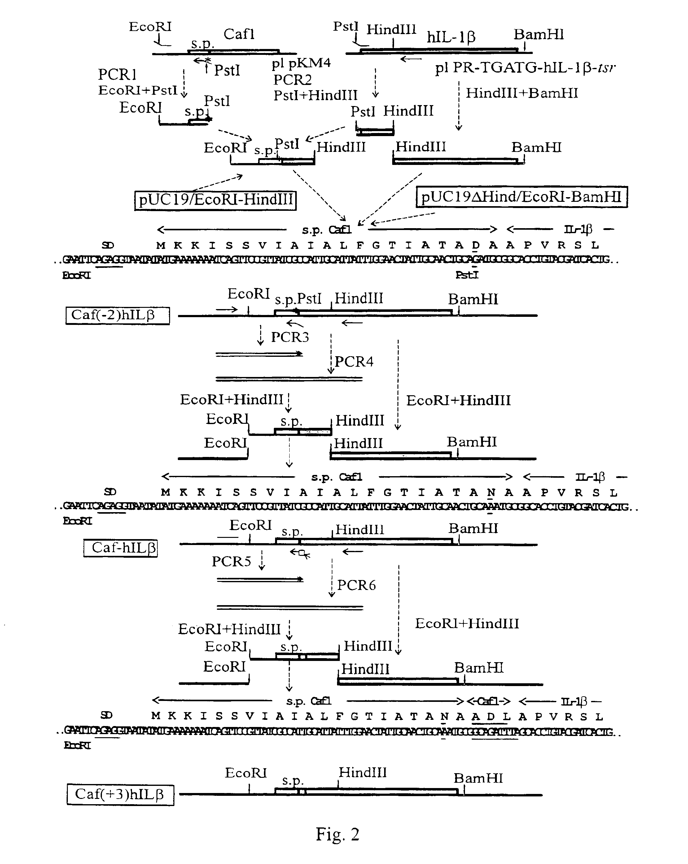
Fusion or hIL-1β with the Signal Peptide of the Caf1 Protein

[0060]The Caf1 signal peptide (s.p.Caf1) was fused with hIL-1β in three variants (FIG. 2). 1. s.p.Caf1-hIL-1β was the straight fusion where the last amino acid of the Caf1 signal peptide was joint to the first amino acid of hIL-1β. 2. s.p.Caf1(−2)-hIL-1β was the straight fusion with a mutation in the Caf1 signal peptide Asn(−2)Asp. 3. s.p.Caf1(+3)hIL-1β was the fusion containing in the joint region three N-terminal amino acids from mature Caf1 to preserve the natural processing site.

[0061]Here and thereafter DNA manipulations and transformation of E. coli were accomplished according to Maniatis, T., et al. (1989) Molecular Cloning: A Laboratory Manual, 2nd edn., Cold Spring Harbor Laboratory Press, Cold Spring Harbor. Restriction enzymes, mung-bean nuclease, and T4 DNA ligase were purchased from Promega (USA). Taq DNA polymerase (Hytest, Finland) was used for polymerase chain reaction (PCR) experiments. Nucleotide sequencin...

example 2

Fusion of s.p.Caf1(−2)hIL-β with the Caf1 Protein Sequence and Expression of Fused Protein in the Presence of Chaperone and Usher Proteins

[0085]The CIC (s.p.Caf-IL-1β-Caf) protein was designed where the s.p.Caf1(−2)hIL-1β amino acid sequence (N-terminus of the fusion) was linked to the Caf1 protein sequence (C-terminus of the fusion) through a spacer GlyGlyGlyGlySer repeated three times. The spacer was inserted to minimise possible conformational problems of the two proteins fused in CIC.

[0086]The pCIC expression vector was constructed according to the scheme on FIG. 6. The IL part of CIC was obtained by PCR using IL-Pst and IL-BamHI oligonucleotides as primers and the pUC19ΔHindIII / s.p.Caf1(−2)hIL-1β plasmid as a template. The Caf1 part of CIC was obtained by PCR using BamHI-Caf1 and Caf1-SalI oligonucleotides as promers and the pKM4 plasmid as a template. PCR products were digested with restrictases as shown in FIG. 3 followed by triple ligation with the pUC19ΔHindIII / s.p.Caf1(−2)...

example 3

Co-expression of hGMCSF-Caf1 Fusion Protein and the Chaperone

[0096]The expression plasmid for the hGM-CSF-Caf1 fusion protein was based on pFGM13 described in: Petrovskaya L. E. et al. (1995) Russian Journal of Bio-organic Chemistry, 21, 785-791. The pFGM13 plasmid contained a synthetic gene coding for hGMCSF with Caf1 signal sequence (scaf1) cloned into HindIII and XbaI sites of pUC19. Transcription was controlled by the lac promoter and was inducible with IPTG. Translation of the scaf1-gmcsf gene in pFGM13 initiated at the first methionine codon of lacZ gene and utilises its Shine-Dalgarno sequence. A primary structure of the signal sequence (scaf1) encoded by this gene differed from wild-type one at its N-terminus, which contained seven extra residues from β-galactosidase N-terminus.

[0097]The construction of the pFGMF1 plasmid coding for the gmcsf-caf1 gene is shown in FIG. 11. The scaf1-gmcsf gene of pFGM13 was modified for the following cloning of the fragment from pCIC contain...


PUM

PropertyMeasurementUnit
pHaaaaaaaaaa
molecular weightaaaaaaaaaa
optical densityaaaaaaaaaa

Abstract

The present invention provides bacterial strains for secretion of soluble biologically active recombinant heterologous proteins into periplasm or on a surface/into a cultivation medium of bacteria. The invention exploits the secretion system of Gram-negative bacteria including periplasmic chaperones and usher/secretin proteins. For accomplishing the aim, the bacterial strains simultaneously express the fusion protein (signal peptide)-(mature hecterologous protein)-(subunit of a bacterial surface structure, Caf1), periplasmic chaperone specific for the subunit, and outer membrane usher/secretin protein specific for the subunit. Secretion of fusion proteins: (signal peptide of Caf1)-(mature human IL-1β)-(mature Caf1), (signal peptide of Caf1)-(mature human GM-CSF)-(mature Caf1), and (signal peptide of Caf1)-(mature human IL-1ra)-(mature Caf1) that were expressed in Escherichia coli simultaneously with the periplasmic chaperone Caf1M and the usher/secretin protein Caf1A are examples of the use of the invention.

Description

[0001]This application is the national phase under 35 U.S.C. § 371 of PCT International Application No. PCT / FI00 / 00387 which has an International filing date of May 3, 2000, which designated the United States of America and was published in English.FIELD OF INVENTION[0002]This invention is related to biotechnology and more specifically to production of recombinant heterologous proteins by microbes. In particular, this invention concerns secretion of soluble biologically active heterologous proteins into periplasm and / or on a surface / into a cultivation medium of Gram-negative bacteria. The invention exploits the secretion system of Gram-negative bacteria including periplasmic chaperones and usher / secretin proteins of the system.BACKGROUND OF INVENTION[0003]Commercial production of various medically and industrially valuable recombinant proteins by microbes is one the key challenges of modem biotechnology. Even though such systems are known, there are severe technical problems which a...

Claims


Application Information

Patent Timeline
Patent Type & Authority Patents(United States)
IPC IPC(8): C12P21/02C12N15/70C12N15/62
CPCC12N15/62C12N15/70C12P21/02C07K2319/00C07K2319/02C07K2319/035C07K2319/35C07K2319/75
Inventor KORPELA, TIMOMACINTYRE-AYANE, SHEILAZAVIALOV, ANTONBATTCHIKOVA, NATALIAPETROVSKAYA, LADAZAV'YALOV, VLADIMIR
Owner BIOTECNOL SA
Why Eureka
Try Eureka