
Composite tissue adhesive

a tissue adhesive and composite technology, applied in the field of composite tissue adhesives, can solve the problems of high level of surgeon skill required, pernicious effects, and methods that have not yet gained clinical acceptance, and achieve the effects of eliminating peripheral tissue damage, reducing treatment time, and eliminating the damage of stay sutures

Inactive Publication Date: 2005-09-06
CONVERSION ENERGY ENTERPRISES
View PDF49 Cites 85 Cited by
  • Summary
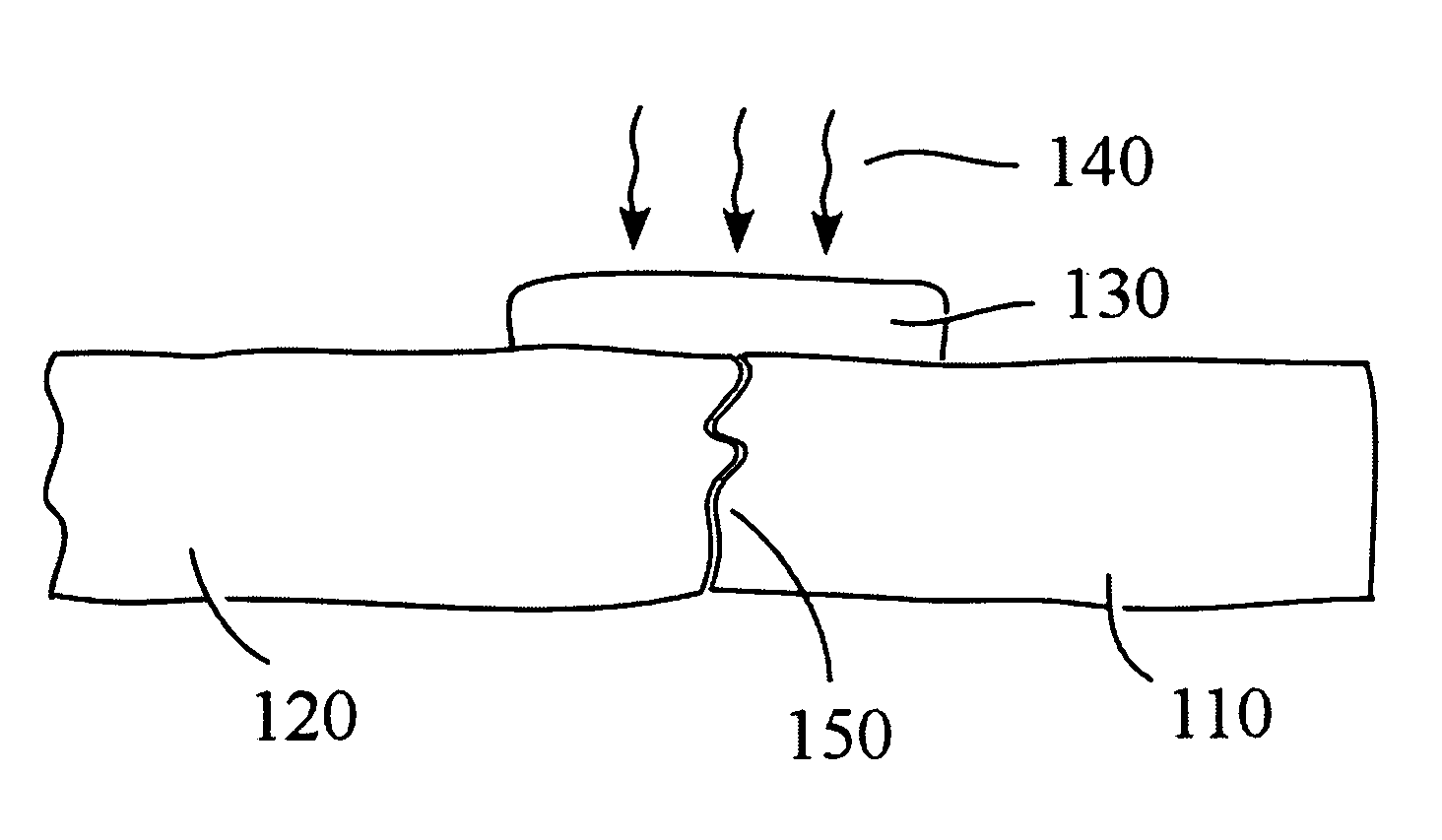
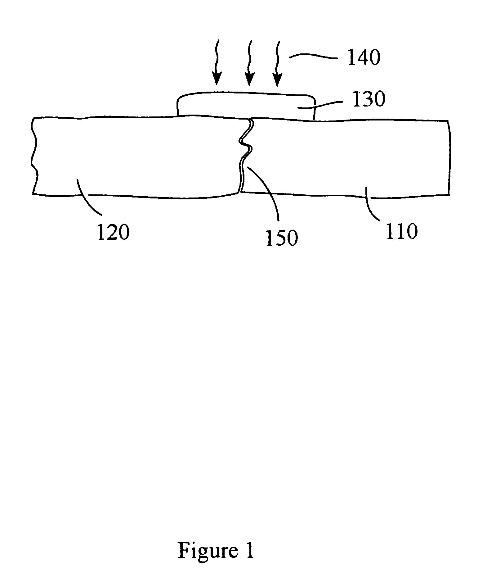
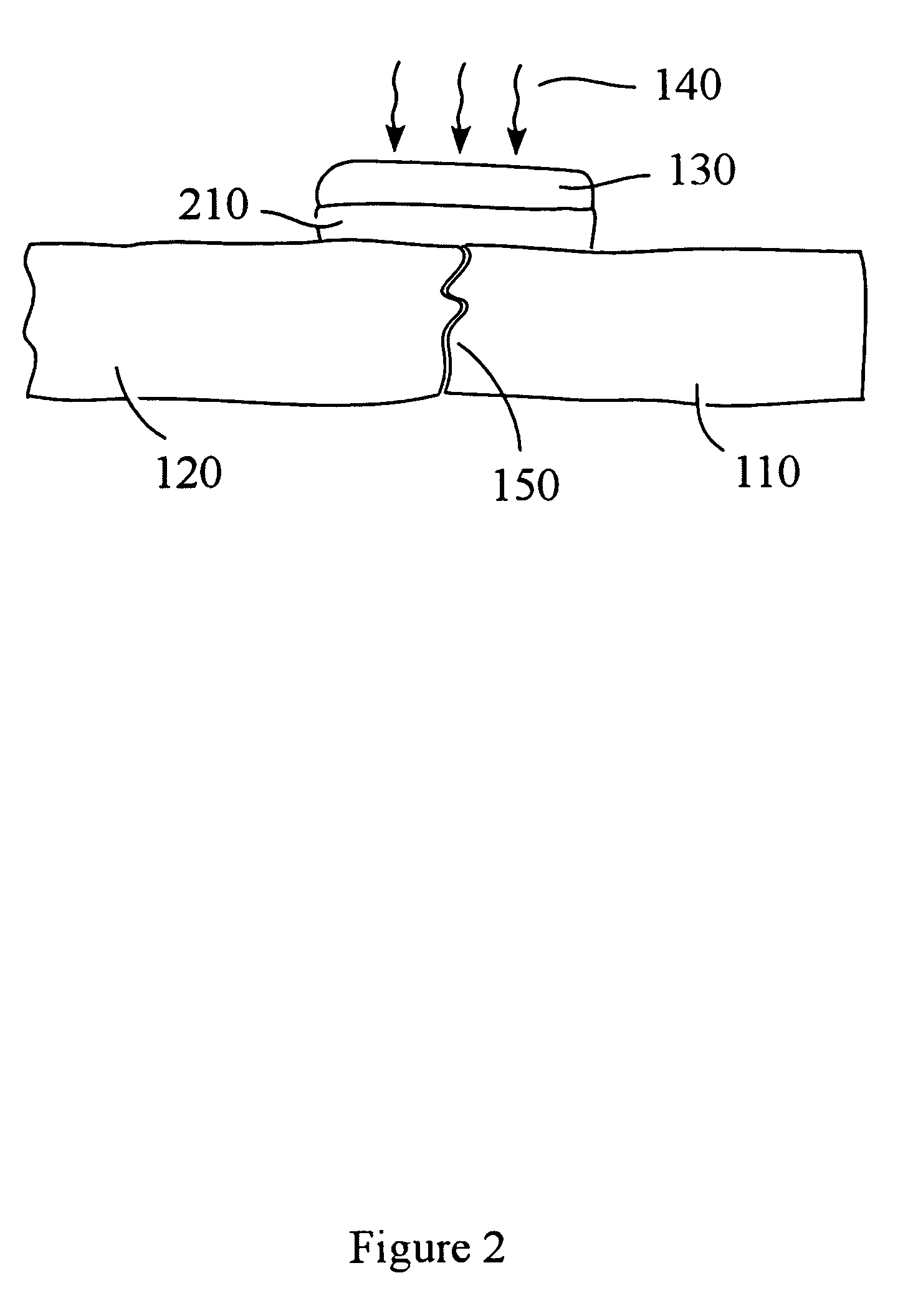
  • Abstract
  • Description
  • Claims
  • Application Information

AI Technical Summary

Benefits of technology

[0004]Consistent with the present invention, a suitable solder and associated laser welding system are provided which avoid the shortcomings of conventional systems described above. For example, the biological solders and sealants consistent with the present invention are biodegradable and do not require chormophores or dyes to promote adhesion. Further, consistent with the present invention, the laser system provides accurate temperature control to eliminate peripheral tissue damage, damage to stay sutures (if required), and large area exposure to reduce treatment time. Further, the use of a feedback controller reduces required surgeon skill. The laser system can be comprised of inexpensive off-the-shelf components and has been designed to be compact, nonintrusive in a surgical setting, inexpensive to manufacture and user friendly.
[0005]It is believed that the laser energy disrupts the three-dimensional structure of collagen fibers found in tissues, promoting tissue crosslinking and improving cohesive strength. The films or gels are easy to apply and fix to the tissue surface.
[0006]Further consistent with the present invention, a method is provided for preparing suitable protein compositions for use with the laser system. The compositions are comprised of chemically derivatized soluble collagen, which is formulated to concentrations ranging from 300 mg / ml (30%) to 800 mg / ml (80%) collagen protein. In particular, Type I collagen, for example, is first prepared by extraction from animal hide, skin or connective tissue and purified. The collagen preparations are then chemically derivatized with sulfhydryl reagents to improve cohesive strength and with secondary derivatizing agents, such as carboxyl groups, to improve the adhesive strength of the solder to the tissue. The compositions are then formed into liquid, gels or solid films which when exposed to energy generated from an infrared laser, for example, undergo thermally induced phase transitions. Solid or semi-solid protein compositions become less viscous enabling the high concentration protein to penetrate the interstices of treated biological tissue or to fill voids in tissue. As thermal energy is released into the surrounding environment, the protein compositions again become solid or semi-solid, adhering to the treated tissue or tissue space.
[0007]In accordance with an additional feature of the present invention, minute quantities of any derivative of commercially available, medical grade cyanoacrylate can be applied to fix and appose tissue edges. Next a layer of the high concentration collagen can be applied adjacent to and on the surface of the tissue to be bonded. This top layer is typically exposed to an infrared laser to melt, denature, and mix the two components to promote both chemical and mechanical bonds to the tissue as well as enhancing intrinsic strength of the composite solder. Infrared laser exposure increases temperature at the bonding site. Feedback control of the laser can be used to adjust solder temperature for optimizing the bonding mechanism to the tissue while preventing thermal damage to adjacent healthy tissue.

Problems solved by technology

These additives, however, may be toxic and cause pernicious effects.
Methods reported to date have not gained clinical acceptance.
The major reasons include the high level of surgeon skill that is required, the strength, toxicity and resorbability of the tissue solder, the potential for irreparable laser damage and cost of the laser system.

Method used


Image

  • Composite tissue adhesive
  • Composite tissue adhesive
  • Composite tissue adhesive

Examples


example 1

[0052]Descending aorta sections were collected from juvenile pigs and stored frozen. Prior to use the tissue, contained in a sealed bag, was thawed 15–20 min in a water bath at room temperature (23° C.). The thawed aorta were cut with a standard single-edged razor blade into 1 cm×1 cm coupons. Excess fascia and loose tissue were removed. The coupons were transferred to a PBS 1X solution that was placed in an ice bath until use.

[0053]The coupons were cut into two 0.5 cm×1 cm pieces and assembled in a butt joint configuration. The incision was first warmed with the laser operating in the 1.4–1.5 micron wavelength range. Laser power density was approximately 10 W / cm2. A minute quantity of medical grade cyanoacrylate is applied sparingly to the apposed tissue edges and thick collagen gels or films were then applied directly to the cut surfaces intended for the joint and subsequently exposed to the laser at a controlled temperature of 55° C. The laser beam was scanned over the entire gel...

example 2

[0054]Several ⅜″ diameter articular cartilage plugs were cut with a tubular harvester in the vicinity of bovine carpel joints. The defects were warmed with the laser operating in the 1.4–1.5 micron wavelength range. Laser power density was approximately 10 W / cm2. Thick collagen-based gels were immediately troweled into the defect by a spatula or dispensed with a microsyringe. Care was taken to apply the collagen gel to all surfaces of the implant prior to insertion into the defect. In most cases the implant surfaces were exposed to the laser to liquefy the collagen-based gel then immediately transferred to the defect. The surface temperature was controlled in the 55–60° C. range. After implant placement additional gel was applied and melted with the laser at temperature controlled in the 55–60° C. range. Rapid cooling was observed once the laser beam was withdrawn and the solder formed a solid and adherent seal around the implant. Lift and torque tests were performed immediately fol...


PUM


Abstract

Consistent with the present invention, tissue adhesive compositions and an associated laser exposure system are provided for bonding or sealing biological tissues. The compositions are comprised of chemically derivatized soluble collagen which is formulated to concentrations ranging from 300 mg / ml (30%) to 800 mg / ml (80%) collagen protein. In particular, Type I collagen, for example, is first prepared by extraction from bovine or porcine hide and purified. The collagen preparations are then chemically derivatized with sulfhydryl reagents to improve cohesive strength and with secondary derivatizing agents, such as carboxyl groups, to improve the adhesive strength of the solder to the tissue. The compositions are then formed into viscous solutions, gels or solid films, which when exposed to energy generated from an infrared laser, for example, undergo thermally induced phase transitions. Solid or semi-solid protein compositions become less viscous enabling the high concentration protein to penetrate the interstices of treated biological tissue or to fill voids in tissue. As thermal energy is released into the surrounding environment, the protein compositions again become solid or semi-solid, adhering to the treated tissue or tissue space.

Description

BACKGROUND OF THE INVENTION[0001]Tissue joining and sealing utilizing sutures and staples is the keystone in modem surgical procedures. An alternative approach utilizes a laser to expose a glue or solder to thereby join adjacent tissues. In many respects, such laser assisted tissue soldering is superior to conventional suture or staple-based methods. The advantages are speed, reduced infection, acceleration in healing, better cosmetic appearance and ease of use particularly in laparoscopic surgery where water tightness and limited access or small size of the repair are important. Laser assisted tissue welding often requires glues or solders for promoting strong bonds, forming a bridge between two apposed tissue sections. The ideal solder is chemically and mechanically matched to the tissue. As a result the solder is strong but stretches and grows with the tissue because of good modulus match. Chromophores mixed with the solder can assist in confining the deposited energy and reduce ...

Claims


Application Information

Patent Timeline
IPC IPC(8): A61B17/03A61B17/00A61B18/22
CPCA61B17/00491A61B18/22A61B2017/0007A61B2017/00084A61B2017/005A61B2017/00508A61B2017/00517
Inventor SOLTZ, BARBARA A.DEVORE, DALE P.DEVORE, BRADEN P.SOLTZ, ROBERTSOLTZ, MICHAEL A.
Owner CONVERSION ENERGY ENTERPRISES
Learn More