
Methods for the isolation of nucleic acids and for quantitative DNA extraction and detection for leukocyte evaluation in blood products

Inactive Publication Date: 2005-10-25
GLOBAL LIFE SCI SOLUTIONS USA LLC
View PDF33 Cites 163 Cited by
  • Summary
  • Abstract
  • Description
  • Claims
  • Application Information

AI Technical Summary

Benefits of technology

[0050]According to the present invention there is provided a substrate for lysing cells and purifying nucleic acid having a matrix and a coating and an integrity maintainer for maintaining the purified nucleic acid. Also provided is a method of purifying nucleic acid by applying a nucleic acid sample to a substrate, the substrate physically capturing the nucleic acid, bonding the nucleic acid to the substrate and generating a signal when the nucleic acid bonds to the substrate indicating the presence of the nucleic acid. A kit for purifying nucleic acid containing a coated matrix and an integrity maintenance provider for preserving the matrix and purifying nucleic acid is also provided.

Problems solved by technology

While relatively rapid and convenient procedures for the purification of nucleic acid (such as DNA) from agarose have been developed, it remains a relatively difficult operation to extract nucleic acid directly from more complex starting samples such as cells and cell lysates.
On the whole, the procedures currently practiced to purify nucleic acid from nucleic acid-containing samples comprising cells or cell lysates remain time consuming and labor intensive.
The method described in EP 0389063 is disadvantageous in that it is a manually intensive, multi-step procedure.
In view of the fact that the method involves a number of centrifugation and vessel transfer steps, this method is unsuitable for automation.
Some of the disadvantages of covalent attachment of biomolecules onto activated membranes are:a) Molecule immobilization is often slow, requiring 20-180 minutes for reaction completion.b) High ligand and biomolecule concentration is needed for fast immobilization.c) Constant agitation is needed during the immobilization process, which may result in biomolecule denaturation and deactivation.d) Once the immobilization process is complete, often a blocking (capping) step is required to remove residual covalent binding capacity.e) Covalently bound molecules can not be retrieved from the filter membrane.
This is a major disadvantage for applications where several downstream processes are required from the same sample, such a STR profiling and genotyping.
Multiple punching is very time consuming, and as yet, has not lent itself to simplified automation.
Despite the increasing need for transfusions and the use of transfusion products, such use involves a number of risks.
Further problems stem from Human Leukocyte Antigen (HLA) alloimmunization, in which the recipient is sensitized to antibodies contained in the transfusion product which can react, for example, to the recipient's leukocytes (HLA sensitization).
Other serious risks of the use of transfusion products include transmission and / or reactivation of cytomegalovirus (CMV), occurrence of graft-versus-host disease (GVHD), and the risk of viral transmissions.
Recent methods have limitations for proposed centralization of testing services for leukoreduced blood products in terms of sample storage time and their integrity.
Both manual and automated approaches require fresh blood, because these described parameters change with blood storage, or are sensitive to sample preparation techniques.
All traditional methods are very sensitive to blood storage time because cells' deterioration and changing of size and / or shape during storage or the freezing-thawing process.
However, deterioration of samples as a result of prolonged sample shipment remains an obstacle to centralized testing service.” (Walter H. Dzick, Von Sang 2000, 78, 223:226).
“The requirement of fresh samples for processing is particularly problematic in connection with applications such as Quality control of WBC-reduced blood components by blood centers.” (T.-H.
Other problems with such conventional methods include timing of the analysis.
For example, the time required for one sample analysis using flow cytometry (sampling and analysis of histogram) is too long (3-10 min for a single sample analysis) to permit large scale testing.
Previous approaches to this technology claimed substantial, but not total, DNA recovery from whole blood samples.
However, a Quantitative PCR protocol takes many hours to process.
Further, PCR methods are very expensive.
They require unique real time PCR devices and expensive PCR reagent kits.
The preparation of calibration standard samples is labor consuming and very sophisticated.

Method used


Image

  • Methods for the isolation of nucleic acids and for quantitative DNA extraction and detection for leukocyte evaluation in blood products
  • Methods for the isolation of nucleic acids and for quantitative DNA extraction and detection for leukocyte evaluation in blood products
  • Methods for the isolation of nucleic acids and for quantitative DNA extraction and detection for leukocyte evaluation in blood products

Examples


example 1

Preparation of Purified Product Using an Elution Method

[0209]DNA extraction from 200 μl of human whole blood was carried out using the present method. The protocol was as follows:[0210]1) Add 200 μl of whole blood to the column, add 1000 μl of red blood cell lysis solution (RBCL), filter to waste.[0211]2) Add 1000 μl RBCL, filter to waste.[0212]3) Add 1000 μl 0.5% SDS, filter to waste.[0213]4) Add 1000 μl TE−1, filter to waste.[0214]5) Add 100 μl TE−1, filter to waste.[0215]6) Incubate at 90° C. for ten minutes.[0216]7) Add 100 μl TE−1, filter to capture DNA solution.

[0217]The mean DNA yield for the present method is 30-40 μg per ml of blood. About 80% of the DNA product is greater than 40 kb. The approximate time per cycle is 20 minutes, i.e. significantly faster than presently known methods.

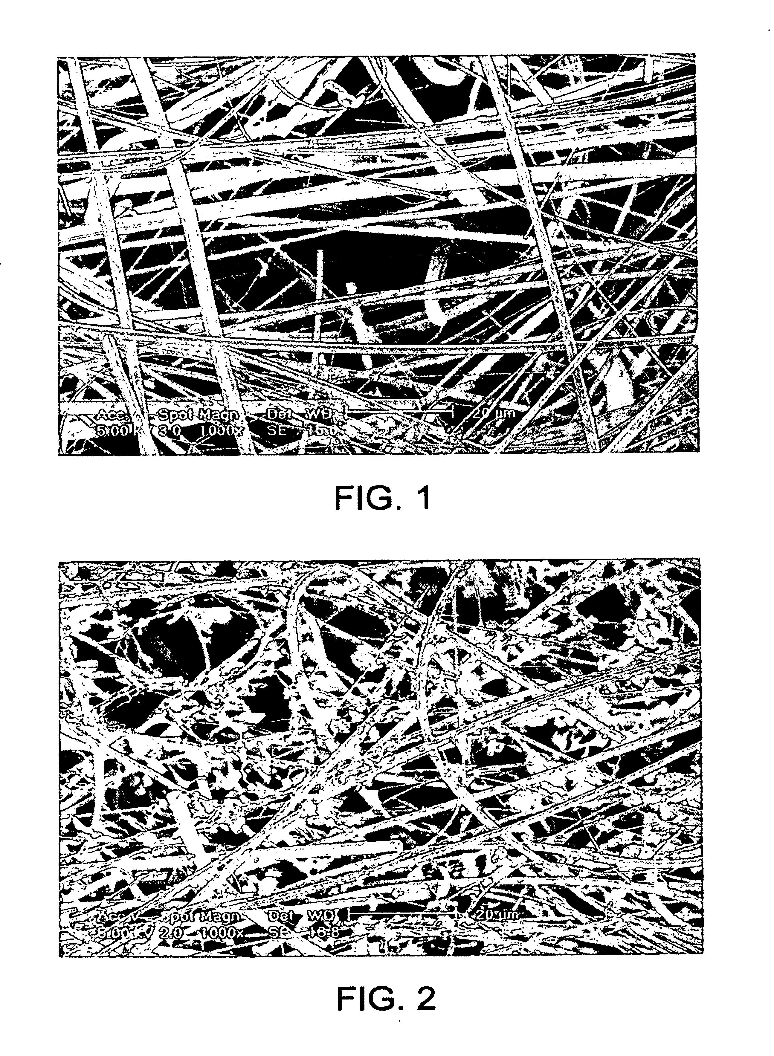
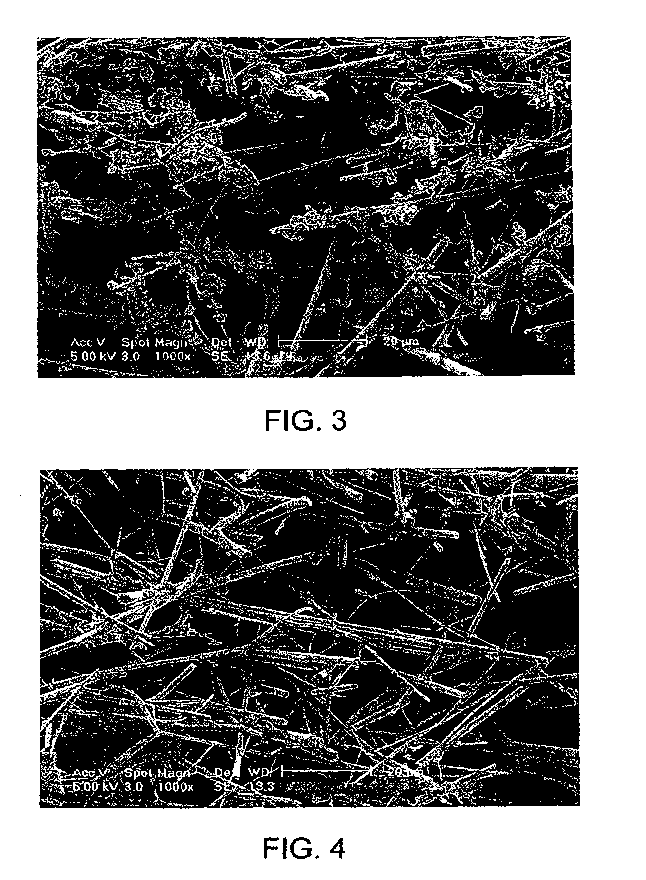
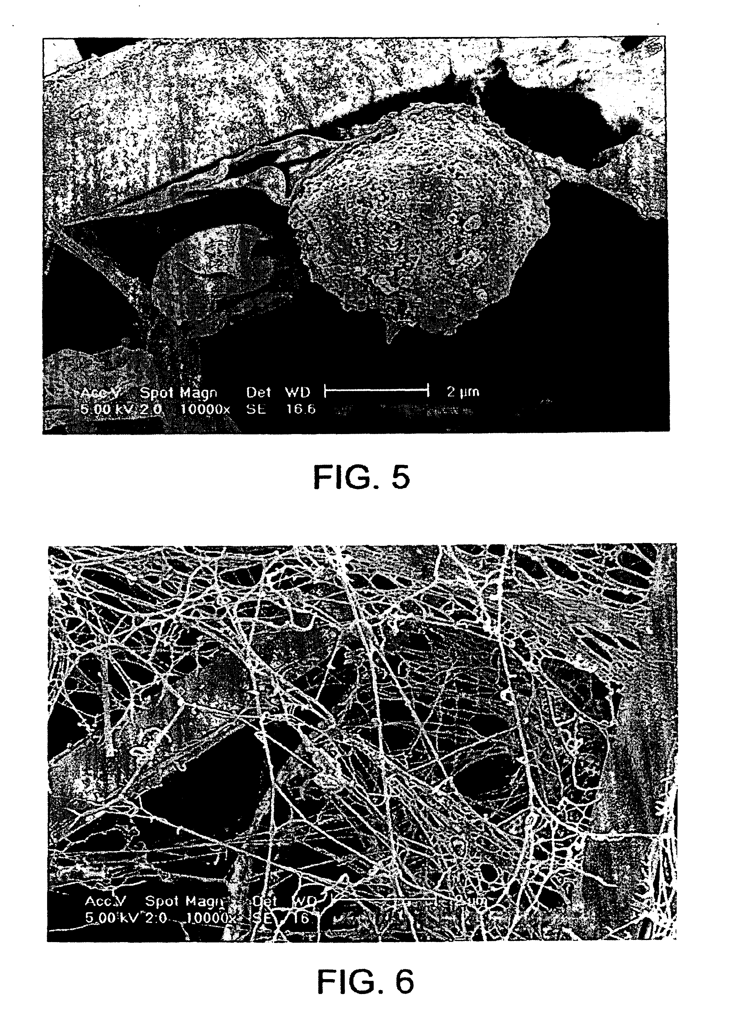
[0218]At various stages of the method, the filter and the retentate were analyzed by scanning electron microscopy (SEM) in order to reveal the mechanism of cell retention, DNA retention and DNA...

example 2

Filter Depth for Filter Used in Elution Method

[0227]It has hitherto been unknown to use a filter in the dual role of white cell capture and DNA capture. Once the white cells are lysed, it is believed that the filter acts as a depth filter to the DNA. In order to explore this, the following investigation was undertaken.

[0228]A number of filters were stacked in an extraction column and DNA was isolated from 500 μl of whole blood in accordance with the following protocol:[0229]1) Add 500 μl of red blood cell lysis solution to the blood and filter to waste.[0230]2) Add 500 μl 0.5% SDS solution and filter to waste.[0231]3) Add 500 μl mM Tris-HCl pH 8.5 and filter to waste.[0232]4) Add 500 μl mM Tris-HCl pH 8.5 and incubate for 50 min at 82° C.[0233]5) Filter and capture DNA eluant.

[0234]Prior to the final incubation and elution step the filters were removed and each one eluted and incubated individually. FIG. 10 shows that there seems to be a gradient of DNA capture from the top filter t...

example 3

Filter Depth for Filter Used in Elution Method

[0236]Analysis has shown that DNA recovery can be improved within the system by increasing the number of filters within the column. An experiment was performed according to the protocol below to assess the percentage of DNA captured by each subsequent layer of filter by processing whole blood using a 4-layered extraction column.

Protocol

[0237]1) Add 200 μl of whole human blood to the 2 ml-extraction vessel.[0238]2) Add 1 ml of RBCL and filter directly to waste.[0239]3) Add a further 1 ml of RBCL and filter directly to waste.[0240]4) Add 1 ml of 0.5% SDS and filter directly to waste.[0241]5) Add 1 ml of TE−1 and filter directly to waste.[0242]6) Add 100 μl of TE−1 and incubate at 90° C. for 8 minutes.[0243]7) Add 200 μl of TE−1 and collect.

[0244]The filters were removed prior to the elution step and the DNA collected off each filter separately. The most DNA was recovered from the uppermost filter and the least DNA was recovered from the lo...


PUM

PropertyMeasurementUnit
temperatureaaaaaaaaaa
diametersaaaaaaaaaa

Abstract

A method for isolating nucleic acid which comprises:(a) applying a sample comprising cells containing nucleic acid to a filter, whereby the cells are retained as a retentate and contaminants are removed;(b) lysing the retentate from step (a) while the retentate is retained by the filter to form a cell lysate containing the nucleic acid;(c) filtering the cell lysate with the filter to retain the nucleic acid and remove remaining cell lysate;(d) optionally washing the nucleic acid retained by the filter; and(e) eluting the nucleic acid, wherein the filter composition and dimensions are selected so that the filter is capable of retaining the cells and the nucleic acid.Additionally, there is provided a substrate for lysing cells and purifying nucleic acid having a matrix and a coating and an integrity maintainer for maintaining the purified nucleic acid. Also provided is a method of purifying nucleic acid by applying a nucleic acid sample to a substrate having an anionic detergent affixed to a matrix, the substrate physically capturing the nucleic acid, bonding the nucleic acid to a substrate and generating a signal when the nucleic acid bonds to the substrate indicating the presence of the nucleic acid. A kit for purifying nucleic acid containing a coated matrix and an integrity maintenance provider for preserving the matrix and purifying nucleic acid is also provided. Further, there is provided a method for quantifying DNA, such as double-stranded or genomic DNA, isolated from cells, such as leukocytes to determine the numbers of leukocytes in a sample of leukoreduced blood.

Description

CROSS-REFERENCE TO RELATED APPLICATIONS[0001]This application is a Continuation-in-Part of U.S. patent application Ser. No. 09 / 736,659, filed Dec. 14, 2000, now U.S. Pat. No. 6,746,841 which is a Continuation-in-Part of PCT application No. PCT / US00 / 10230, filed Apr. 14, 2000, which claims the benefit of priority under 35 USC Section 119(e) of U.S. Provisional Patent Application No. 60 / 129,191, filed on Apr. 14, 1999; U.S. Provisional Patent Application No. 60 / 180,353, filed on Feb. 4, 2000; and U.S. Provisional Patent Application No. 60 / 193,556, filed on Mar. 31, 2000, all of which are incorporated herein by reference; this application is also a Continuation-in-Part of a CPA application filed Mar. 21, 2001, which is a CPA of U.S. patent application Ser. No. 09 / 414,533, filed Oct. 8, 1999, which claims the benefit of priority of United Kingdom application 9918193.5, filed on Aug. 2, 1999, and United Kingdom application 9822141.9, filed on Oct. 9, 1998, all of which are incorporated h...

Claims


Application Information

Patent Timeline
Patent Type & Authority Patents(United States)
IPC IPC(8): C12N15/10
CPCC12N15/1017
Inventor FOMOVSKAIA, GALINA N.SMITH, MARTIN A.FOMOVSKY, MIKHAIL A.BUTT, NEIL J.
Owner GLOBAL LIFE SCI SOLUTIONS USA LLC
Learn More