Methods and compositions usable in cataract surgery

a cataract surgery and composition technology, applied in the field of cataract surgery, can solve the problems of severe visual impairment or vision loss, decrease the quality of life, and loss of visual ability, and achieve the effect of facilitating access to the interior of the capsul

Inactive Publication Date: 2006-01-17
PHACO TREAT
View PDF27 Cites 48 Cited by
  • Summary
  • Abstract
  • Description
  • Claims
  • Application Information

AI Technical Summary

Benefits of technology

[0020]In view of the problems and drawbacks associated with prior art methods it is an object of the present invention to provide methods and compositions that facilitates access to the interior of the capsule during intraocular surgery in general.

Problems solved by technology

A cataract is defined as every form of opacity in the lens or its capsule; the lens becomes cloudy, resulting in a loss of visual ability A cataract is a painless phenomenon, but decreases the quality of life if the lens is not surgically extracted and replaced by an artificial lens.
However, YAG laser capsulotomy exposes patients to the risk of complications that can lead to severe visual impairment or loss of vision, such as retinal detachment, pupillary block glaucoma and cystoid macular edema Other complications associated with YAG laser capsulotomy include damage to implanted intraocular lenses resulting in glare and photophobia, dislocation of intraocular lenses, iritis, vitritis, corneal edema, iris damage and rupture of the anterior hyaloid.
However, none of them has been proved to be practical, effective, and safe enough for routine clinical practice.
Capsular polishing, aspiration of residual lens epithelial cells, ultrasound combined with aspiration, cryocoagulation, and osmolysis are examples of methods that have been developed and shown to remove or destroy remaining lens epithelial cells, but none of these methods have been proven to be efficient in PCO prophylaxis.
However, in vivo animal studies have shown that there are toxic side effects in the tissues of the eye when anti-metabolites are used in sufficiently high concentration to inhibit lens epithelial cells proliferation.
However, no conclusive results have been obtained Another pharmacological approach is to separate lens epithelial cells from the lens capsule.
EDTA seems not to be more efficient than other agents evaluated in PCO prophylaxis.
However, the zonules were damaged by the trypsin solution.
The use of an active enzyme can be a problem even when an enzyme solution is introduced into the lens capsule because it can damage the zonules bound to the lens capsule.
A drawback when using this method is that the efficacy of the treatment may be strongly limited, if the natural lens is not removed before administrating the cell-killing substance.
The natural lens may absorb or decrease the efficacy of the substance due to the huge number of lens epithelial cells within the lens.
However, physically and technically, it seems to be difficult to efficiently remove the substance introduced into the lens capsule before capsulorhexis without damaging the lens capsule.
The remaining substance may escape from the lens capsule and damage the cells and tissues facing the anterior chamber during and after capsulorhexis.
An important problem in connection with all methods relating to cataract surgery is the difficulty of observing the interior of the lens capsule, especially behind the iris, in order to ascertain that measure taken were successful, such as the removal of residual lens epithelial cells.
However, this patent is silent in regard of evaluating the capsular inside in a turbulent flow of irrigation solution and lens materials flowing around the end of the fiber optic during the process of phacoemulsification and irrigation-aspiration, and such evaluation appears very difficult given the premises in the patent.
Additionally, it seems difficult to use the technique of closing off the capsule with a patch for performing different procedures within the capsule, as the patch would block introduction of devices into the capsule and performance of different methods within the capsule.

Method used


Image

  • Methods and compositions usable in cataract surgery
  • Methods and compositions usable in cataract surgery
  • Methods and compositions usable in cataract surgery

Examples


example 1

Evaluation of a Preferred Embodiment of the Invention

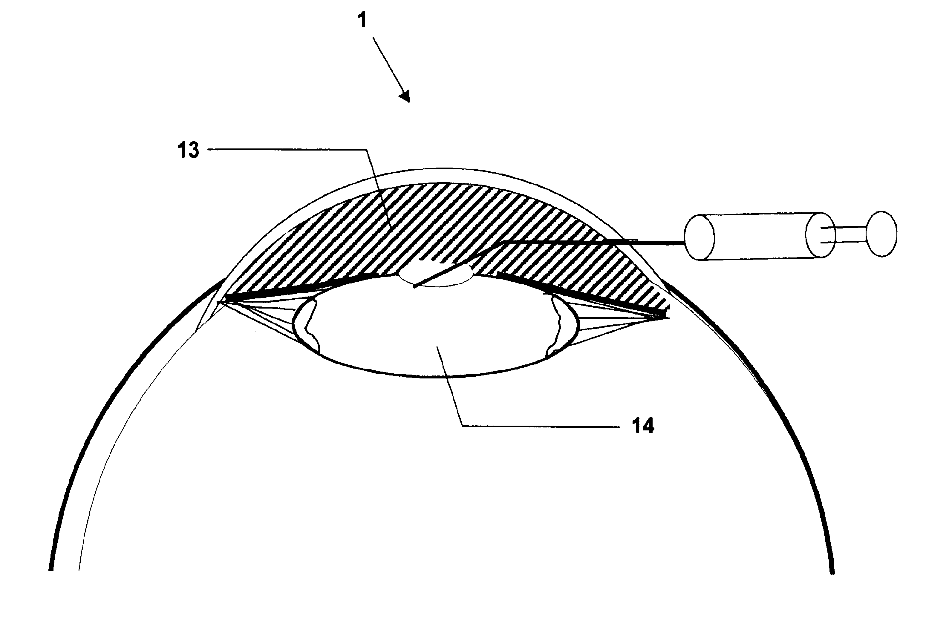
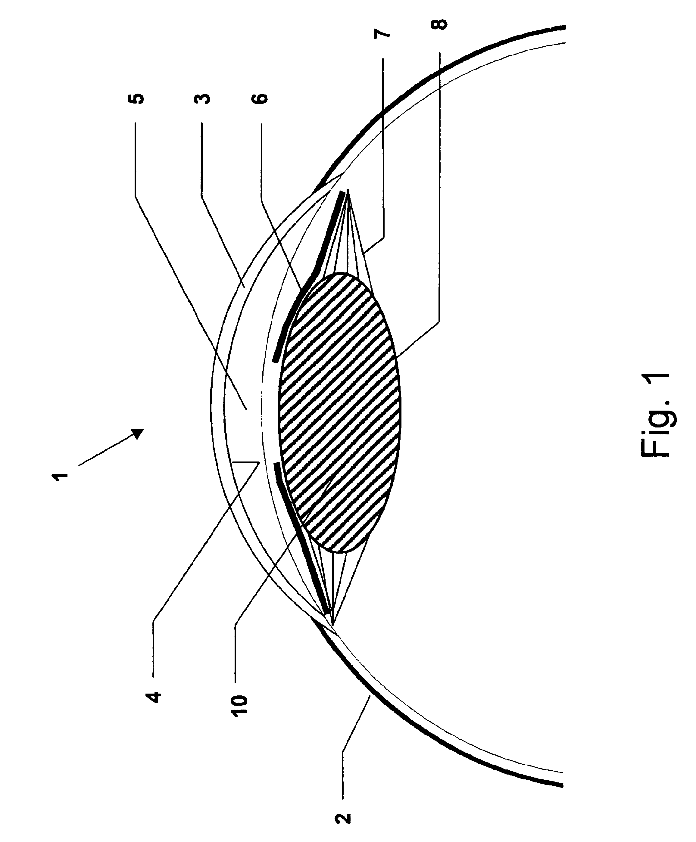
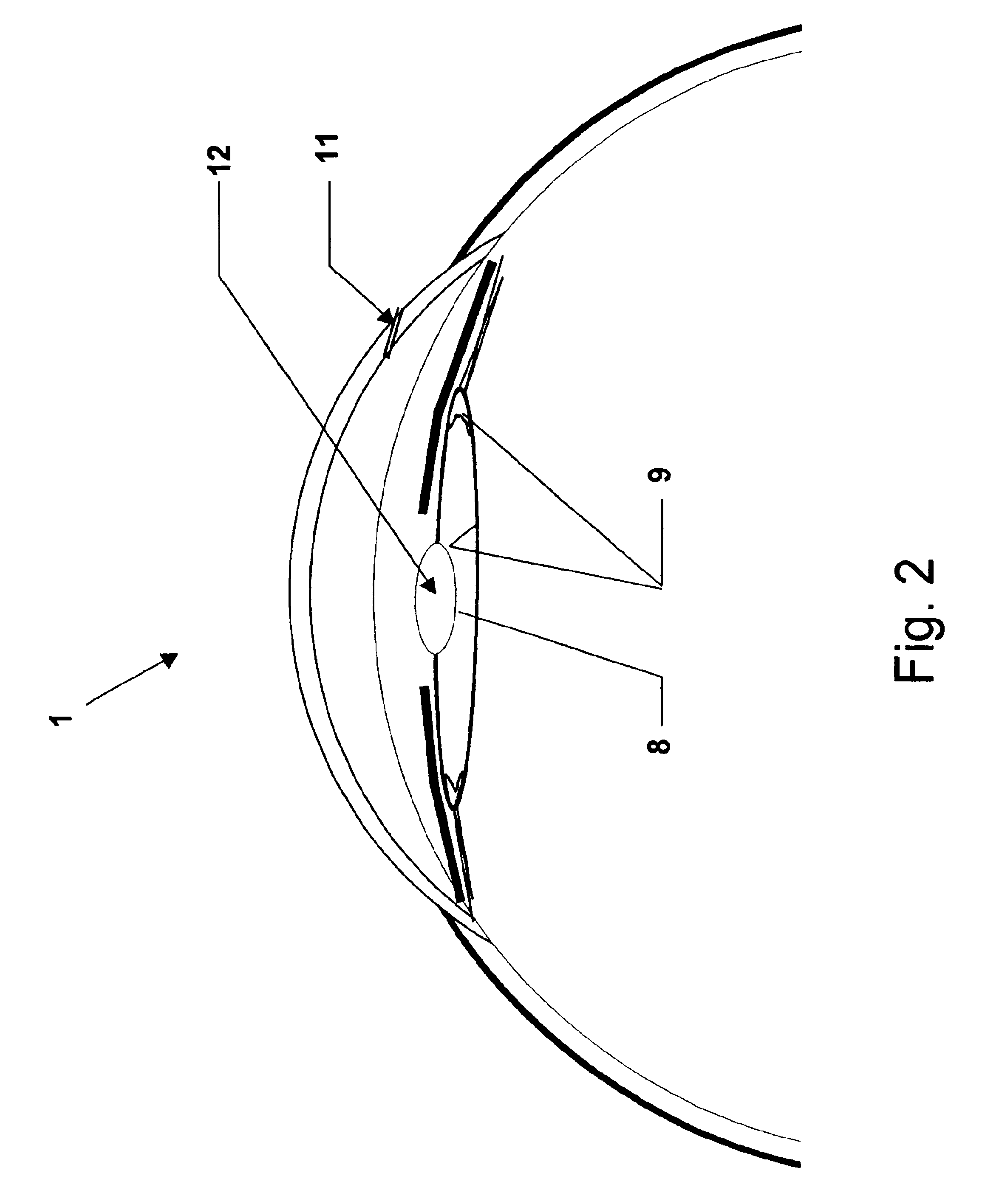
[0190]Cataract surgery was simulated in porcine cadaver eyes An eye was fixed on a plastic holder while maintaining a standardized intraocular pressure of 10-15 mm Hg before surgery. Further details of the model are published elsewhere (Holmen J B, Ekesten B & Lundgren B (2001) Anterior chamber depth estimation by Scheimpflug photography. Acta Ophthalmol Scand Vol. 79:576-579.) The example was performed as follows:[0191]1) A corneal incision 11 was performed at the limbus of the cornea 3.[0192]2) The anterior chamber 5 was filled with a viscoelastic solution 13 (Healon5, Pharmacia AB, Uppsala, Sweden) by injection.[0193]3) A continuous circular capsulorhexis was created.[0194]4) Phacoemulsification was performed by an anterior segment operating system (Oertli Quinto, Oertli Instrumente A G, Berneck, Switzerland) with complete removal of the cataractous lens 10.[0195]5) Additional viscoelastic solution 13 (Healon5) was injected int...

example 2

[0207]Example 2 was performed using the method described in Example 1, except for using trypan blue instead of fluorescein in the agent solution. The distribution of the active agent solution 15 was studied by free preparation of the lens capsule 8.

The results were equal to the ones in Example 1

example 3

[0208]Example 3 is performed using the method described in Example 1, but using a plurality of known active substances in the agent solution, e.g. doxorubicin, EDTA, indomethacin, 5-fluorouracil (5-FU), FGF-saporin, methotrexate, mitomycin, colchicine or daunomycin / daunorubicin.


PUM


Abstract

The invention relates to a method of performing ocular surgery, after an anterior capsulotomy has been made, by forming a sealed expanded capsular bag. The method includes sealing the capsular bag with a viscoelastic material to provide a gas tight seal to prevent leakage into the anterior chamber of the eye during the surgical process; expanding the capsular bag by introducing a gas capable of exerting a pressure on the inner surface of the capsular bag wall; inspecting and / or treating the capsular bag with one or several devices and / or agents suitable for performing inspection and / or treatment. The inspection and / or treatment can comprise any of visual inspection, estimation of capsular bag volume; labeling any residual epithelial cells to detect the presence thereof; removing residual epithelial cells, implanting one or more intracapsular implants; injecting a lens forming material for molding a lens in situ; drying the lens capsule; alone or in any combination. Use of a viscoelastic compound for the preparation of a temporary intraocular seal capable of sealing the capsular bag is also provided.

Description

PRIOR RELATED APPLICATIONS[0001]This application is a continuation-in-part of application Ser. No. 09 / 848,784 filed on May 3, 2001, U.S. Pat. No. 6,533,769.FIELD OF THE INVENTION[0002]The present invention relates generally to cataract surgery (i.e. lensectomy), and more specifically to methods and compositions for treatments inside the lens capsule, preferably directed to treat lens epithelial cells, and methods of providing intraocular implants into the lens capsule, preferably injectable intraocular lenses molded inside the lens capsule, by the use of an ophthalmic composition of sodium hyaluronate with a specifically defined molecular mass and concentration.BACKGROUND OF THE INVENTION[0003]The crystalline lens of the human eye is located in the posterior chamber between the posterior iris surface and the vitreous body It is a biconvex transparent tissue without nerves and blood vessels, weighing approximately 0.2 g. The lens is enveloped in a capsule, a structureless, transparen...

Claims


Application Information

Patent Timeline
Patent Type & Authority Patents(United States)
IPC IPC(8): A61M31/00A61B19/00A61F2/16A61F9/00A61F9/007A61K9/00A61P27/02A61P27/12
CPCA61F9/0017A61K9/0048A61F9/00745A61P27/02A61P27/12
Inventor HOLMEN, JORGEN
Owner PHACO TREAT
Why Eureka
Try Eureka