Lentiviral vector-mediated gene transfer and uses thereof

a technology of lentiviral vectors and gene transfer, applied in the field of lentiviral vectormediated gene transfer and its use, can solve the problems of loss of visual axis clarity or separation of retina, permanent loss of vision, and compromising retinal function

Inactive Publication Date: 2006-10-17
RES DEVMENT FOUND
View PDF8 Cites 29 Cited by
  • Summary
  • Abstract
  • Description
  • Claims
  • Application Information

AI Technical Summary

Benefits of technology

[0017]The present invention also provides a method of preventing neovascularization and corneal transplant failure by transducing corneal tissue ex vivo with a lentiviral vector comprising a therapeutic gene that inhibits neovascularization.

Problems solved by technology

One of the most common causes of human blindness is abnormal, intraocular cellular proliferation that often results in a loss of clarity of the visual axis or in a separation of the retina from the retinal pigment epithelium due to tractional forces applied directly to the retinal surface.
Proliferative retinal detachment, whether it is related to proliferative diabetic disease, retinopathy of prematurity, proliferative vitreoretinopathy, or neovascular age-related muscular degeneration, ultimately will result in permanent loss of vision if left untreated.
While these three clinical entities are distinct and affect different groups of patients, they share a final common pathway that involves uncontrolled division of endothelial cells leading to formation of new blood vessels that ultimately compromise retinal function.
Contraction of the posterior vitreous surface results in traction at the sites of vitreo-fibrovascular adhesions and ultimately detaches the retina.
It is this process which results in blindness in millions of diabetics, premature infants or the aged in our society.
Current treatments for intraocular neovascular diseases are invasive and destructive.
Currently it is difficult to perform gene transduction in mammalian cells with great degree of effectiveness.
It is also problematic to introduce these vectors into the eye without induction of a strong inflammatory response.
Presently, there is a lack of means of transducing terminally differentiated or proliferating human cells within or derived from the eye.

Method used


Image

  • Lentiviral vector-mediated gene transfer and uses thereof
  • Lentiviral vector-mediated gene transfer and uses thereof
  • Lentiviral vector-mediated gene transfer and uses thereof

Examples


example 1

Cells and Tissue

[0112]Primary explants of human choroidal fibroblasts (HCF), human umbilical vein endothelial cells (HUVEC) and human fetal retinal pigment epithelial cells (HRPE) were established and were plated in conditions which either did or did not promote mitotic activity. Stable photoreceptor-derived cells (Y-79 and Weri-Rb-1) were also cultured.

[0113]Human retina and RPE, obtained at the time of enucleation for retinoblastoma were used to demonstrate the ability of lentiviral vectors to transduce these mitotically inactive cells and induce the expression of an exogenous human peripherin transgene. Human corneas obtained at the time of corneal transplant surgery were used to demonstrate the ability of lentiviral vectors to transduce these mitotically inactive cells with the marker gene enhanced green fluorescence protein gene.

example 2

Lentivirus Vector

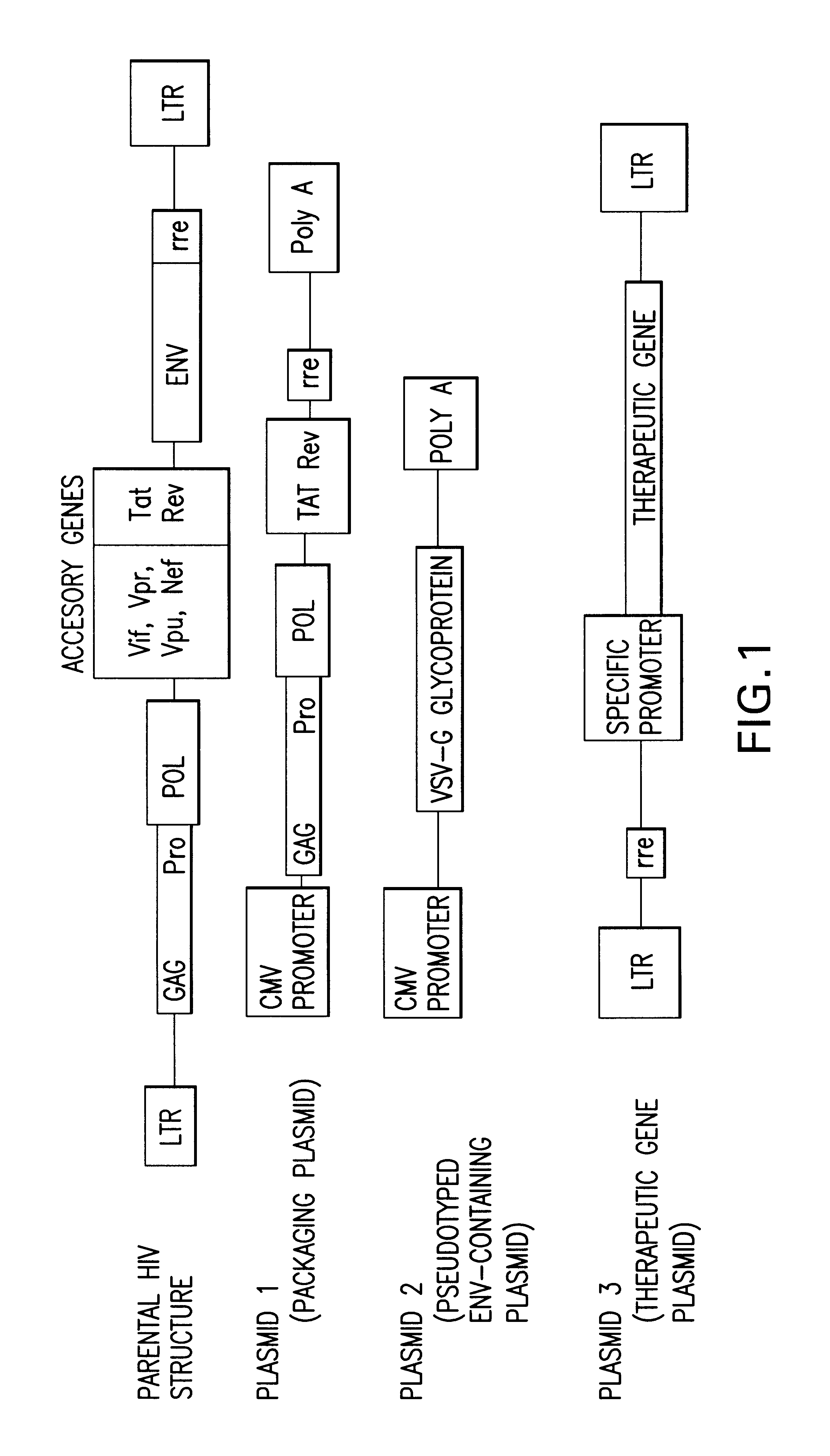
[0114]A three plasmid-based lentiviral vectoring system pseudotyped with the vesicular stomatitis virus (VSV) envelope and which contained the green fluorescent protein (GFP) gene as a marker was used (FIG. 1). Recombinant lentiviruses were produced as described by Naldini et al. The cytomegalovirus (CMV) immediate-early gene promoter directed expression of eGFP in the plasmid pHR′-CMV-eGFP. Stocks of virus were generated as follows. Human kidney 293T cells (5×10⁶) were plated on 10 cm plates, and were cotransfected the following day with 10 ug of pCMVΔR8.91 (packaging function plasmid), 10 ug of pHR′-CMV-eGFP (marker gene plasmid), and 2 ug of pMD.G (the VSV-G envelope containing plasmid) by calcium phosphate precipitation in D10 growth medium (high glucose DMEM with 10% fetal bovine serum) and antibiotics. After 12-16 h at 37° C., the medium was removed and fresh D10 growth medium was added. Cells were cultured for an additional 10 hours. Fresh D10 medium containi...

example 3

Lentivirus Vector Transduction

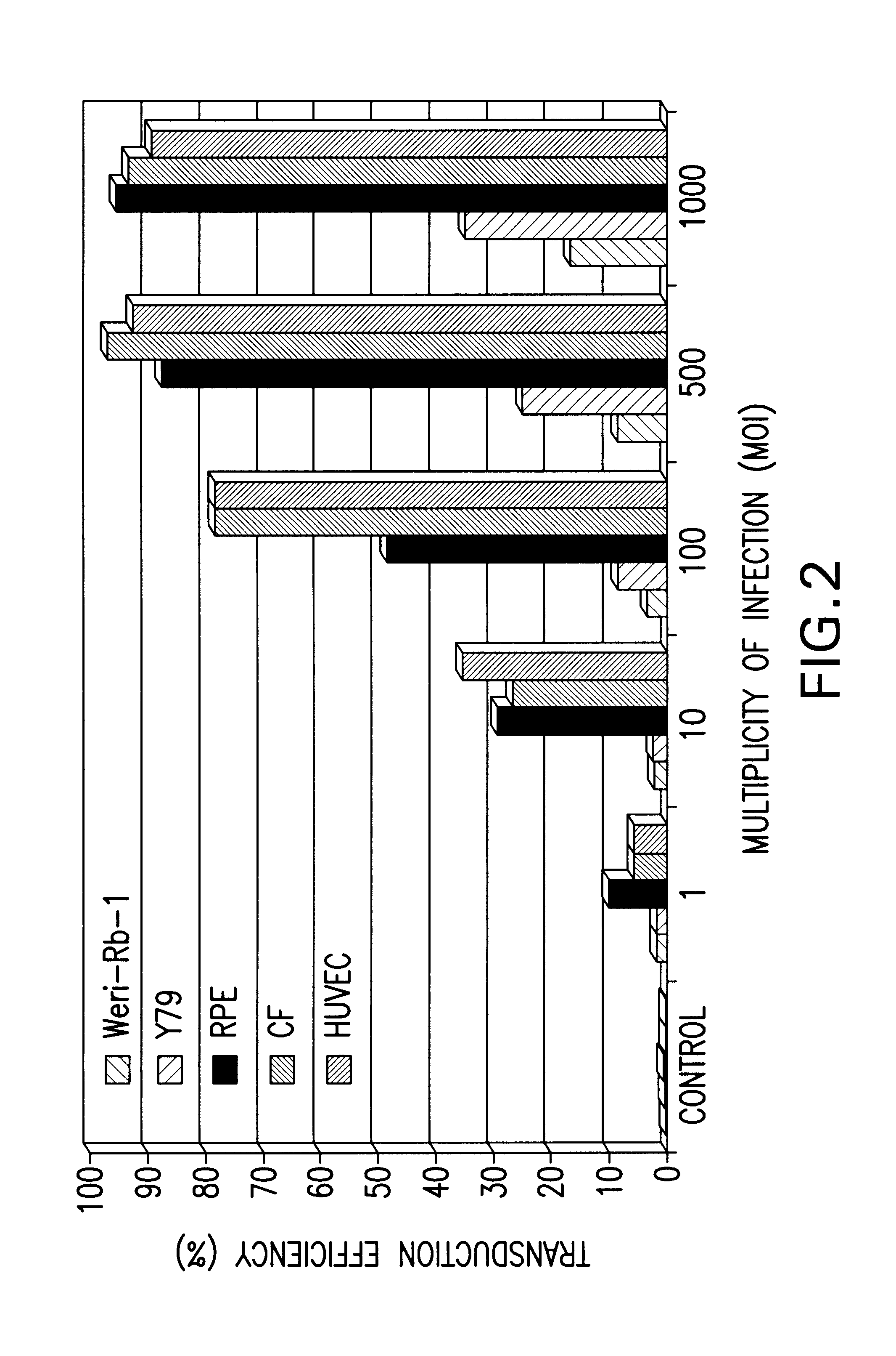
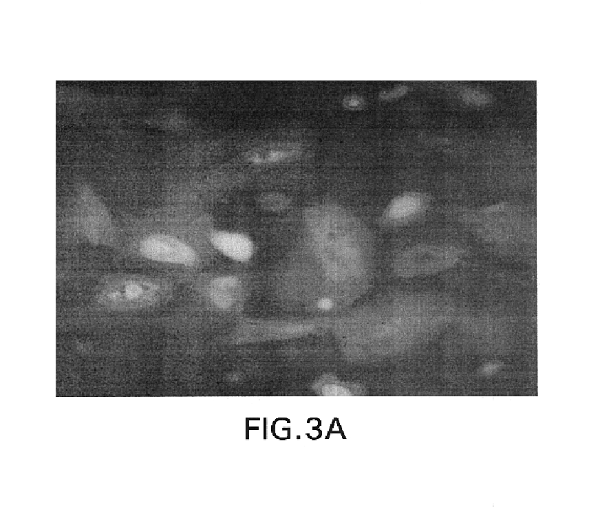
[0117]Supernatants containing 2×10⁶ replication-deficient lentiviral particles / ml were generated by the transfection of 293T cells with the lentivirus vector described above. Cells were cultured with the viral particles for 24 hours and then recovered in normal media for four days prior to the determination of GFP expression by fluorescent-activated cell sorting (FIGS. 2-3).

[0118]Transduction efficiency was measured as a function of multiplicity of infection with MOIs ranging from 1 to 1000. Results of in vitro transduction of a number of human cell lines demonstrate a positive correlation between MOI and transduction efficiency as more cells were transduced with increasing number of lentiviral particles (FIG. 2).

[0119]The ability of the lentiviral vector to transduce non-dividing cells was examined. Human retinal pigment epithelial cells were transduced by lentiviral or murine leukemia viral vectors. Cells were mitotically inactive (confluent) or mitot...


PUM

PropertyMeasurementUnit
green fluorescent proteinaaaaaaaaaa
concentrationaaaaaaaaaa
fluorescentaaaaaaaaaa

Abstract

The present invention provides lentiviral vectors that are useful in human gene therapy for inherited or acquired proliferative ocular disease. It furnishes methods to exploit the ability of lentiviral vectors to transduce both mitotically active and inactive cells so that eye diseases may be treated.

Description

CROSS-REFERENCE TO RELATED APPLICATION[0001]This is a continuation-in-part application of U.S. Ser. No. 10 / 025,264, filed Dec. 19, 2001 now abandoned, which claims benefit of provisional patent application U.S. Ser. No. 60 / 256,701, filed Dec. 19, 2000, now abandoned.BACKGROUND OF THE INVENTION[0002]1. Field of the Invention[0003]The present invention relates generally to the fields of gene delivery vectors and gene therapy. More specifically, the present invention relates to lentiviral vectors useful in human gene therapy for inherited and proliferative ocular disease.[0004]2. Description of the Related Art[0005]One of the most common causes of human blindness is abnormal, intraocular cellular proliferation that often results in a loss of clarity of the visual axis or in a separation of the retina from the retinal pigment epithelium due to tractional forces applied directly to the retinal surface. Proliferative retinal detachment, whether it is related to proliferative diabetic dise...

Claims


Application Information

Patent Timeline
Patent Type & Authority Patents(United States)
IPC IPC(8): A01N63/00A01N43/04C12N15/00C12N15/63C12N5/00C12N15/09A61K35/76A61K38/00A61K38/21A61K38/22A61K38/55A61K48/00A61P27/02A61P27/06C07K14/47C07K14/52C12N15/867D21B1/08D21C5/02
CPCA61K48/0075C07K14/47C07K14/522C12N15/86A61K48/00C12N2740/15043C12N2740/16043C12N2740/16045C12N2830/42C12N2840/203A61P27/02A61P27/06C12N15/867
Inventor STOUT, J. TIMOTHYAPPUKUTTAN, BINOY
Owner RES DEVMENT FOUND
Why Eureka
Try Eureka