Method of cryopreservation of tissues by vitrification

a tissue and cryopreservation technology, applied in the field of cryopreservation of tissues by vitrification, can solve the problems of limited study, inability to obtain viable tissues and cells, and many patients without suitable autologous veins, and achieve direct protective effects on the cell membrane and rapid rate

Inactive Publication Date: 2007-01-02
LIFELINE SCI
View PDF18 Cites 37 Cited by
  • Summary
  • Abstract
  • Description
  • Claims
  • Application Information

AI Technical Summary

Benefits of technology

[0046]After being immersed in a cryoprotectant-free solution, the tissue or organ is immersed in a solution containing cryoprotectant with or without perfusion. (An advantage of the present invention is that perfusion is not necessarily required.) A cryoprotectant concentration sufficient for vitrification generally contains from 6.0 to 9.5M cryoprotectant, preferably from 7 to 9M cryoprotectant, more preferably from 8 to 9M cryoprotectant. The cryoprotectant solution may contain any combination of cryoprotectants sufficient for vitrification. Cryoprotectants include, but are not limited to, dimethyl sulfoxide, formamide, 1,2-propanediol, 2,3-butanediol, glycerol, ethylene glycol, n-dimethyl formamide and 1,3-propanediol.
[0047]Impermeable cryoprotectant agents such as polyvinylpyrrolidone or hydroxyethyl starch may be more effective at protecting biological systems cooled at rapid rates. Such agents are often large macromolecules, which effect the properties of the solution to a greater extent than would be expected from their osmotic pressure. Some of these non-permeating cryoprotectant agents have direct protective effects on the cell membrane. However, the primary mechanism of action appears to be the induction of extracellular glass formation. When such cryoprotectants are used in extremely high concentrations, ice formation may be eliminated entirely during cooling to and warming from cryogenic temperatures. Impermeable chemicals with demonstrated cryoprotective activity include agarose, dextrans, glucose, hydroxyethylstarch, inositol, lactose, methyl glucose, polyvinylpyrrolidone, sorbitol, sucrose and urea.
[0048]In a particular embodiment of the present invention, the cryoprotectant solution contains dimethyl sulfoxide, formamide, and 1,2-propanediol. For example, the cryoprotectant solution may be a solution called VS55. VS55 is a solution containing 24.2% w / v (3.1M) dimethyl sulfoxide, 16.8% w / v (2.2M) 1,2-propanediol, and 14.0% w / v (3.1M) formamide in a vehicle solution, such as Euro-Collins solution. Thus, the solution contains about 55% w / v cryoprotectant or 8.4M cryoprotectant. The amount of dimethyl sulfoxide may be varied from 20 to 30% w / v. Similarly, the amount of 1,2-propanediol and formamide may each be varied from about 10 to 20% w / v. However, the total amount of cryoprotectant in the full strength solution should be between 45% w / v to 60% w / v, preferably from about 50% w / v to 55% w / v.
[0049]VS55 may also be modified with conventional cryoprotectants and / or natural or synthetic ice-blocking molecules, for example, acetamide, agarose, alginate, alanine, albumin, ammonium acetate, anti-freeze proteins, butanediol, chondroitin sulfate, chloroform, choline, cyclohexanediols, cyclohexanediones, cyclohexanetriols, dextrans, diethylene glycol, dimethyl acetamide, dimethyl formamide, erythritol, ethanol, ethylene glycol, ethylene glycol monomethyl ether, glucose, glycerol, glycerophosphate, glyceryl monoacetate, glycine, glycoproteins, hydroxyethyl starch, inositol, lactose, magnesium chloride, magnesium, sulfate, maltose, mannitol, mannose, methanol, methoxy propanediol, methyl acetamide, methyl formamide, methyl ureas, methyl glucose, methyl glycerol, phenol, pluronic polyols, polyethylene glycol, polyvinylpyrrolidone, proline, pyridine N-oxide, raffinose, ribose, serine, sodium bromide, sodium chloride, sodium iodide, sodium nitrate, sodium nitrite, sodium sulfate, sorbitol, sucrose, trehalose, triethylene glycol, trimethylamine acetate, urea, valine and / or xylose.
[0050]In addition, in further preferred embodiments of the invention, 1,2-propanediol may be replaced by similar concentrations of 2,3-butanediol. Similarly, dimethyl sulfoxide may be replaced by similar concentrations of glycerol or ethylene glycol or combinations thereof.
[0051]The vehicle for the cryoprotectant solution may be any type of solution that maintains cellular integrity under in vitro conditions. In particular, the vehicle generally comprises slowly penetrating solutes. In VS55, the vehicle solution is a Euro-Collins solution containing 10 mM HEPES. HEPES is included as a buffer and may be included in any effective amount. In addition, other buffers, as well as no buffer, may be used. Alternative vehicles include, but are not limited to, the solutions discussed in Tables 2 and 3 above.

Problems solved by technology

The supply of viable tissues and cells for autologous implantation and heterologous transplantation (hereinafter jointly referred to as transplantation) and study is limited in part by the time a tissue or organ can be maintained in a viable state.
Many of these patients do not have autologous veins suitable for grafts due to pre-existing vascular disease, vein stripping or use in prior vascular procedures.
It has been estimated that as many as 30% of the patients who require arterial bypass procedures will have saphenous veins unsuitable for use in vascular reconstruction.
Over time, most full-thickness defects deteriorate and cause significant joint impairment.
Since cartilage is avascular, the recruitment of cells to aid healing of partial thickness defects is difficult.
Unfortunately, however, these procedures generally result in mechanically inferior fibrous scars.
The limited availability of fresh allograft tissues, however, necessitates the use of osteoarticular allograft banking for long-term storage.
Although cryopreservation involving freezing is a preferred method for storing tissue until needed, conventional protocols result in death of 80–100% of the chondrocytes and damage to the extracellular matrix due to ice formation.
These detrimental effects are the main obstacles preventing successful clinical outcome.
Such results greatly limit the possibilities for transplantation or grafting harvested cartilage.
If the organ or tissue is frozen, ice crystals may form within the organ or tissue that may mechanically disrupt its structure and thus damage its ability to function correctly when it is transplanted into a recipient.
Organized tissues and organs are particularly susceptible to mechanical damage from ice crystals formed during freezing.
Even when all cryopreservation variables are controlled, there is a limit, which is largely a function of tissue volume and geometry (including any associated fluids and packaging), beyond which traditional cryopreservation methods do not consistently work.
Cryopreservation can also be effective for isolated islets of Langerhans, but preservation of islets in bioengineered capsules can be technically difficult.
It appears that thawing of skin-like products, however, can be technically difficult due to the narrow window for error during warming, outside of which ice growth resulting in tissue damage can occur.
Owing to the kinetics of nucleation and crystal growth, it is effectively impossible for water molecules to align for crystal formation at temperatures much below Tg.
In addition, on cooling most dilute aqueous solutions to the glass transition temperature, Th is encountered before Tg, and ice nucleation occurs, which makes it impossible to vitrify the solution.
While it is generally known that high hydrostatic pressures raise Tg and lower Th, vitrification of most dilute solutions by the application of pressure is often impossible or impractical.
In particular, for many solutions vitrifiable by the application of pressure, the required pressures cause unacceptably severe injury to unprotected biomaterials during vitrification thereof.
While it is also known that many solutes, such as commonly employed cryoprotectants like DMSO, raise Tg and lower Th, solution concentrations of DMSO or similar solutes high enough to permit vitrification typically approach the eutectic concentration and are generally toxic to biological materials.
One type of damage caused by cryoprotectants is osmotic damage.
Despite efforts to control the deleterious osmotic effects of cryoprotectants, limits of tolerance to cryoprotectants are still observed.
Some of these techniques are in potential conflict with need to control osmotic forces.
However, this patent does not describe a particular method to be used for blood vessels, cartilage or other tissues for which vitrification offers a potential technique for improved crypreservation.
In addition, this patent does not discuss any protocols for cooling or warming the organ or tissue.

Method used


Image

  • Method of cryopreservation of tissues by vitrification
  • Method of cryopreservation of tissues by vitrification
  • Method of cryopreservation of tissues by vitrification

Examples


example 1

Blood Vessels

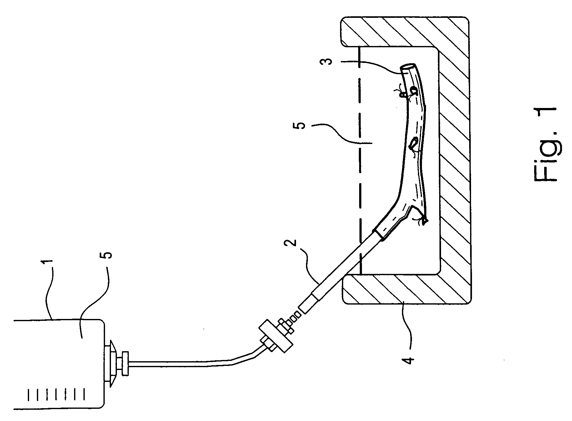
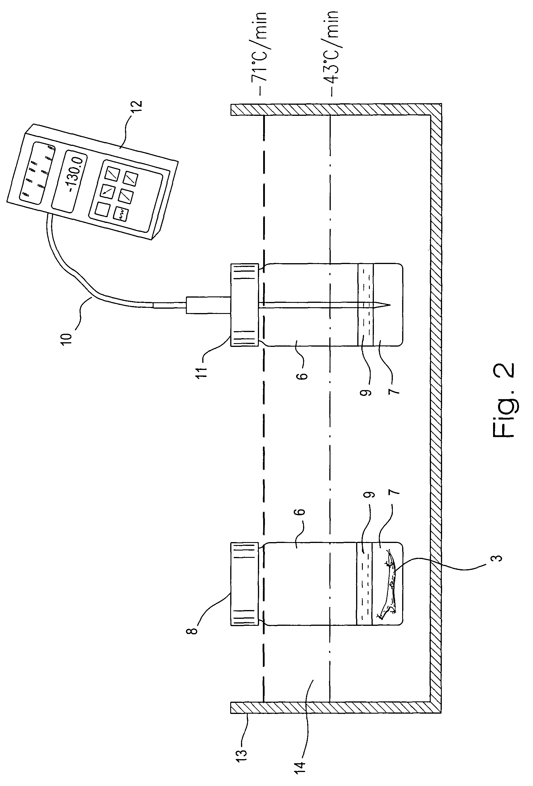
[0073]The external jugular vein was obtained from New Zealand white rabbits. The distal site of each vein segment above the bifurcation was cannulated in situ with a silicone tube. The proximal site was left open for fluid outflow. The vein segments, which were about 40–60 mm long, were perfused at 0° C. to 4° C.

[0074]To perfuse the veins, the perfusion system of FIG. 1 was used. The perfusion system comprises a reservoir 1 (a 60 cc syringe) containing perfusion solution 5 connected to the cannula 2 with a 3-way stopcock. The reservoir 1 was adjusted to physiologic pressure. The vein 3 was placed in a petri dish 4 (Dia.×H, 50×15 mm) containing perfusion solution 5. The perfusion solution 5 in both reservoir 1 and petri dish 4 was the same and was pre-cooled (0° C.–4° C.) and the petri dish 4 was placed in ice (0° C.–4° C.) during the perfusion process.

[0075]The vitrification solution used was VS55. The full strength VS55 solution was introduced in six serial steps. In t...

example 2

Cartilage

[0095]Pig cartilage was used in this study. The sample size was varied from 100–300 mg, and >300 mg. The experiments included fresh control and vitrification groups. The tissue viability was tested using Alamar Blue assay.

[0096]Outbred pigs of either sex, weighing 30–60 kg, were selected in this pilot study. The animal was weighed and then anesthesia was induced with 33 mg / kg ketamine and 1.1 mg / kg acepromazine, and isoflurane was delivered in oxygen via a face mask. After intubation, anesthesia was maintained using isoflurane delivered in oxygen. Pig knee cartilage was harvested for subsequent studies.

[0097]Prior to subzero cooling, the cartilage tissue samples were exposed to the cryoprotectant mixture by adding the vitrification solution VS55 in six steps, for 15 minutes each, on ice (4° C.). This was accomplished in a glass scintillation vial (Dia.×H, 25 mm×60 mm) containing Euro-Collins solution, with successive steps increasing the concentration of cryoprotectant in t...

example 3

Additional Tissues

[0101]Pigs were selected as an animal model for a source of various tissues for testing the feasibility of vitrification for long-term storage of tissues. The sample size was varied from <100 mg and 100–300 mg. These pilot experiments included fresh control and vitrification groups. The tissue viability was tested using the Alamar Blue assay.

[0102]Outbred pigs of either sex, weighing 30–60 kg, were selected in this experiment. The animal was weighed and then anesthesia was induced with 33 mg / kg ketamine and 1.1 mg / kg acepromazine, and isoflurane was delivered in oxygen via a face mask. After intubation, anesthesia was maintained using isoflurane delivered in oxygen. Pig heart valves, myocardium, skin and corneas were harvested for subsequent studies.

[0103]Prior to subzero cooling, the tissue samples were exposed to the cryoprotectant mixture by adding the vitrification solution VS55 in six steps, for 15 minutes each, on ice (4° C.). This was accomplished in a glass...


PUM


Abstract

A method for vitrification of a tissue or organ includes immersing the tissue or organ in increasing concentrations of cryoprotectant solution at a temperature greater than −15° C. to a cryoprotectant concentration sufficient for vitrification; cooling the tissue or organ at an average rate of from 2.5–100° C. per minute to a temperature between −80° C. and the glass transition temperature; and further cooling the tissue or organ at an average rate less than 30° C. per minute to a temperature below the glass transition temperature to vitrify the tissue or organ. After the vitrified tissue or organ has been stored, the tissue or organ may be removed from vitrification by warming the tissue or organ at an average rate of from 20–40° C. per minute to a temperature between −80° C. and the glass transition temperature; further warming the tissue or organ at a rate greater than 80° C. per minute to a temperature above −75° C.; and reducing the concentration of the cryoprotectant. Tissues or organs treated in this manner exhibit near normal functions, for example, blood vessels exhibit near normal smooth muscle contractility and normal graft functions.

Description

[0001]This application is a division of U.S. patent application Ser. No.09 / 691,197, filed Oct. 19, 2000 now U.S. Pat. No. 6,740,484, which in turn is a continuation-in-part of U.S. patent application Ser. No. 09 / 289,977, filed Apr. 13, 1999, now U.S. Pat. No. 6,194,137. The entire contents of these prior applications are hereby incorporated by reference.[0002]The U.S. Government has a paid-up license in this invention and the right in limited circumstances to require the patent owner to license others on reasonable terms as provided by the terms of cooperative agreement No. 70NANB7H3071 awarded by NIST.BACKGROUND OF THE INVENTION[0003]The supply of viable tissues and cells for autologous implantation and heterologous transplantation (hereinafter jointly referred to as transplantation) and study is limited in part by the time a tissue or organ can be maintained in a viable state. Increasing the length of time that a tissue or organ remains viable may drastically increase the likeliho...

Claims


Application Information

Patent Timeline
Patent Type & Authority Patents(United States)
IPC IPC(8): A01N1/02A61K35/12A61K35/44
CPCA01N1/02A61K35/44A01N1/0284A01N1/0221
Inventor KHIRABADI, BIJAN S.SONG, YING C.BROCKBANK, KELVIN G. M.
Owner LIFELINE SCI
Why Eureka
Try Eureka