Methods for structural analysis of proteins

Inactive Publication Date: 2007-10-30
THE BOARD OF TRUSTEES OF THE LELAND STANFORD JUNIOR UNIV
View PDF2 Cites 8 Cited by
  • Summary
  • Abstract
  • Description
  • Claims
  • Application Information

AI Technical Summary

Benefits of technology

[0038]One advantage of the present invention is that the (MPAX) protein footprinting method overcomes many of the limitations of traditional protein footprinting by allowing for use of multiple probes in a single molecule, non-uniform cleavage methods, and lack of data at single amino acid resolution.
[0039]Another advantage of the present invention is that the MPAX method overcomes many of the limitations as to, for example, protein size, solubility, and the requirement for expensive, specialized equipment, which are often required in traditional structure determination methods such as X-ray crystallography and nuclear magnetic resonance.
[0040]Another advantage of the invention is that cysteine residues can be misincorporated at specific amino acid types at different positions within the amino acid sequence. The misincorporation efficiency at each amino acid type can be adjusted using genetic selection to select for mutant misincorporator plasmids.
[0041]Another advantage is the method of the invention requires only microgram quantities of material, is not limited by protein size, and, as such, is ideally suited to proteome-wide structural studies.
[0042]These and other advantages, and features of the invention will become apparent to those persons skilled in the art upon reading the details of the method of protein analysis as more fully described below.

Problems solved by technology

Characterization of the abundance, post-translational modification, structure, and function of these proteins represents a major challenge for genomic analysis.
Defining protein structure in molecular detail constitutes a particularly difficult task.
While X-ray crystallographic and nuclear magnetic resonance techniques are extremely powerful, they require large investments of time and material, and are limited in their application to large protein complexes, membrane proteins and other insoluble or partially folded polypeptides.
Many proteins and protein complexes prove unsuitable for NMR and X-ray work.
In principle, protein footprinting is an option for studying protein structure, but it has been far less successful than the corresponding techniques developed for nucleic acids (Galas D. J., A. Schmitz, Nucleic Acids Res.
Furthermore, protein separation techniques such as SDS-PAGE do not provide the single-monomer resolution of the urea-acrylamide gels used for the separation of nucleic acids, thus complicating the analysis of observed cleavage patterns.
Finally, because of the cooperative nature of protein unfolding, proteolytic cleavage at one site often leads to a global loss of structure and to increased cleavage at other sites in the same molecule, resulting in artifactual data.
In general, these studies have been limited in scope, however, as they examine only a few naturally-occurring residues, or require extensive site-directed mutagenesis to introduce additional structural probes.

Method used


Image

  • Methods for structural analysis of proteins
  • Methods for structural analysis of proteins
  • Methods for structural analysis of proteins

Examples


example 1

Misincorporation of Cysteine for Protein Footprinting

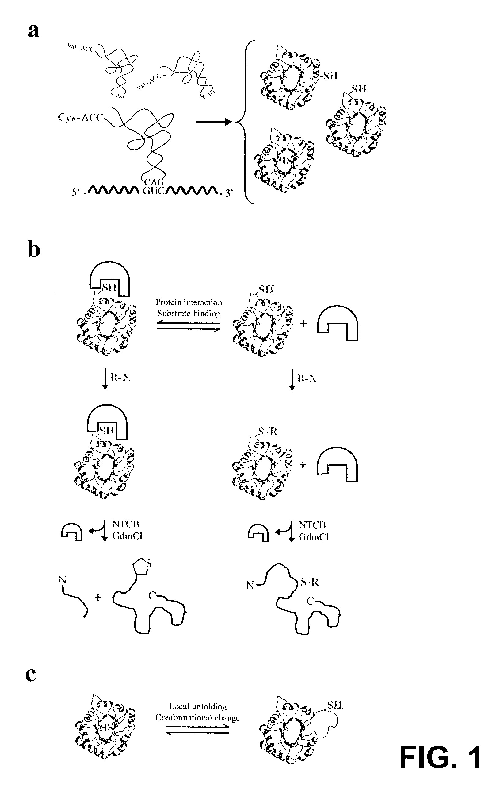
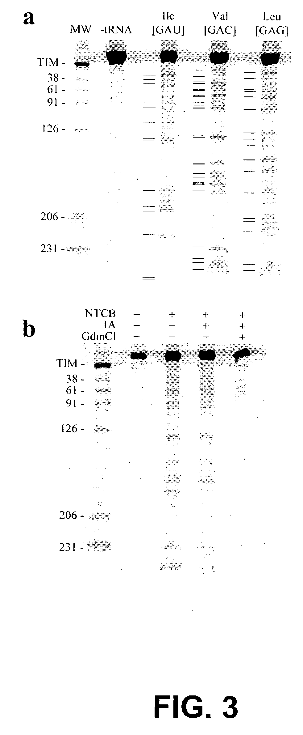
[0225]A cysteine-free variant of yeast triosephosphate isomerase (TIM) was used as a model system to investigate the MPAX technique. TIM is a dimeric (β / α)8 barrel protein. The TIM construct used here includes a C-terminal protein kinase A tag to allow labeling with radioactive phosphate, and an N-terminal 6×His tag on a 30 amino acid linker for purification. This linker shifts the full-length, 290-residue protein away from the fainter cleavage products during gel electrophoresis.

[0226]The misincorporation of cysteine at specific amino acid positions was first verified by expressing TIM in the presence or absence of a series of cysteine misincorporator tRNAs. Cleavage of TIM by NTCB was observed only when TIM was co-expressed with a misincorporator tRNA (FIG. 3, panel A). FIG. 3, panel A is an autoradiograph of an electrophoretic gel which shows that the TIM protein was expressed in the presence of the indicated misincorporator tR...

example 2

Mapping Binding Sites

[0229]Strategies for chemically mapping protein interaction sites fall into three broad categories: interference, crosslinking and protection (Creighton, T. E., Proteins (W. H. Freeman and Company, New York, 1993) pp. 333-334). Interference is based on scanning mutagenesis of a protein. If a residue substitution interferes with a physical interaction, it is inferred that the residue plays a role in binding. In the chemical crosslinking approach, residues that participate in crosslinks are identified, and the existence of the crosslink implies spatial proximity to the binding partner. Finally, protection methods are based on chemical modification of a protein in the presence and absence of a binding partner. Binding sites are identified as residues protected from modification by the presence of the partner. The three techniques provide distinct and complementary information. For example, protection and interference analysis identifies regions of a protein that un...

example 3

Mapping Protein Topology

[0234]Current de novo protein structure prediction algorithms yield multiple reasonable structural models given an input sequence. The inclusion of sparse experimental NMR data in the prediction process significantly improves the accuracy and convergence of the computed models (Bowers, P. M., et. al., J. Biomolecular NMR 18:311, 2000). To address the possibility that data derived from the MPAX method might also be useful for guiding computational structure prediction, the inventors investigated whether the MPAX method could be used to map protein topology.

[0235]The alkylation rates of cysteine residues misincorporated at 61 positions in the TIM sequence were measured. Solvent exposed residues are expected to alkylate more rapidly than buried residues, and thus alkylation rates should be useful for assigning sequence positions to interior or exterior environments. The observed alkylation rates can be interpreted using a kinetic model derived from hydrogen exch...


PUM

PropertyMeasurementUnit
Tertiary structureaaaaaaaaaa
Stabilityaaaaaaaaaa

Abstract

The invention provides methods and compositions for protein structure analysis, including substrate binding sites, sites of protein-protein interactions, three dimensional structure analysis, and stability, all with single amino acid resolution. In general, the subject methods involve introduction of cysteine residues, which serve as probes for physical analysis, into a protein by translational misincorporation in vivo. In many embodiments, proteins containing misincorporated cysteine residues are reacted with a crosslinking agent that covalently links misincorporated cysteine residues to a proximal amino acid in the folded protein. These methods, termed “MXLINK” methods, may be used for protein tertiary structure analysis. In other embodiments, cysteine-misincorporated proteins are used in protein footprinting methods, termed “MPAX” or “MSX” methods.

Description

FIELD OF THE INVENTION[0001]The present invention relates to the field of protein analysis, structural analysis and topology mapping.BACKGROUND OF THE INVENTION[0002]In the last decade, the genomes of over 50 organisms have been sequenced, resulting in a vast increase in the number of known proteins. Characterization of the abundance, post-translational modification, structure, and function of these proteins represents a major challenge for genomic analysis. Understanding protein conformation, interactions and ligand binding is essential to all biological inquiry. Defining protein structure in molecular detail constitutes a particularly difficult task. Accurate measurement of these properties currently requires high-resolution physical methods. While X-ray crystallographic and nuclear magnetic resonance techniques are extremely powerful, they require large investments of time and material, and are limited in their application to large protein complexes, membrane proteins and other i...

Claims


Application Information

Patent Timeline
IPC IPC(8): G01N33/53C07K14/435C12N5/06C12P21/02C12N15/67G01N33/68
CPCC12N15/67G01N33/6803G01N33/6848G01N33/6842G01N33/6818
Inventor HARBURY, PEHR A. B.SILVERMAN, JOSHUA A.MARTIN, WARHAM LANCE
Owner THE BOARD OF TRUSTEES OF THE LELAND STANFORD JUNIOR UNIV
Why Eureka
Try Eureka