Artificial tissue constructs comprising alveolar cells and methods for using the same

a technology of artificial tissue and alveolar cells, which is applied in the field of artificial or manmade models of mammalian lung tissue, can solve the problems of emphysema permanently enlarge, easy damage to alveoli, and inability to fully realize the effect of oxygen-carbon dioxide diffusion,

Active Publication Date: 2014-02-11
SANOFI PASTEUR VAX DESIGN
View PDF59 Cites 3 Cited by
  • Summary
  • Abstract
  • Description
  • Claims
  • Application Information

AI Technical Summary

Problems solved by technology

However, because of the expense of development, coupled with uncertainty regarding efficacy, only about 2% of the global pharmaceutical budget is allocated to development of new prophylactic vaccines (Kiney & Girard (2005) Vaccine 23, 5705-7).
Alveoli have fragile, thin walls, which are easily damaged.
Breakage of these walls makes the oxygen-carbon dioxide diffusion much less efficient.
Once damaged, the bronchioles tend to collapse, trapping stale air in the isolated sacs and not letting fresh air in, leading to atelectasis.
Emphysema permanently enlarges and irreversibly damages the alveoli.
It damages the ends and walls of the smallest bronchioles (the tiny breathing tubes that branch off from the trachea and bronchi), and diminishes pulmonary elasticity.
This need for more air eventually leads to lung over-inflation.
As the lung over-expands, it gradually enlarges, completely filling the maximum thoracic cage volume and causing a sense of shortness of breath.
Because the lung can no longer expand or contract as completely as before, stale air left in the lung is never completely replaced with fresh air, resulting in poor gas exchange.
The combination of a larger, less elastic lung and damaged, non-functioning tissue means that the air flow out of the lung is much slower, resulting in the feeling of an obstructed airway.
In addition to the irreversible damage smoking causes to lung tissue, it causes inflammation of the lungs, which resolves only when smoking stops.
Additionally, irritants contained in tobacco smoke tend to inhibit activity of the cilia of the airways.
Without their activity, it becomes difficult or impossible to cough up the mucus that accompanies pneumonia and other lung infections.
Cigarette smoke can temporarily paralyze the cilia.
However, our limited understanding of the mechanistic details of the respiratory drug delivery has hampered the development of aerosolized therapeutics.
While in vivo animal models have been developed, variations in animal size, species, and differences in therapeutic distribution have resulted in inconsistencies in reported findings by various research groups using such models.
However, the intrinsic differences between the functionality of these cell lines and native primary cells make these systems less than ideal.
However, undesirable changes in cell line characteristics over passage culture have been reported, such as morphology, growth rates, protein expression, permeability, and signaling (ATCC Technical Bulletin no.
However, differences between tissue responses between species have been noted, a possible drawback to using primary animal lung cells (Denham et al.

Method used


Image

  • Artificial tissue constructs comprising alveolar cells and methods for using the same
  • Artificial tissue constructs comprising alveolar cells and methods for using the same
  • Artificial tissue constructs comprising alveolar cells and methods for using the same

Examples


example 1

Fabrication of Bilayer Artificial Tissue Constructs

[0102]In an embodiment of the present invention, artificial tissue constructs were prepared by creating a bilayer cellular structure on top of a collagen and laminin-coated porous plastic membrane. Primary cell isolations from normal human tissue biopsy samples were performed to isolate autologous epithelial and endothelial cells. The tissue was stored in preservation media for up to ˜24 h before cell isolation. The cell isolation protocol was similar to that in previous reports (Bur et al. (2006) Eur. J. Pharm. Sci. 28, 196-203; Elbert et al. (1999) Pharm. Res. 16, 601-8). Enzyme-based tissue digestion was performed with elastase and trypsin, followed by IgG panning to isolate different cell populations.

[0103]Experiments were performed with a series of extracellular matrix proteins and chitosan, a natural polysaccharide, as examples. Specifically, collagen, fibronectin, laminin, Matrigel™, collagen, and chitosan-blended extracellul...

example 2

Characterization of the Constructs

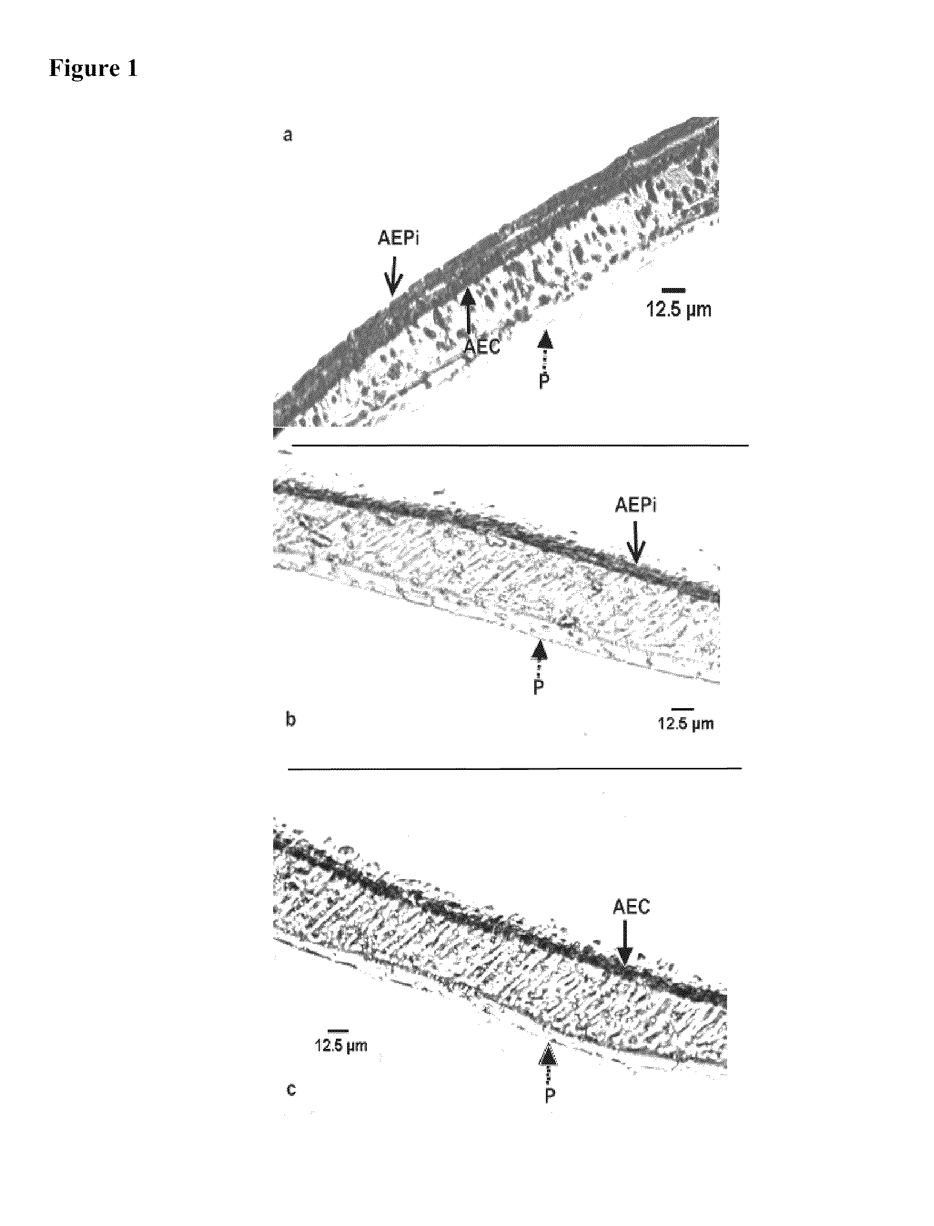
[0114]Immunohistochemical staining of the mucosal construct was used to identify the presence and location of the epithelial and endothelial cells in the bilayer structure. Epithelial cells in the construct were identified by immunohistochemical staining of a number of alveolar cell-specific cytokeratins, including CK-1, 4, 5, 6, 8, 10, 13, 18, 19.

[0115]In the staining method formalin-fixed tissue was embedded in paraffin wax and sectioned at 5 μm. The paraffin wax was then dissolved with xylene and sections were blocked with ˜10% goat serum. A primary cytokeratin antibody cocktail was then added, followed by the addition of the biotinylated secondary antibody. Streptavidin-peroxidase enzyme conjugate was then applied and aminoethyl carbazole (AEC) chromagen was later added to the sections. The samples were counterstained with hematoxylin and rinsed thoroughly with dH2O and incubated until color developed. Light microscopy was used to image the cyto...

example 3

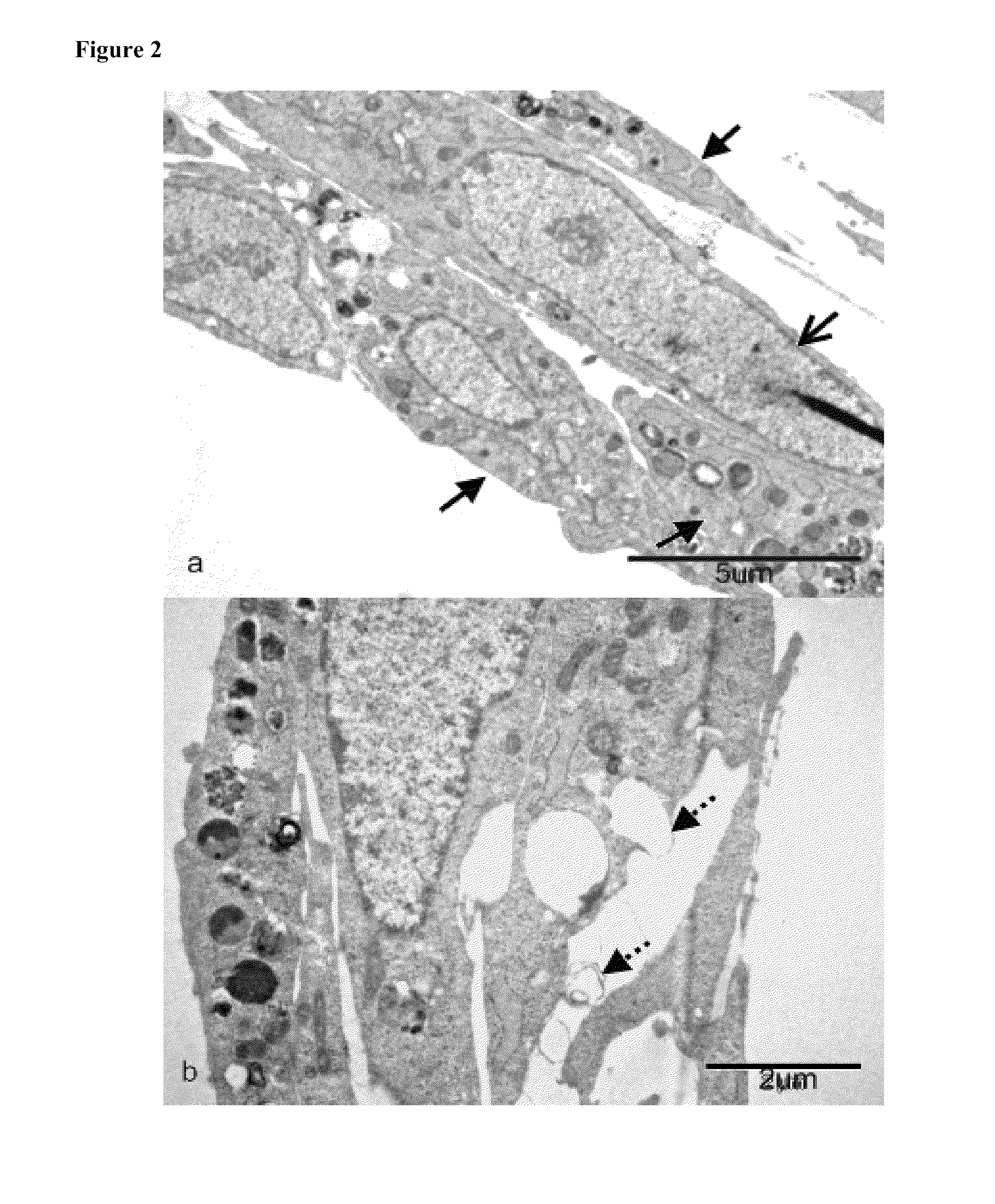
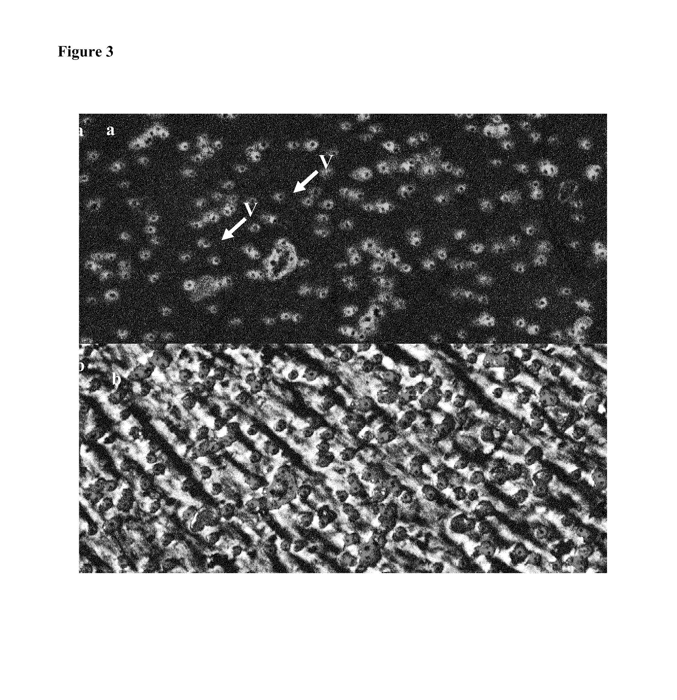
Type II Alveolar Epithelial Cell

[0118]After successfully expanding and co-culturing human epithelial and endothelial cells on laminin-coated membranes and porous scaffolds, cell morphology and activity was examined. In particular, the presence of Type II alveolar epithelial cells was studied. These cells participate in vivo in primary inflammatory and immune reactions in lung mucosa towards external contaminants and pathogens. Type II cells produce a lung surfactant substance that comprises lipoproteins and surfactant proteins able to bind polysaccharides of bacterial membranes, and enhance protective release of oxygen radicals by alveolar macrophages.

[0119]The morphology of Type I and Type II epithelial cells was examined by transmission electron microscopy of epoxy-embedded and microtome-sectioned samples of the mucosal tissue constructs of the present invention. Briefly, cultured epithelial cells were fixed with 2.5% gluteraldehyde and were later exposed to osmium tetroxide. Seri...


PUM

PropertyMeasurementUnit
surface areaaaaaaaaaaa

Abstract

The present invention comprises artificial tissue constructs that serve as in vitro models of mammalian lung tissue. The artificial tissue constructs of the present invention comprise functionally equivalent in vitro tissue scaffolds that enable immunophysiological function of the lung. The constructs can serve as novel platforms for the study of lung diseases (e.g., interstitial lung diseases, fibrosis, influenza, RSV) as well as smoke- and smoking-related diseases. The artificial tissue constructs of the present invention comprise the two components of alveolar tissue, epithelial and endothelial cell layers.

Description

CROSS REFERENCE TO RELATED CASES[0001]This application claims the benefit of U.S. Provisional Application Ser. No. 60 / 949,944, filed Jul. 16, 2007, which is incorporated by reference herein in its entirety.FIELD OF THE INVENTION[0002]The present invention comprises artificial or man-made models of mammalian lung tissue and methods of using the same. The artificial tissue constructs of the present invention are functionally equivalent to in vitro lung tissue scaffolds that enable reliable and predictable modeling of human immunophysiological responses to particular conditions, environments and / or factors. The artificial tissue constructs can serve as novel platforms for the study of lung diseases (e.g., interstitial lung diseases, fibrosis, influenza, RSV, tuberculosis, anthrax, allergies), smoke- and smoking-related diseases, and the effects of particulate inhalation. The artificial tissue constructs of the present invention provide a structural basis for two components of alveolar ...

Claims


Application Information

Patent Timeline
Patent Type & Authority Patents(United States)
IPC IPC(8): G01N33/53C12Q1/02C12Q1/68
CPCC12N5/0688C12N2502/28C12N2533/54G01N2800/12G01N33/5082A61K35/36A61K35/17A61K9/70
Inventor MAHMOOD, AYESHAKACHURIN, ANATOLYWARREN, WILLIAMHIGBEE, RUSSELL
Owner SANOFI PASTEUR VAX DESIGN
Why Eureka
Try Eureka