

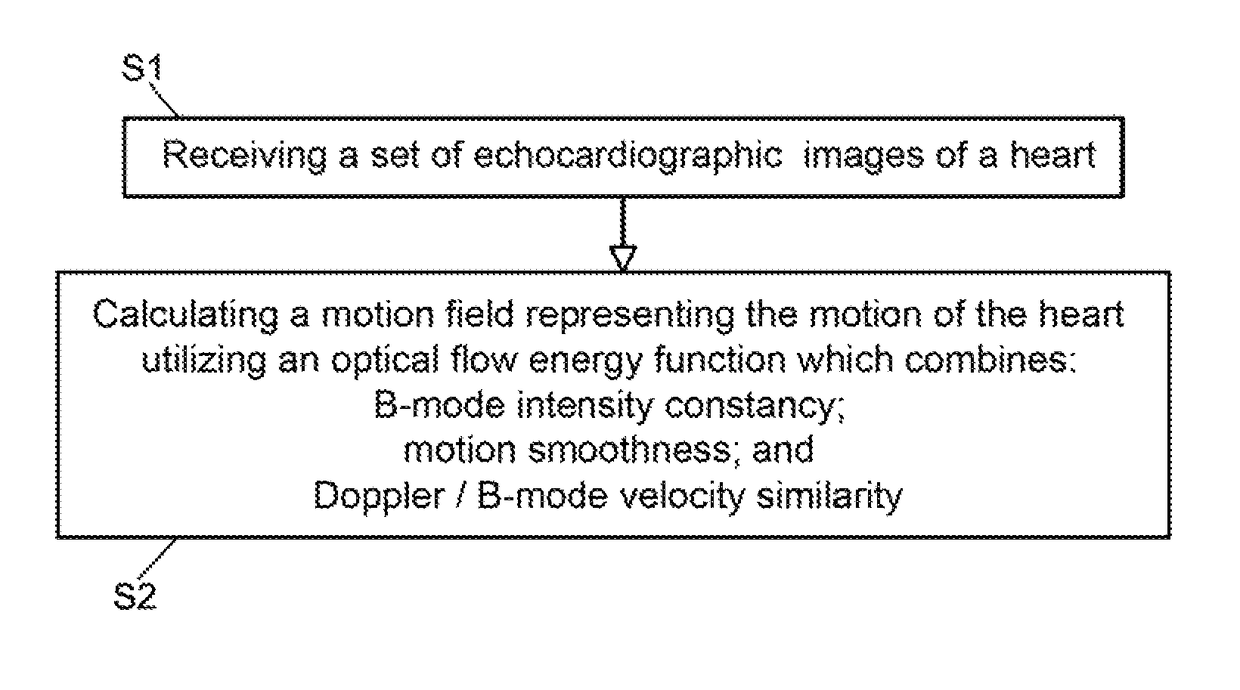
Combined B-mode / tissue doppler approach for improved cardiac motion estimation in echocardiographic images
a tissue doppler and echocardiographic image technology, applied in the field of cardiac motion estimation in echocardiographic images and to a combined bmode/tissue doppler approach for improving cardiac motion estimation performance, can solve the problems of discarded speckle tracking data in the beam direction, and the inability to improve the motion estimation performan
- Summary
- Abstract
- Description
- Claims
- Application Information
AI Technical Summary
Benefits of technology
Problems solved by technology
Method used
Image

Examples
example 1
n Using Simulated Computerized Phantom
[0115]Echocardiographic images are the result of the mechanical interaction between the ultrasound field and the contractile heart tissue. Previously, development and use of an ultrasound cardiac motion simulator was reported [30]. The COLE convolution based simulation technique [31] is currently utilized. The significance of an Ultrasound cardiac motion simulator is the availability of both echocardiographic images as well as the actual ground-truth vector field of deformations.
[0116]A moving 3D heart was modeled based on a pair of prolate-spheroidal representations and used for the ultrasound simulation. The 3D forward model of cardiac motion was simulated using 13 time-dependent kinematic parameters of Arts et al. [32] (see Table 2). The evolution of the 13 kinematic parameters was previously derived by Arts following a temporal fit to actual location of tantalum markers in a canine heart. In Arts' model, seven time-dependent parameters are a...
example 2
n Using Physical Cardiac Phantom
[0120]A physical cardiac phantom was built in-house, suitable for validation of echocardiographic motion estimation algorithms [36]. Here, a brief description of this phantom is provided. To manufacture the phantom, a cardiac computerized model was used to build an acrylic based cardiac mold. A 10% solution of Poly Vinyl alcohol (PVA) and 1% enamel paint were used as the basic material. PVA has the ability to mimic cardiac elasticity, ultrasound and magnetic properties. The solution was heated up to 90 deg. C. Consequently, it was poured into the cardiac mold and gradually exposed to the temperature of −20 C until it froze. The mold and the solution were kept in that temperature for 24 hours. Finally, the mold and the frozen gel were gradually exposed to the room temperature. At this point, the normal heart phantom has passed one freeze-thaw cycle.
[0121]An additional model consisting of the left and right ventricles but with a segmental thin wall in t...
example 3
tudies Validation: Set A: Echocardiography Studies
[0127]Two separate sets of data were utilized for in vivo validations (sets A and B). Set A contained 15 patients and was used for manual tracking validation.
[0128]Data from fifteen subjects who had already undergone echocardiographic imaging as part of their diagnostic evaluation were deidentified and transferred to the laboratory following IRB approval. The data included 13 male, 4 female, average age 52.9 (SD: 7.3). 2D echocardiography (short-axis, long-axis, four-chamber, two-chamber B-mode with TDI. At the University of Louisville Hospital's echocardiography laboratory, Echocardiographic images are acquired with a commercially available system (iE33, Philips Health Care, Best, The Netherlands) using a S5-1 transducer (3 MHz frequency) and the operator is free to change the gain and filter as needed. The full data set included two-chamber, three-chamber, four-chamber, and long-axis views.
PUM
 Abstract
Description
Claims
Application Information