
HIV protease substrate molecular with evolution immune globulin binding molecular, preparation method and application thereof

An immunoglobulin and protease substrate technology, applied in the fields of botanical equipment and methods, biochemical equipment and methods, and applications, can solve problems such as large sequence gaps, unfavorable screening, and decline in drug effectiveness, and achieve improved screening. Efficiency, improved sensitivity, high positive rate

Inactive Publication Date: 2007-10-03
SECOND MILITARY MEDICAL UNIV OF THE PEOPLES LIBERATION ARMY
View PDF0 Cites 4 Cited by
  • Summary
  • Abstract
  • Description
  • Claims
  • Application Information

AI Technical Summary

Problems solved by technology

Its shortcomings are also very clear: (1) Since it competes with existing protease inhibitors for the binding site, only congeners of known protease inhibitors can be screened out. For proteases that cannot bind known protease inhibitors with different sequences Drug screening with drug-resistant proteases is not effective
(2) It is not conducive to the screening of biological macromolecules and other types of drugs such as traditional Chinese medicine
However, due to the limitation of the method itself, the enzyme cleavage site substrates simulated by the above two methods need a short enough distance to work, resulting in the fragment containing the enzyme cleavage site can only be very short and cannot be retained. The natural structural domain around the complete HIV protease target sequence, so the obtained conformation is far from the actual sequence around the HIV cleavage site. When the sequence around the cleavage site affects the protease action, some of the screened out Protease inhibitor candidates may not be effective in the actual environment in vivo, resulting in a decrease in the effectiveness of the screened drug in the HIV cell model

Method used


Image

  • HIV protease substrate molecular with evolution immune globulin binding molecular, preparation method and application thereof
  • HIV protease substrate molecular with evolution immune globulin binding molecular, preparation method and application thereof
  • HIV protease substrate molecular with evolution immune globulin binding molecular, preparation method and application thereof

Examples


Embodiment 1

[0097] Example 1 A novel immunoglobulin-binding molecule with evolved

[0098] Method for producing phage displaying HIV protease target molecule

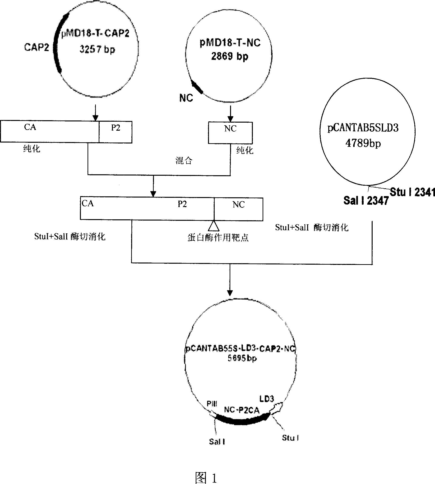
[0099] The construction process of the phage vector pCANTAB5S-LD3-CAP2NC is shown in Figure 1

[0100] Recombinant phagemid vector pCANTAB5S-LD3-CAP2NC containing a cDNA sequence clone displaying the HIV protease target molecule encoding the evolved immunoglobulin binding molecule LD3 by cloning the cDNA sequence of CAP2NC into the StuI and SalI of the phage vector PCANTAB5S-LD3 The specific steps for the preparation of pCANTAB5S-LD3 constructed between the sites are in "Phage Display SpA and PpL Ig Binding Single Domain Random Combinatorial Library and Affinity Screening", Advances in Biophysics and Biochemistry, Volume 32, Issue 6, 2005 It is fully disclosed on pages 535-543.

[0101] 1 Primer Synthesis

[0102] The CAP2NC cDNA sequence was synthesized in vitro by the overlap extension PCR method. Acc...

Embodiment 2

[0180] Example 2 Prokaryotic Preparation and Application of Novel HIV Protease Target Molecules with Evolved Immunoglobulin Binding Molecules

[0181] 1 Construction of prokaryotic expression vectors for protease targets with evolved immunoglobulin-binding molecules

[0182] The construction process of prokaryotic expression vectors expressing prokaryotic target molecules with evolved immunoglobulin binding molecules is shown in Figure 4

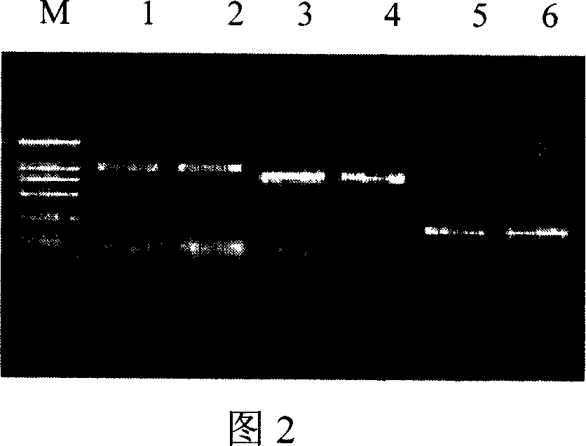
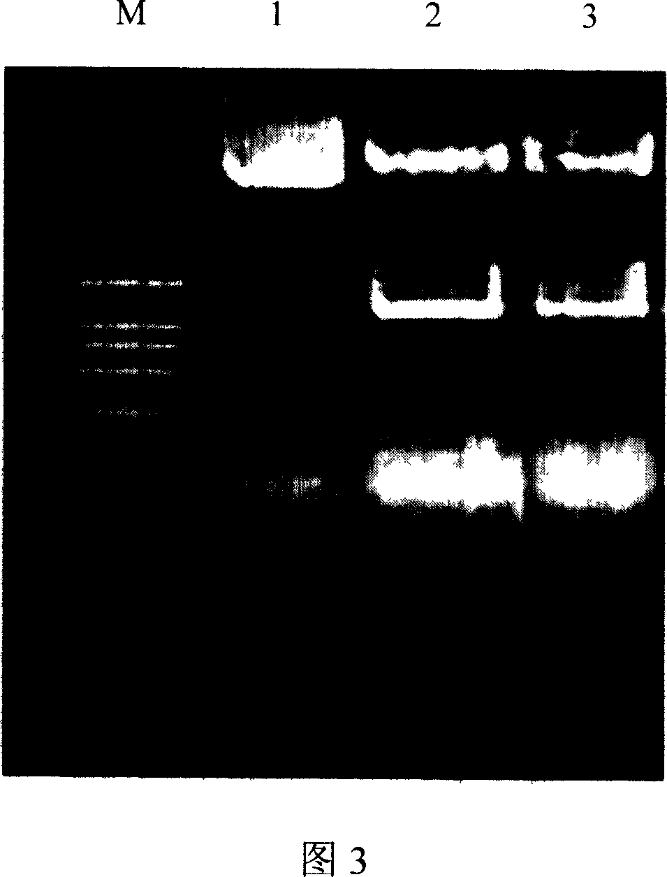
[0183] Using the pCANTAB5S-LD3-CAP2NC plasmid as a template, LCu-bam, C1Ld-6 (LCu-bam (SEQ ID NO: 32): 5'-gggg GGA TCC CCG GCC TCT AGA GAG-3', Shanghai Sangon Biotech Co., Ltd.) The upstream and downstream primers amplify the LD3-CAP2NC DNA fragment respectively, 94°C for 40s; 55°C for 40s; 72°C for 120s; Taq enzyme 0.3μl, 50μl reaction system, 30 cycles. The PCR product was analyzed by 1.0% agarose gel electrophoresis, and the kit solution was recovered, as shown in Figure 5; the recovered product Bam H I and Sal I were double-enzymaticall...

Embodiment 3

[0193] Example 3 HIV Protease Cleaves Phages Displaying HIV Protease Target Molecules with Evolved Immunoglobulin Binding Molecules

[0194] body

[0195] Horseradish peroxidase-labeled mouse anti-M13 phage monoclonal antibody (anti-M13-HRP monoclonalconjugate) was purchased from Amersham Pharmacia.

[0196] Adjust pCANTAB5S-LD3-CAP2NC and pCANTAB5S-LD3 recombinant phage to 1.0×10 with 2×YT culture medium 12 After TU / ml, take 100 μl of bacteriophage respectively, put them in the reaction wells of human IgG antibody coated with the solid phase, incubate at 37°C for 3 hours and wash 10 times. Add different dilutions of HIV SF2 protease to each well, incubate at 37°C for 4h and wash 10 times. After the reaction, add anti-M13 phage enzyme-labeled antibody to each well of the sample well, incubate at 37°C for 45 minutes, add TMB for color development, and measure the D450 value.

[0197] As shown in Table 1 and Figure 10, pCANTAB5S-LD3-CAP2...


PUM

PropertyMeasurementUnit
Titeraaaaaaaaaa

Abstract

This invention relates to system construction, preparation method and application of a new type of HIV proteolytic enzyme cutting model. This invention discloses a recomposed HIV proteolytic enzyme substrate molecule that carry with evolution immunoglobulin binding molecule, it is the protein which amino acid sequential showed as SEQ ID NO: 1 or has conservatism variation. This invention also discloses the preparation method and application of above protein, and the preparation method and application of its bacteriophage. This HIV proteolytic enzyme cutting model be able to used in judging HIV proteinase activity, judging sensibility of HIV proteolytic enzyme to proteolytic enzyme depressor drug, and screening HIV proteolytic enzyme depressor drug.

Description

technical field [0001] The present invention relates to HIV protease substrate molecule, its preparation method and application, in particular to recombinant HIV protease substrate molecule with evolved immunoglobulin binding molecule, its preparation method and application. Background technique [0002] Acquired Immunodeficiency Syndrome (AIDS) caused by human immunodeficiency virus (HIV) infection has become the most serious infectious disease that endangers human health in the world. [0003] Human immunodeficiency virus type 1 (HIV-1) protease comprises 99 amino acids and is active as a homodimer. This enzyme promotes the maturation of virus particles by cleaving HIV gag and gag-pol fusion protein. The cleavage of Gag protein causes the change of virion shape and the concentration of core protein, and the cleavage products are closely related to the formation of virion structure and RNA packaging. The packaging of the protease into the virion and the cleavage event occ...

Claims


Application Information

Patent Timeline
IPC IPC(8): C07K14/435C07K14/16C12N15/12C12N15/48C12N15/63C12N1/21G01N33/68G01N33/573C12N7/01
Inventor 潘卫贾建安周波蒋少华陈秋莉曹洁赵平潘欣温宗梅戚中田
Owner SECOND MILITARY MEDICAL UNIV OF THE PEOPLES LIBERATION ARMY
Learn More