Preparation method of nano biosensor for differentiating different sensitive and drug-resistant cancer cell

A biosensor and cancer cell technology, applied in the field of biosensors, can solve the problems of speeding up the screening and research of tumor markers, poor repeatability of research results, limited chip capture ability, etc., and achieve the effect of easy operation, simple method and stable properties

Inactive Publication Date: 2007-12-12
SOUTHEAST UNIV
View PDF0 Cites 3 Cited by
  • Summary
  • Abstract
  • Description
  • Claims
  • Application Information

AI Technical Summary

Problems solved by technology

SELDI protein chip technology (also known as SELDI-TOF-MS technology) has greatly accelerated the screening and research of tumor markers, and has very important clinical application value in the early diagnosis of tumors, but there are also some shortcomings, such as operation The process is cumbersome and complicated, the capture ability of the chip is limited, the difference between chip batches and the low resolution of mass spectrometry make the reproducibility of the research results poor, and the obtained peptide peaks are few.

Method used


Image

  • Preparation method of nano biosensor for differentiating different sensitive and drug-resistant cancer cell
  • Preparation method of nano biosensor for differentiating different sensitive and drug-resistant cancer cell
  • Preparation method of nano biosensor for differentiating different sensitive and drug-resistant cancer cell

Examples


Embodiment 1

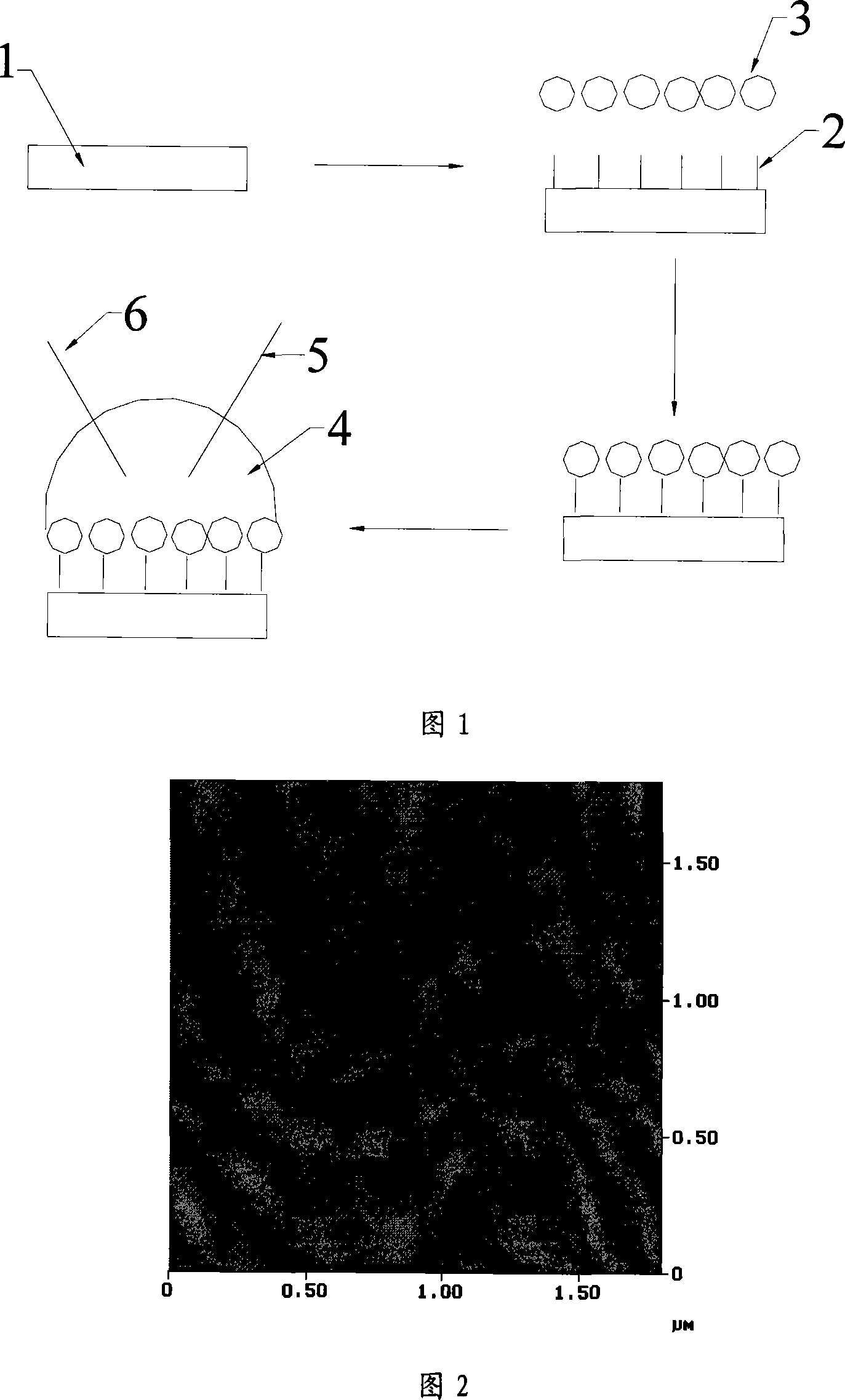
[0022] A method for preparing a nano-biosensor for differentiating sensitive and drug-resistant cancer cells, comprising the following steps:

[0023] The first step is to generate hydroxyl groups on the surface of the electrode: first, the glassy carbon electrode is polished to a mirror surface on the suede with 0.05 μm aluminum powder, cleaned in acetone, absolute ethanol, and ultrapure water, and dried, and then placed in a pH In the 5.0 phosphate buffer solution (PBS), the voltage was set at 1.70V, and the electrochemical multi-potential step method was used for 280s. After the treatment, the cyclic voltammetry was scanned continuously for 20 circles in the positive potential range of 0.3V-1.25V. Then cyclic voltammetry scans continuously for 20 laps in the negative potential range of -1.3V ~ 0.3V. After scanning, take it out, rinse with distilled water, and dry it; the second step, nanoparticle modification: the aqueous solution of functionalized gold nanoparticles Mix wi...

Embodiment 2

[0025] A method for preparing a nano-biosensor for differentiating sensitive and drug-resistant cancer cells, comprising the following steps:

[0026] The first step is to generate hydroxyl groups on the surface of the electrode: first, the glassy carbon electrode is polished to a mirror surface on the suede with 0.05 μm aluminum powder, cleaned in acetone, absolute ethanol, and ultrapure water, and dried, and then placed in a pH In the 5.0 phosphate buffer solution (PBS), the voltage is set at 1.80V, and the electrochemical multi-potential step method is used for 320s. After the treatment, the cyclic voltammetry scans continuously for 20 circles in the positive potential range of 0.3V ~ 1.25V. Then cyclic voltammetry scans continuously for 20 laps in the negative potential range of -1.3V~0.3V. After scanning, take it out, rinse it with distilled water, and dry it; the second step, nanoparticle modification: the aqueous solution of polylactic acid nanofibers Mix volume ratio 1...

Embodiment 3

[0028]A method for preparing a nano-biosensor for differentiating sensitive and drug-resistant cancer cells, comprising the following steps:

[0029] The first step is to generate hydroxyl groups on the surface of the electrode: first, the gold electrode is polished to a mirror surface on the suede with 0.05 μm aluminum powder, washed in acetone, absolute ethanol, and ultrapure water, dried, and then placed in a pH 5.0 In the phosphate buffer solution (PBS), the voltage is set at 1.70V, and the electrochemical multi-potential step method is used for 280s. After the treatment, the cyclic voltammetry scans continuously for 20 circles in the positive potential range of 0.3V-1.25V, and then Continuously scan 20 laps by cyclic voltammetry within the negative potential range of -1.3V to 0.3V, take it out after scanning, rinse with distilled water, and blow dry;

[0030] The second step, nanoparticle modification: mix the aqueous solution of polylactic acid nanofibers in a volume rat...


PUM


Abstract

The making of nanometer biological sensor separating sense and drug fast cancer comprises the electrode surface generating with hydroxyl group, polishing the electrode into mirror plane, cleaning in acetone, water free alcohol and pure water, drying and processing with multi electric potential step phase by putting it into the PBS solution with PH of 5.0, modifying the nanometer grain, mixing the functional metal nanometer grain or polymer fiber in the dimension of 1:1 and PH=7.2, adding 0.5mg carbamate, dipping the pretreated electrode in the modifying solution to get the nanometer sensor after 10 hours. It can be used to test various cancer cell to separate the sensitive and drug fast cancer cell.

Description

technical field [0001] The invention relates to a biosensor based on a nano-interface, in particular to a preparation method of a nano-biosensor for differentiating sensitive and drug-resistant cancer cells. Background technique [0002] Cancer has become one of the biggest killers that endanger human health today, and how to effectively diagnose and treat it has always been a difficult problem that the medical field has been working hard to overcome. After more than half a century of development and improvement, surgery, radiotherapy, chemotherapy and other methods have become the main means of comprehensive treatment of malignant tumors, but they often cause serious side effects in clinical application, and the therapeutic effect is not very good. A large number of clinical facts have proved that the cure rate of early cancer is quite high. Therefore, the key to treating cancer lies in effective early diagnosis and treatment. At present, the most effective means of diagn...

Claims


Application Information

Patent Timeline
Patent Type & Authority Applications(China)
IPC IPC(8): G01N27/327
Inventor 王雪梅何芳周謇郭大东陈晨
Owner SOUTHEAST UNIV
Why Eureka
Try Eureka