Antineoplastic drug evaluation and screening method based on cell microscopic image information

An anti-tumor drug, microscopic image technology, applied in biochemical equipment and methods, microbial determination/inspection, particle and sedimentation analysis, etc., can solve the problems of low degree of automation, limited applicable cell types, poor sensitivity, etc.

Inactive Publication Date: 2008-03-26
ZHEJIANG UNIV
View PDF0 Cites 24 Cited by
  • Summary
  • Abstract
  • Description
  • Claims
  • Application Information

AI Technical Summary

Problems solved by technology

At present, MTT and SRB methods are the most common and commonly used cytotoxicity test methods. Many researchers have adopted this method for drug screening. However, these methods have defects such as slow speed, poor sensitivity, and low degree of automation. The need for throughput drug screening
Other methods for cytotoxicity detection include HPLC detection, isotope labeling detection, and flow cytometry, etc. These methods have disadvantages such as cumbersome means, radioactive contamination, and limited applicable cell types.

Method used


Image

  • Antineoplastic drug evaluation and screening method based on cell microscopic image information
  • Antineoplastic drug evaluation and screening method based on cell microscopic image information
  • Antineoplastic drug evaluation and screening method based on cell microscopic image information

Examples


Embodiment 1

[0055] Example 1 Drug Screening and Evaluation System Design of Cell Microscopic Image Information

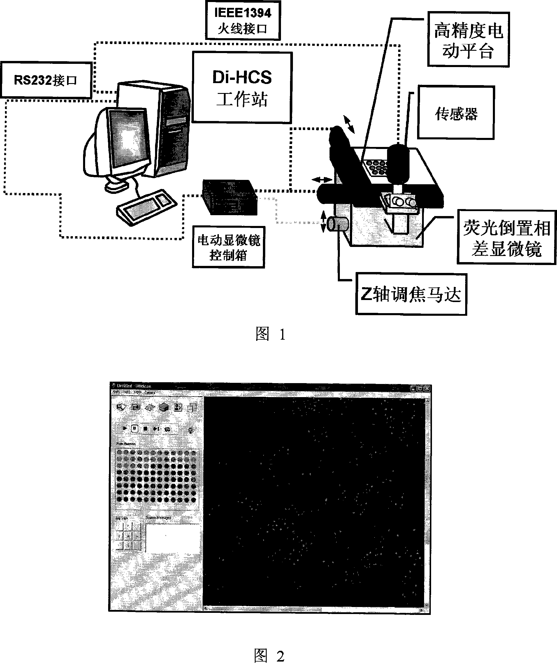
[0056] Referring to Fig. 1, it is the framework of the screening and evaluation system of the present invention, which mainly includes: a high-precision electric water cloud platform, a fluorescent vision system, an image acquisition and processing system, and a workstation.

[0057] Referring to Figure 1 and Figure 2, the main functions and performance indicators of each part of the drug screening and evaluation system based on cell microscopic image information are:

[0058] (1) The high-precision electric water cloud platform is used to load multiple cell sample objects treated by different drugs required for the test. The platform is controlled by a stepping motor and can move precisely with a specified step in the X-Y direction. , which can realize the program control, and send the observation samples in the designated holes to the microscope observation place. The minimu...

Embodiment 2

[0062] Example 2 FDA staining method for measuring the number of living cells

[0063] 1. Experimental method

[0064] Accurately weigh 1mg of FDA and dissolve it in 0.1ml DMSO to make FDA stock solution, dispense it into 0.5ml centrifuge tubes, and store at -20°C. Before use, dilute the FDA stock solution 1000 times with PBS for use.

[0065] a) FDA staining to measure the linear range of viable cell number method

[0066] Take KB cells in the logarithmic growth phase, digest with EDTA-trypsin mixed solution, and divide 5×10 4 , 1×10 4 , 5×10 3 , 1×10 3 , 2×10 2 , 4×10 1 The density of cells / well was seeded on a 96-well culture plate, each well contained 100 μL of culture solution, and 6 replicate wells were set up for each cell density, at 37°C, 100% humidity, containing 5% CO 2 and 95% air conditions for 24h. Discard the culture medium in the wells before the test, add 50 μL of FDA PBS solution to each well, incubate at 4°C for 30 minutes, and use the screening and...

Embodiment 3

[0086] Example 3 Hoechst 33342 and propidium iodide (PI) double staining method to measure drug-induced apoptosis

[0087] 1. Experimental method

[0088] Take KB cells in the logarithmic growth phase, digest with EDTA-trypsin mixed solution, and dilute 2×10 3 The density of cells / well was seeded on a 96-well culture plate, each well contained 100 μL of culture solution, at 37°C, 100% humidity, 5% CO 2 After culturing for 24 hours under the condition of 95% air, discard the culture medium. Different concentrations of vincristine (2×10 -7 , 2×10 -6 M) Adding to the wells, 4 parallel wells were set up for each drug concentration, and 200 μL of culture solution was added to each well. After incubation for 48 h, the culture medium was discarded, and 50 μL of apoptosis and necrosis detection reagents (50 μL of C1056-1 cell staining buffer, 0.25 μL of C1056-2 Hoechst 33342 staining solution, and 0.25 μL of C1056-3 PI staining solution) were added to each well. After incubating ...


PUM


Abstract

The invention provides the appraisal and selective method for the antineoplastic drug based on the cell micrograph information, which uses a sort of selection and appraisal hardware system to appraisal and select the antineoplastic drug by different fluorescence dye marking and measuring the multicellular parameter change in cell. The hardware system is made up of the high precision electric hydrous platform, the fluorescence vision system, the image collecting and processing system and working station. The diacetoxyl fluoresceine dyeing measures the active cell number; the double dyeing method of Hoechst33342 and iodized pyridine appraise the drug inducing the cell die; the three dyeing method of FDA, Hoechst33342 and PI analyzes the die mode induced by drug. The invention can measure at least two kinds of single cell or cell subgroup which expresses different drone cell organ. The method is in reason and can be used in study of drug action mechanism and selecting the high hedonic drug and toxicity analyzing, which can be used in selecting drug and appraising the drug toxicity.

Description

technical field [0001] The invention belongs to the field of drug screening methods, and relates to the application of automatic technology of cell microscopic image information, optical marking and optical measurement technology to record cell images in a porous plate, and to obtain indicators for screening and evaluating anti-tumor compounds by analyzing image information. Background technique [0002] Cancer seriously threatens people's health, among which lung cancer, liver cancer, breast cancer, and leukemia are several cancers with high incidence rates. Lung cancer has become the largest cancer in my country, and its morbidity and mortality are increasing most rapidly. The incidence of liver cancer is second only to lung cancer, and the incidence of liver cancer in some areas is increasing year by year. About 260,000 patients die of liver cancer every year in the world, and nearly 100,000 in my country. Statistics show that the incidence of breast cancer in almost all...

Claims


Application Information

Patent Timeline
Patent Type & Authority Applications(China)
IPC IPC(8): G01N15/10G01N21/64C12Q1/18
Inventor 程翼宇周晨光黄欣
Owner ZHEJIANG UNIV
Why Eureka
Try Eureka