Method for detecting blood serum tumor markers

A tumor marker and detection method technology, applied in biological tests, measurement devices, material inspection products, etc., can solve the problem of lack of specific tumor markers for early screening of pancreatic cancer, and achieve improved cure rate, reasonable design, The effect of high specificity and sensitivity

Inactive Publication Date: 2009-02-11
ZHEJIANG UNIV
View PDF0 Cites 5 Cited by
  • Summary
  • Abstract
  • Description
  • Claims
  • Application Information

AI Technical Summary

Problems solved by technology

There is still a lack of specific tumor markers for early screening of pancreatic cancer

Method used


Image

  • Method for detecting blood serum tumor markers
  • Method for detecting blood serum tumor markers
  • Method for detecting blood serum tumor markers

Examples


Embodiment 1

[0019] Preparation of serum samples|:

[0020] Main instruments and reagents: CM10 protein chip

[0021] 96-well protein chip working support is Bio-proeessor

[0022] Analysis of pure urea, sodium acetate, acetonitrile, DTT and cHAPS buffer salt (sigma company)

[0023] Energy absorbing molecule SPA,

[0024] Implementation steps: take 1ml of whole blood, centrifuge at 4°C low temperature (4000 rpm) for 10 minutes, take the supernatant, centrifuge again for 10 minutes, and take the supernatant serum. Add 5 μL of serum to 10 μl of U9 serum treatment solution, shake on a shaker to mix thoroughly. U9-treated serum was diluted to 200 μl with Binding Buffer for loading.

[0025] Take 100 μl of the above-mentioned U9-treated and diluted serum and add it to the chip, and remove the sample after the chip fully reacts with the serum for 1 hour. Add 200 μl of corresponding Binding Buffer to each well, shake on a shaker for 5 minutes (600 rpm, room temperature), and then remove the...

Embodiment 2

[0027] Embodiment 2 On-machine detection:

[0028] Main instruments and software: PBS-II surface enhanced laser desorption ionization-time-of-flight mass spectrometer (SELDI-TOF-MS),

[0029]Analysis software ProteinChipSoftware3.0 (Ciphergen, USA): Support vector machines (supportveetor machines, SVM), discriminant analysis (discriminant analysis) and time-sequence analysis (time-sequence analysis) software are developed using Matlab6.5 data processing software (MathworkS company) ;

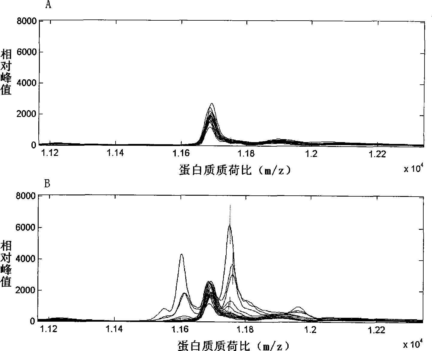
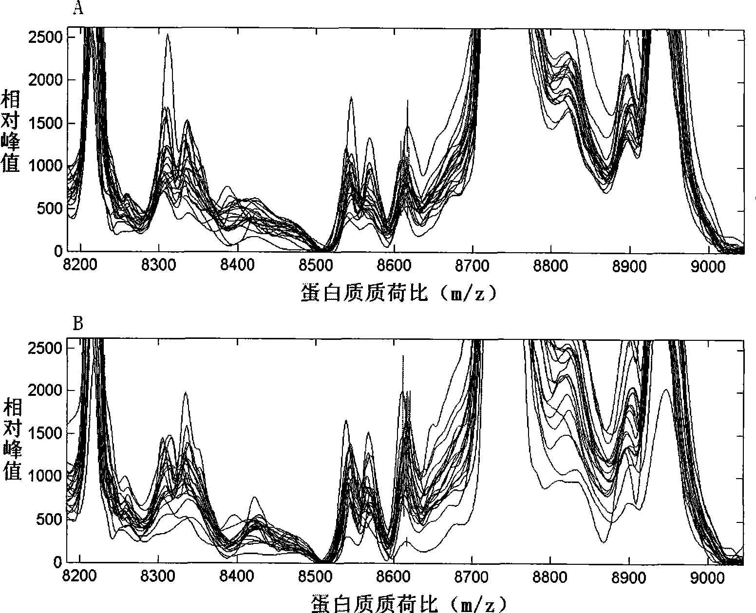
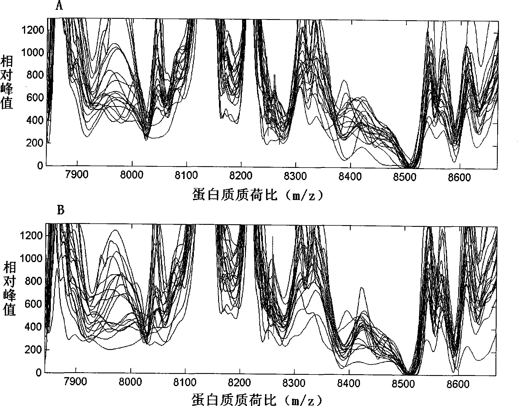
[0030] Implementation steps: mass spectrometer parameter setting: laser intensity 165, sensitivity 7, data collection range 2000-30000m / z (protein mass and charge ratio, ie mass-to-charge ratio), molecular weight is corrected with a standard protein chip before each data collection. Serum samples were detected by surface-enhanced laser desorption ionization-time-of-flight mass spectrometry to obtain serum protein profiles.

Embodiment 3

[0031] Example 3 Data Collection and Bioinformatics Analysis

[0032] The original data was first corrected with Proteinchip Software3.0 software to make the total ionic strength and molecular weight uniform, and to filter the noise. The initial noise filtering value was 5, and the secondary noise filtering value was 2. Clustering was performed with 10% as the minimum threshold value. After the above data preprocessing, the Mann-Whitney rank test was used to compare the serum protein mass spectrometry data of each group (completed by the Biomarker Wizard3.2.0 software that comes with Proteinchip Software3.0), to find protein peaks with differences in expression among the groups.

[0033] The data of serum protein mass spectrometry comparison in each group were imported into the SVM and discriminant analysis software of Matlab6.5 data processing software. The 10 protein peak data with the most significant difference between the two groups were selected for analysis. The combin...


PUM


Abstract

The invention provides a method for detecting the totem of serum tumor. The composition difference of serum protein is analyzed by utilizing the surface-enhancement laser desorption ionization-flight time mass-spectrometric technique, and a serum protein finger print of pancreatic cancer is established. A mass spectrometric model is formed by the serum protein with three mass / charge ratios at 11753Da, 8615Da and 8256Da, and the tumor totem of pancreatic cancer is analyzed by the bioinformatics method. The tumor totem of pancreatic cancer with high diagnostic efficiency is screened by utilizing the SELDI-TOF-MS technique and the bioinformatics method. The method provides an animal experiment base for clinically diagnosing pancreatic cancer, has high sensitivity, specificity and accuracy, and can improve and modify the protein finger print technique. The method has a reasonable design, and is used for reducing the national fatality rate of pancreatic cancer, improving the curative ratio of pancreatic cancer and further screening pancreatic cancer.

Description

technical field [0001] The invention belongs to biological technology, and relates to a method for detecting serum of a nude mouse pancreatic cancer model by using proteomics to obtain serum tumor markers, which provides animal experiment basis for early diagnosis of pancreatic cancer. Background technique [0002] Pancreatic cancer is one of the most malignant tumors characterized by its insidious onset, high degree of malignancy, low surgical resection rate, and extremely poor prognosis. It ranks sixth in the cancer mortality rate in my country, and its 5-year survival rate is only 1%-4%. Radical surgery is currently the only effective method for the treatment of pancreatic cancer. Combined with chemotherapy after surgery, the 5-year survival rate can reach 15%-40%. But clinically, only 10%-15% of patients have the chance of radical resection, and about 85% of pancreatic cancer patients have lost the chance of radical surgery due to metastasis and other reasons when they ...

Claims


Application Information

Patent Timeline
Patent Type & Authority Applications(China)
IPC IPC(8): G01N33/48G01N27/64
Inventor 曹利平刘达人汪亮王贵锋阙日升
Owner ZHEJIANG UNIV
Why Eureka
Try Eureka