A medical equipment carrying extracellular matrix and its production method

A technology of medical devices and extracellular matrix, which is applied in the field of medical devices to achieve the effect of promoting endothelialization, reducing restenosis rate, and accelerating surface endothelialization

Active Publication Date: 2009-10-07
LEPU MEDICAL TECH (BEIJING) CO LTD
View PDF2 Cites 17 Cited by
  • Summary
  • Abstract
  • Description
  • Claims
  • Application Information

AI Technical Summary

Problems solved by technology

[0010] To sum up, how to choose a suitable extracellular matrix and how to firmly load an effective amount of extracellular matrix on the surface

Method used


Image

  • A medical equipment carrying extracellular matrix and its production method
  • A medical equipment carrying extracellular matrix and its production method
  • A medical equipment carrying extracellular matrix and its production method

Examples


Embodiment 1

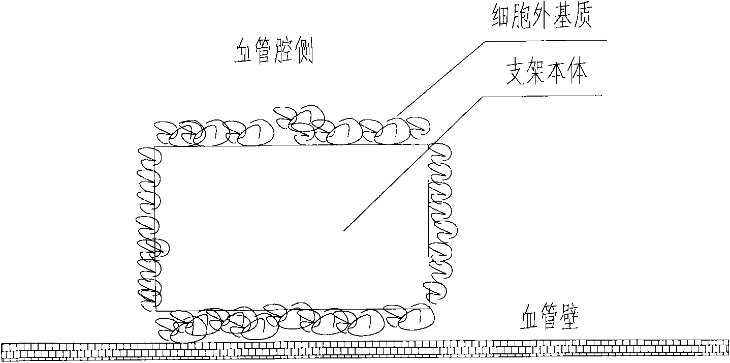
[0049] Preparation of scaffolds carrying extracellular matrix (collagen I and laminin 1), the cross-sectional view of which is shown in figure 2 Shown:

[0050] (1) The bare 316L stainless steel stent was cut, slag-removed, polished, placed in 75% medical alcohol, cleaned with ultrasonic waves at a frequency of 30khz for 15 minutes, set at 40°C, dried for 60 minutes, and then taken out.

[0051] (2) Put the scaffold into a 5mL glass test tube (1 support corresponds to 1 test tube), then add 1mL collagen I (concentration: 150μg / mL, solvent: 12M HCl) to the glass test tube, and add 1mL laminin at the same time 1 (the concentration is 10 μg / mL, the solvent is 0.1 MCBC buffer solution, pH 9.0), shake at 4°C for 1 hour, and take it out.

[0052] (3) The scaffolds coated with collagen I and laminin 1 were placed in an ultraviolet curing spotlight DymaxTM Bluewave 200 for 30 seconds, then rinsed and dried with deionized water, and dried at room temperature.

Embodiment 2

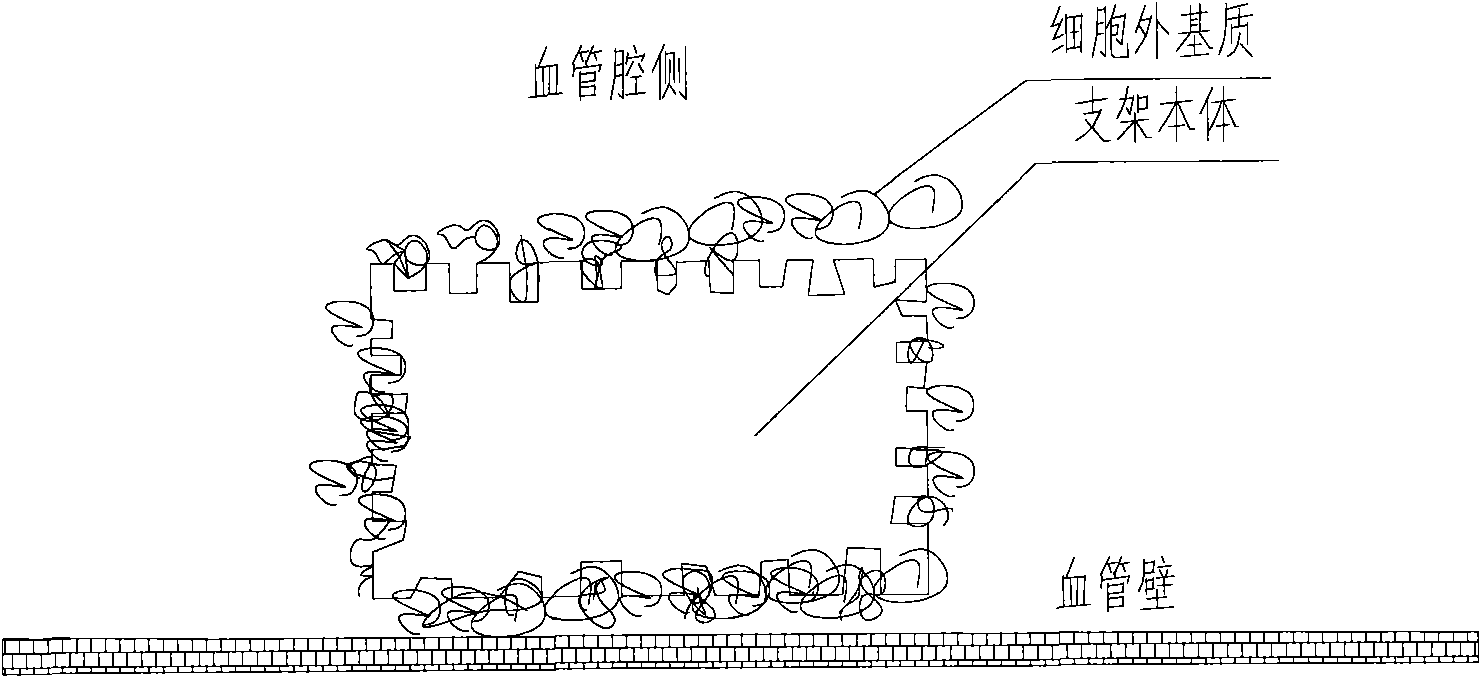
[0054] Preparation of extracellular matrix (collagen I and laminin 5) coated scaffolds, the cross-sectional view of which is shown in image 3 Shown:

[0055] (1) The bare 316L stainless steel stent was cut, slag-removed, polished, placed in 75% medical alcohol, cleaned with ultrasonic waves at a frequency of 30khz for 15 minutes, set at 40°C, dried for 60 minutes, and then taken out. Then place the bracket in ~38% hydrochloric acid for 12 hours of corrosion, connect the bracket body as the anode to the positive pole of the pulse power supply, connect the titanium metal sheet to the negative pole of the pulse power supply as the cathode, and place the bracket body and the cathode metal sheet at the same time. In 28% hydrochloric acid, the current is set to 20A, the frequency is 2000 Hz, and the time is 5 minutes, holes are prepared on the surface of the 316L bare metal stent body.

[0056] (2) Put the scaffold into a 5mL glass test tube (1 support corresponds to 1 test tube),...

Embodiment 3

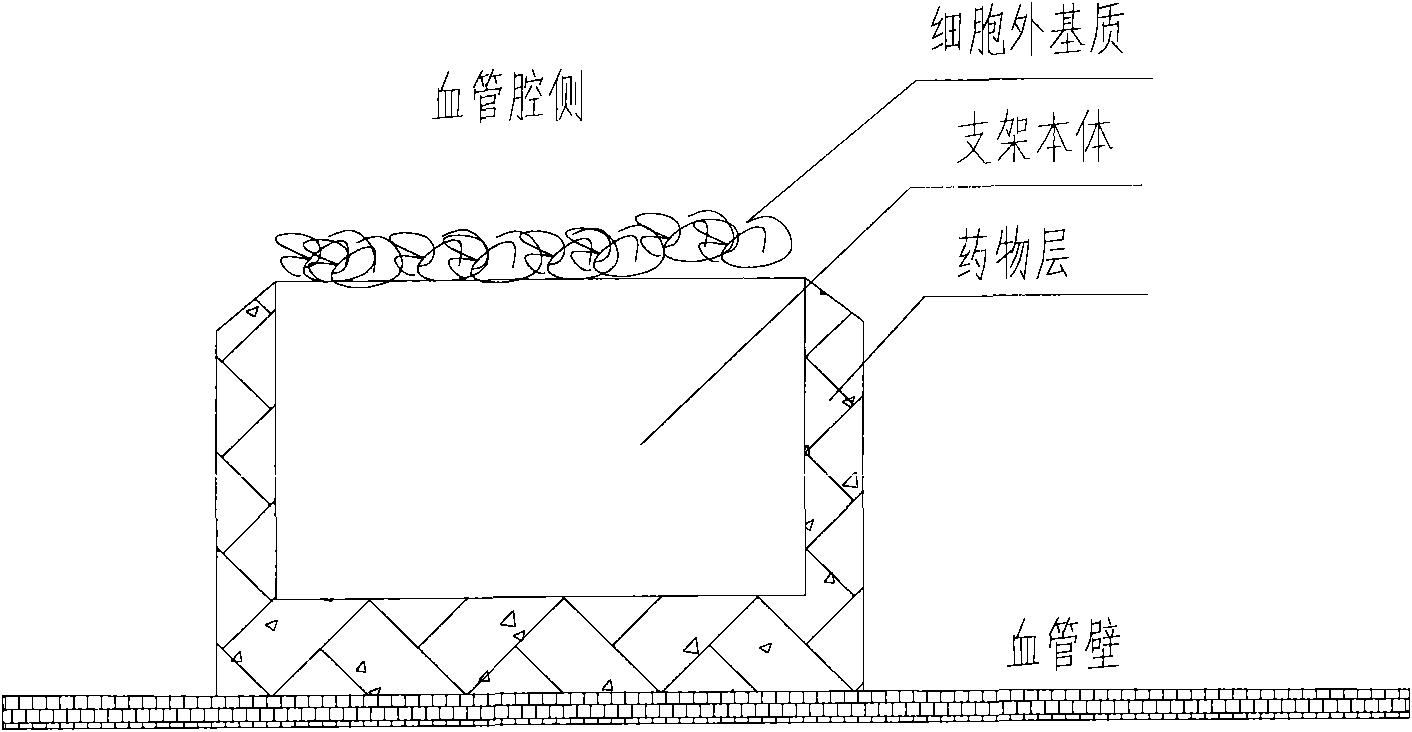
[0058] Prepare a kind of extracellular matrix (collagen I and laminin 1) drug-loaded (rapamycin) coated stent, its cross-sectional view is as follows Figure 4 Shown:

[0059] (1) The bare 316L stainless steel stent was cut, slag-removed, polished, placed in 75% medical alcohol, cleaned with ultrasonic waves at a frequency of 30khz for 15 minutes, set at 40°C, dried for 60 minutes, and then taken out.

[0060] (2) Add 0.8g polylactic acid into 10ml tetrahydrofuran solution, add 0.2g rapamycin after dissolving, then spray on the surface of the stent body, and solidify in the air for 60min, so that the rapamycin drug loading capacity of the stent reaches 1.2 μg / mm2.

[0061] (3) Then put the prepared drug stent into a 5mL glass test tube (one stent corresponds to one test tube), and then add 1mL collagen I (concentration: 150μg / mL, solvent: 12M HCl) into the glass test tube, and at the same time Add 1 mL of laminin 1 (concentration: 10 μg / mL, solvent: 0.1 M CBC buffer, pH 9.0)...


PUM


Abstract

The invention relates to a kind of medical equipment carrying extracellular matrix and its production method. It includes the medical equipment body and the layer of extracellular matrix coating on the surface of the medical equipment body. The described extracellular matrix is collagen, laminin, non-collagenous glycoprotein, GAG and proteoglycan, elastin, and one or multiple endothelial cell extracellular matrixes obtained through cell treatment from the cultured endothelial cells. For the described medical equipment in the invention, the extracellular matrix on the body surface can withstand the wash of blood and other body fluids and advance the vascular endothelialization through slow release; if working together with the drug, it can not only reduce the restenosis rate, but also speed up the surface endothelialization to improve treatment; the preparation method of the invention is simple and may have no carrier. It uses the electrostatic and/or micropore adsorption principle to coat the extracellular matrix directly on the body surface, thus it can prevent the side effects such as the inflammation brought by the carrier.

Description

technical field [0001] The invention relates to a medical device, in particular to a medical device carrying an extracellular matrix and a preparation method thereof. Background technique [0002] In 1987, Sigwart et al. used intravascular metal stents for coronary arteries for the first time, which provided a good way for the treatment of vascular occlusion diseases. However, in-stent restenosis has always been the main reason affecting the efficacy of percutaneous coronary intervention (PCI). With the listing of Johnson & Johnson's Cypher rapamycin drug stent in 2004 and Boston Scientific's Taxus paclitaxel drug stent in 2005, drug-eluting stents have reduced the in-stent restenosis rate from 30% in the bare metal stent era to less than 10%. [0003] However, with the in-depth study of drug stents, the continuous expansion of the scope of application, and the gradual increase in the number of accumulated cases, people should become more rational in their use and have a cl...

Claims


Application Information

Patent Timeline
IPC IPC(8): A61F2/82A61F2/06A61F2/24A61M25/00A61F2/02A61N1/36A61N1/02A61N1/39A61B17/12A61B17/122A61M1/14A61B17/08A61L31/16A61M31/00
Inventor 余占江陈韵岱朱华刚张正才邱笑违
Owner LEPU MEDICAL TECH (BEIJING) CO LTD
Why Eureka
Try Eureka