Method for treating externally-autoplastic autogenous skull sheet

A technology of autologous transplantation and treatment method, which is applied in the field of biomaterials and can solve problems such as rejection, clinical application failure, and inability to fully meet clinical needs.

Inactive Publication Date: 2012-07-18
THE FIRST AFFILIATED HOSPITAL OF HENAN UNIV OF SCI & TECH
View PDF3 Cites 2 Cited by
  • Summary
  • Abstract
  • Description
  • Claims
  • Application Information

AI Technical Summary

Problems solved by technology

The popularization and use of artificial materials is limited due to their deficiencies in shaping, pressure resistance, impact resistance, temperature insulation, rejection, and high price (about 20,000 yuan per repair material).
The advantages of autogenous skull repair are difficult to achieve with other repair materials. The physical characteristics of the skull flap fully meet the anatomical and physiological needs of the human body, and the bone induction activity is good. Comparatively speaking, it is the most ideal repair material, but it can meet the requirements of clinical application. There are large technical obstacles in the preparation of autologous skull flap repair materials, which limit its application; there are two methods of preservation of autologous skull flap: in vivo or in vitro
The disadvantage of in vivo preservation is that the trauma is large, the bone flap can be absorbed and becomes smal

Method used


Image

  • Method for treating externally-autoplastic autogenous skull sheet
  • Method for treating externally-autoplastic autogenous skull sheet
  • Method for treating externally-autoplastic autogenous skull sheet

Examples


Embodiment Construction

[0025] Below in conjunction with accompanying drawing and specific embodiment the present invention is described in further detail:

[0026] The present invention can be explained in more detail by the following examples, and the purpose of disclosing the present invention is intended to protect all changes and improvements within the scope of the present invention, and the present invention is not limited to the following examples;

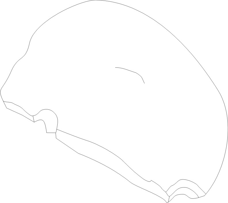
[0027] After the fresh bone flap is taken out in a clean environment, it is placed in the refrigerator at -10°C for 24 hours, and then rewarmed in a natural environment (about 20°C) for 48 hours; Ruptured, cells autolyzed. Use a brush to remove the residual periosteum, muscle, and fascia tissue on the surface of the skull, and rinse thoroughly with running water.

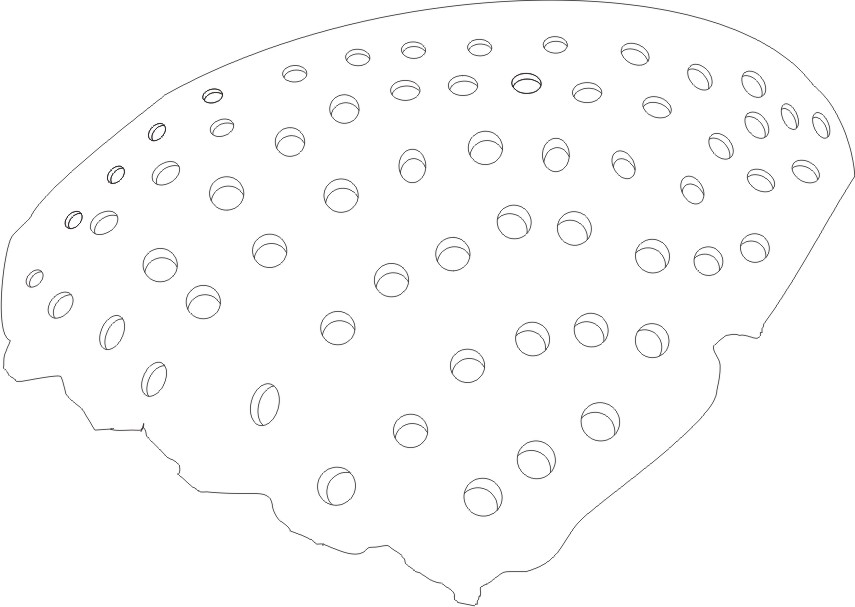
[0028] Use a drill bit with a diameter of 4mm to drill holes in the skull mesh, so that the area between the holes is about 1cm×1cm. When drilling, try to leave the bone suture as muc...


PUM


Abstract

The invention relates to the field of biology materials, in particular to a method for treating an externally-autoplastic autogenous skull sheet, which includes: 1 freezing fresh skull flap for 24 hours at temperature of -10 DEG C, rewarming for 48 hours in natural environment (about 20 DEG C) so as to break integrity of cells and lead the cells to be self-dissolved fully; 2 removing soft tissue on the surface of skull manually; 3 drilling holes on the skull in mesh mode and leading areas among holes to be 1cm*1cm; 4 soaking distilled water and trypsin for 48 hours at the temperature of 37 DEG C, changing the distilled water every 24 hours so as to fully precipitine an autogenous solution; and 5 soaking for 72 hours in medical hydrogen dioxide, changing the hydrogen dioxide every 24 hours to fully remove proteins in diploe; and soaking for 12 hours in the distilled water and then draining when no adverse reactions including infection, effusion and the like appear. Aiming at the problem that ankle joints are movable so that needle heads on the foot portion can move easily, the method combines special structure of the foot portion for fixing the foot portion so as to prevent liquid medicine from permeating into subcutaneous tissues.

Description

[0001] technical field [0002] The invention relates to the field of biomaterials, in particular to a processing method of bone materials that can be implanted in the body. [0003] Background technique [0004] In order to save the patient's life, neurosurgery often removes the patient's skull flap to reduce the patient's intracranial pressure and relieve the state of brain herniation. After the patient's condition is stable, the skull repair operation is performed. Currently commonly used repair materials generally use artificial materials (such as titanium alloy mesh) and autologous bone flap. The popularization and use of artificial materials is limited due to their deficiencies in shaping, pressure resistance, impact resistance, temperature insulation, rejection, and high price (about 20,000 yuan per repair material). The advantages of autogenous skull repair are difficult to achieve with other repair materials. The physical characteristics of the skull flap fully m...

Claims


Application Information

Patent Timeline
IPC IPC(8): A61L27/36
Inventor 李涛郑凤勤付留俊
Owner THE FIRST AFFILIATED HOSPITAL OF HENAN UNIV OF SCI & TECH
Why Eureka
Try Eureka